Download presentation
Presentation is loading. Please wait.

1
Focus on pathogen: T. vaginalis diagnosis/ID Morphology of troph: general overall pear shape, 10-23uM, with protruding axostyle on pointed end, 4 flagella in a group on opposite end near an obvious dark-staining nucleus, and an undulating membrane down the side. Troph may “round-up” in hypotonic urine and resemble WBCs. Very active, rhythmic, jerky, non-directional motility due to 4 anterior flagella and undulating membrane extending from the rounded anterior end to half-way down the body-length. A 5 th flagella may aid in direction. No other protozoa in urogenital specimens to be confused with T. vaginalis

3
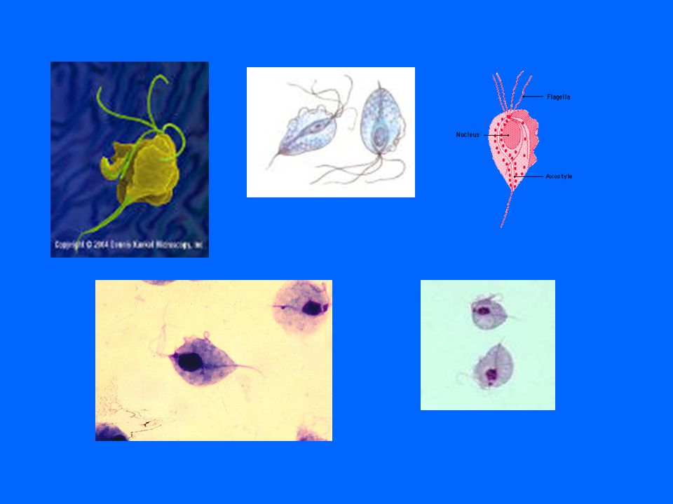
Non-pathogenic flagellates Trichomonas hominis Chilomastix mesnili

4
Trichomonas hominis T. hominis morphologically closely resembles T. vaginalis. The difference lies in the position of the undulating membrane on the side of the cell: a)T. hominis: the undulating membrane begins at the nuclear end and ends near the pointed end. b)T. vaginalis: the undulating membrane begins near the nucleus but ends somewhere midway between the nucleus and the pointed end. This membrane is very thin which makes it difficult at times to use as a diagnostic too, so…...
T. hominis: the undulating membrane begins at the nuclear end and ends near the pointed end. b)T. vaginalis: the undulating membrane begins near the nucleus but ends somewhere midway between the nucleus and the pointed end. This membrane is very thin which makes it difficult at times to use as a diagnostic too, so…....")
5
Trichomonas hominis The similar morphology will not lead to misidentification because T. hominis does not inhabit the urogenital tract and T. vaginalis cannot survive in feces. Motility of the two species are quite different, which almost assures the two will not be confused: a) T. hominis darts rapidly in wet mounts of feces b) T. vaginalis has jerky movements in urogenital specimens
 T. hominis darts rapidly in wet mounts of feces b) T. vaginalis has jerky movements in urogenital specimens.")
6
Trichomonas hominis

7
Chilomastix mesnili Commensal nonpathogenic intestinal flagellate Diagnostic features of troph in permanent stains and wet preparations Size: 6-20uM long, 5-15uM wide Round on one end, tapered to a point on the other Single nucleus on round end –peripheral chromatin may be visible –karyosome may be central or eccentric –may contain radiating chromatin filaments or may have granular appearance orbiting karyosome

8
Chilomastix mesnili MORE Diagnostic features of troph in permanent stains and wet preparations Cytostome emanating from round end near the nucleus and extending over half of body length Cytoplasm usually vacuolated Three flagella emanating from oral end and one in cytostome (difficult to see except in wet preps) Motility (wet prep) stiff and rotary Debbie?
 Motility (wet prep) stiff and rotary Debbie")
9
Chilomastix mesnili Diagnostic features of cyst in permanent stains and wet preparations: Size: 6-10uM long, 4-6uM wide Morphology: Lemon shaped oval with a knob or “handle” on one end Single nucleus similar to one in troph Curved fibril resembling a “shepherd’s crook”

10
Chilomastix mesnili

11
Blood and tissue protozoans Sarcodina: Naegleria fowleri Acanthamoeba species Mastigophora: Trypanosoma (circ), Leishmania (circ),
, Leishmania (circ),")
Similar presentations


 : Protozoa: 1- Protozoa are unicellular (eukaryotic) or acellular organisms. 2- Protozoan is measured in microns;>")


