Download presentation
Presentation is loading. Please wait.

1
PRESSURE ULCER CARE AND PREVENTION
Presented by: Dr. Naeem A. Chaudhry

2
Introduction Pressure ulcers are localized areas of tissue necrosis that tend to develop when soft tissue is compressed between a bony prominence and an external surface for a prolonged period of time.

3
Introduction Failure to provide appropriate pressure ulcer care may expose providers to significant liability; legal judgments against nursing homes in excess of fifty million dollars have occurred when patients develop an ulcer.

4
The consequences of pressure-induced skin injury range from nonblanchable erythema of intact skin to deep ulcers extending down to the bone. The ulcer imposes a significant burden not only on the patient, but the entire health care system.

5
Anatomy Largest organ of the body Weighs 6-8 pounds Varied thickness
Elastic Function Protection Transmits sensations Regulates body temperature Excretes waste Prevents excessive loss of body fluids

6
Physiology Three major levels Epidermis Dermis Subcutaneous tissue

7
Epidermis Thickness <1mm Avascular Keratinocytes Melanocytes
Langerhans cells

8
Dermis Middle layer Hair follicles, blood vessels, nerve endings, sweat glands Fibroblasts

9
Subcutaneous Tissue Innermost layer Mechanical and thermal insulation
Limited bloody supply

10
Aging and the Skin Fewer sweat glands Atrophy & thinning of all layers
Collagen/elastin fibers degenerate Atherosclerosis of cutaneous vessels Decrease in sebaceous glands Decrease in immune response Less elasticity Changes in thermoregulation

11
Staging Proposed by: National Pressure Ulcer Advisory Panel
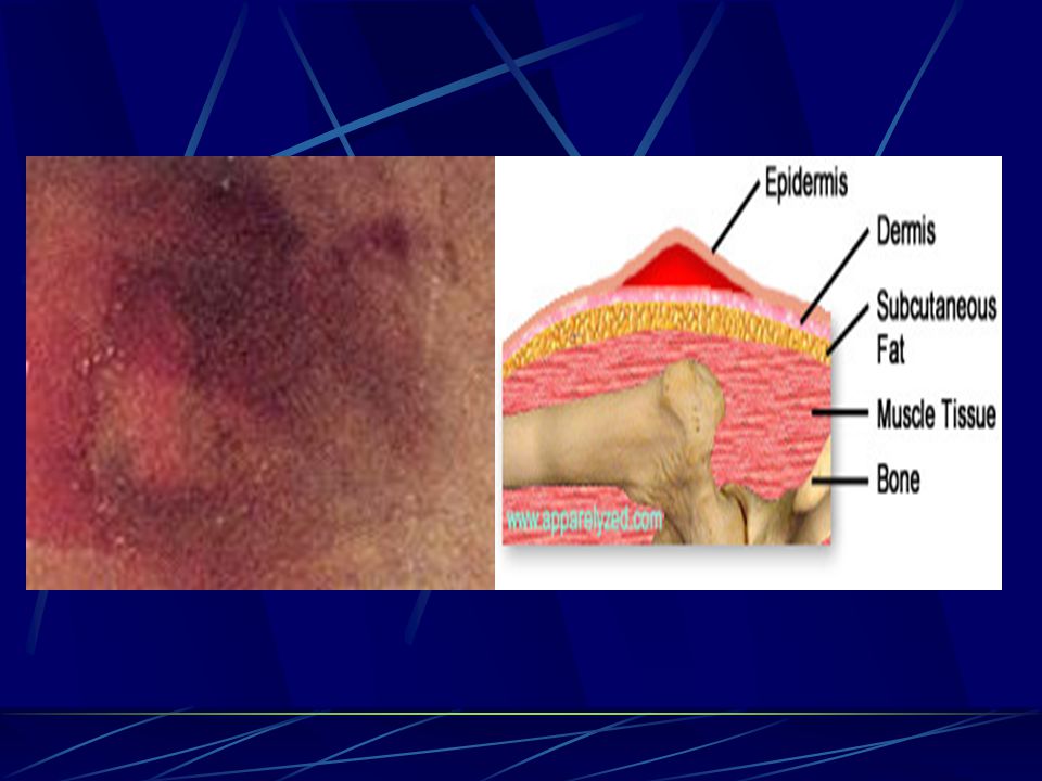
Stage 1-Observable pressure-related alteration of intact skin which when compared to an adjacent or opposite site area on the body Skin Temperature (warmth or coolness) Tissue Consistency (firm or boggy feel) Sensation (pain, itching)
 Tissue Consistency (firm or boggy feel) Sensation (pain, itching)")
12
Stage 1 cont… The ulcer appears as a defined area of persistent redness in lightly pigmented skin; in darker skin tones the ulcer may appear with persistent red, blue, or purple hues.

14
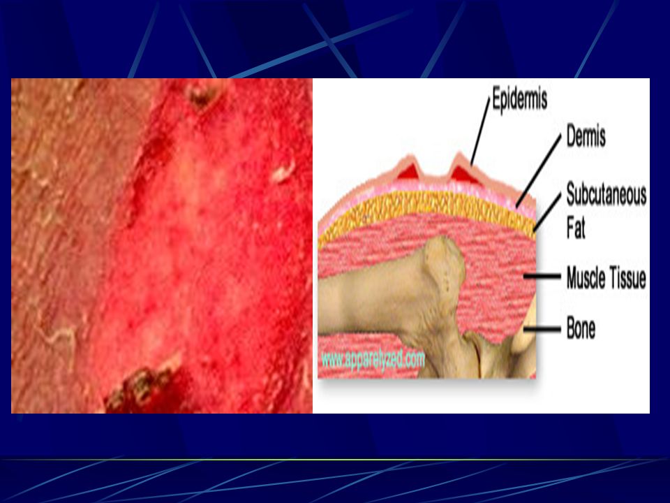
Stage 2 Characterized by a partial thickness skin loss involving the epidermis and/or dermis. The ulcer is superficial and presents clinically as an abrasion, blister, or shallow crater.

16
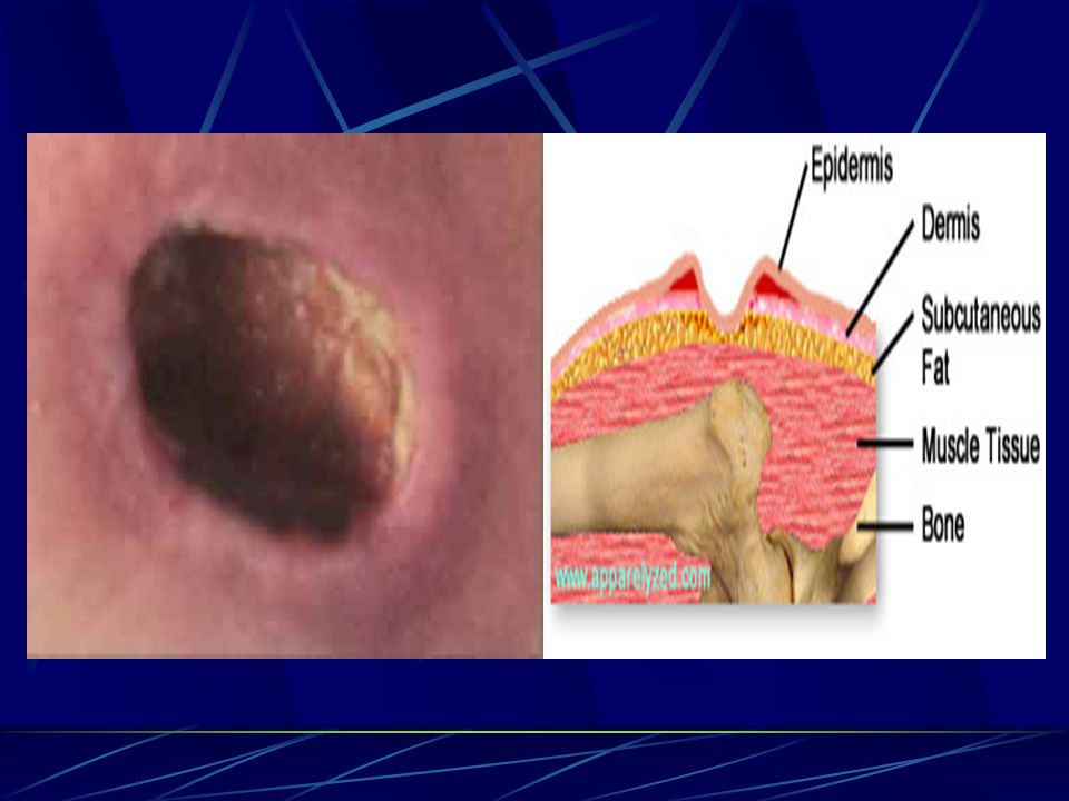
Stage 3 Characterized by a full thickness skin loss involving damage or necrosis of subcutaneous tissue which may extend down to, but not through the underlying fascia. The ulcer presents clinically as a deep crater with or without undermining of the adjacent tissue.

18
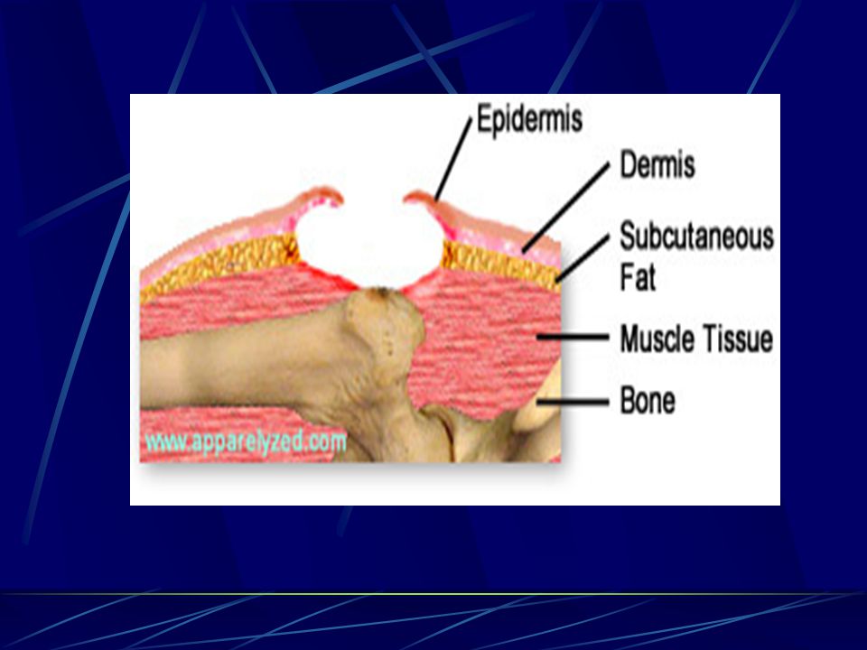
Stage 4 Characterized by a full thickness skin loss with extensive destruction, tissue necrosis, or damage to the muscle, bone, or supporting structures.

21
Staging cont… Pressure ulcers covered by eschar are not stageable.
Controversy exists over the staging and implications of “deep purple lesions,” particularly when localized over the heals. These lesions are believed to be indicative of deep tissue injury.

22
Epidemiology Prevalence rate for pressure ulcers have ranged in most studies from 3-14%, and incidence rates from 1 to 8 during the period of hospitalization. However, the rates may be considerably higher in select groups of hospital patients.

23
Pathogenesis Development of a pressure ulcer is a complex process that requires the application of external forces to the skin. However, external forces alone are not sufficient to cause an ulcer; their interaction with host-specific factors culminates in tissue damage.

24
External Factors Pressure Shearing forces Friction Moisture

25
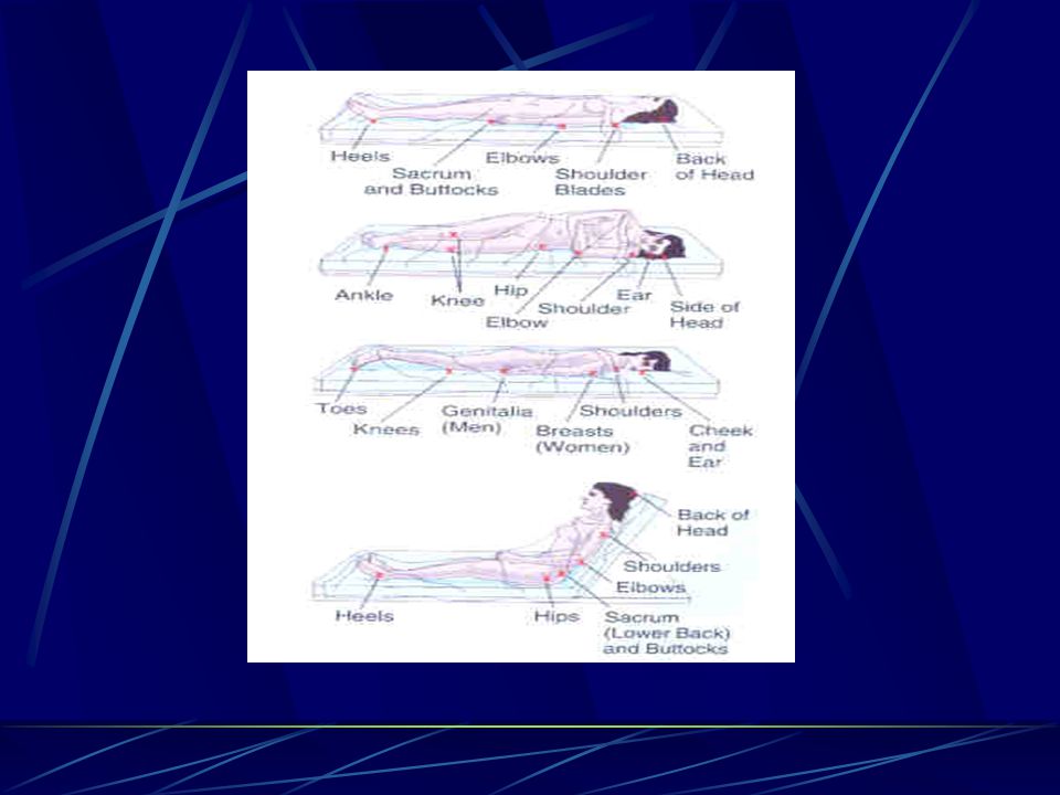
Pressure Pressure applied to the skin in excess of the arteriolar pressure (32mmHg) prevents the delivery of oxygen and nutrients to tissues, resulting in the accumulation of metabolic waste products. Pressures are greatest over bony prominences where weight-bearing points come in contact with external surfaces. A patient lying on a standard hospital mattress may generate pressures of 150mmHg; sitting produces pressures as high as 300 mmHg over the ischial tuberosities.
 prevents the delivery of oxygen and nutrients to tissues, resulting in the accumulation of metabolic waste products. Pressures are greatest over bony prominences where weight-bearing points come in contact with external surfaces. A patient lying on a standard hospital mattress may generate pressures of 150mmHg; sitting produces pressures as high as 300 mmHg over the ischial tuberosities.")
26
Pressure cont…. Pressure in excess of 70mmHg for 2 hours results in irreversible tissue damage in animal models. Pressure over a bony prominence tends to result in a cone-shaped distribution with the most affected tissues located deep, adjacent to the bone-muscle interface.

27
Pressure cont… Tissues vary in their susceptibility to pressure-induced injury; muscle is the most susceptible, followed by subcutaneous fat and then dermis. Thus, extensive deep tissue damage may occur with little or no evidence of superficial tissue injury. A deep necrotic wound may be the first evidence of pressure-induced injury, rather than a gradual progression of an ulcer from stages 1 through 4.

30
Shearing Forces Shearing forces occur when patients are placed on an incline. Deeper tissues, including muscle and subcutaneous fat, are pulled downwards by gravity, while the superficial epidermis and dermis remain fixed through contact with the external surface. The result is stretching and angulation of local blood vessels and lymphatics. Shear forces alone may not cause ulceration, but appear to have an additive effect so that in the presence of pressure, more severe tissue damage will occur.

31
Friction Occurs when patients are dragged across an external surface. This results in an abrasion with damage to the most superficial layer of skin. Friction is most likely to result in stage 2 pressure ulcers since it does not cause the necrotic changes associated with deep tissue injury; limited contribution to the development of stage 3 and 4 ulcers.

32
Moisture Exposure to moisture in the form of perspiration, feces, or urine may lead to skin maceration and predispose to superficial ulceration.

33
Host Factors Immobility Incontinence Nutritional status Skin perfusion
Neurologic diseases Other factors

34
Immobility Immobility is one of the most important host factors that contributes to pressure ulcer development. Immobility may be permanent or transient.

35
Incontinence Urinary incontinence is frequently cited as a predisposing factor for pressure ulcers. Some studies suggest that incontinent patients have up to a five-fold higher risk for pressure ulcer development.

36
Nutritional Status The role of nutritional status in the development of pressure ulcers is uncertain. Animal studies have found that more severe pressure-induced skin destruction occurred in malnourished animals than in well nourished animals exposed to similar amounts of pressure. In addition, cross-sectional studies have suggested that patients with pressure ulcers are more likely to have hypoalbuminemia.

37
Skin Perfusion Contributing factors to the development of tissue ischemia have been postulated to include hypotension, dehydration, vasomotor failure, and vasoconstriction secondary to shock, heart failure, or medications. When vital organs such as the kidneys and gastrointestinal tract are not receiving adequate perfusion, it is likely that blood flow to the skin will also be decreased, which increases the risk for the development of pressure ulcers.

38
Neurologic Diseases Neurologic diseases such as dementia, delirium, spinal cord injury, and neuropathy are important contributors to pressure ulcer development. This may in large part be related to immobility, spasticity, and contractures that are common in these conditions. Sensory loss is also common, suggesting that patients may not perceive pain or discomfort arising from prolonged pressure.

39
Other Factors Other factors identified in some studies are: older age, white race,, and male gender. Specific diagnoses that have been associated with ulcer development include the presence of dry skin, recent lower extremity fractures, diabetes, and cardiovascular disease.

40
Identification of Patients At Risk
Knowledge of factors contributing to the pathogenesis of pressure ulcers allow the identification of patients at risk for ulcer development. Preventive interventions may then be targeted to those specific patients.

41
Identification cont…. Two general approaches have been used when developing prediction rules that allow the identification of patients at high-risk for pressure ulcers. The first approach involves use of clinical judgment to determine patient characteristics and their associated weights. Assessment of risk for pressure ulcer development is not a one time activity. Patients should be reassessed periodically, particularly when there is a change in health status.

42
Braden Scale The Braden scale rates patients in six subscales:
sensory perception moisture activity mobility nutrition friction and shear using scores ranging from 1 to 3 or 4. The maximum total score is 23; a score of 18 or less indicates high-risk.

43
Clinical Manifestation and Diagnosis
Pressure ulcers are usually easy to identify by their appearance and location overlying a bony prominence. The exception may be stage 1 ulcers, which can be difficult to recognize, particularly in patients with darkly pigmented skin. They also may be confused with other conditions that cause erythema such as cellulitis.

44
Clinical Manifestation cont….
Eschar often covers deep ulcers, making it difficult to determine whether lesions are stage 3 or 4. In addition, the extent of stage 4 ulcers is often underestimated due to undermining and fistula formation; a relatively small superficial skin defect may mask extensive deep tissue necrosis.

45
Complications Pressure ulcers may be associated with both medical and psychosocial complications. The medical complications can be life threatening and are more common with stage 3 and 4 ulcers. Psychosocial consequences are not often considered. However, patients with pressure ulcers may suffer pain and feel stigmatized by the development of chronic skin ulcer. This could result in depression, social isolation, and decrements in overall health-related quality of life.

47
Infections Infection is common among patients with pressure ulcers occasionally leading to bacteremia, sepsis, and death. Pressure ulcers may also pose a risk to other hospitalized patients by serving as a reservoir for resistant organisms such as methicillin-resistant Staphylococcus aureus, vancomycin-resistant enterococci, and multiply-resistant gram negative bacilli.

48
Other Complications Sinus tracts may develop that communicate with the deep viscera including the bowel or bladder. Heterotrophic calcification also occasionally occurs. The chronic inflammatory state arising from the ulcer may result in systemic amyloidosis. Cellulitis and Osteomyelitis Squamous cell carcinoma occasionally develops in a pressure ulcer and should always be considered in patients with a non-healing wound.

49
Healing Process Three stages Inflammation Proliferative (regenerative)
Maturation (remodelling)
")
50
Inflammation Vasoconstriction for hemostasis Vasodilation follows
Increases vessel permeability Leakage of plasma and leukocytes

51
Inflammation Neutrophils clean up wound Macrophages digest bacteria
digest necrotic tissue digest dead neutrophils release growth factors

52
Inflammation Clinical signs & symptoms Local erythema Warmth Edema
Pain or tenderness Increased wound drainage

53
Proliferative Granulation tissue by collagen synthesis and angiogenesis Wound contraction by myofibroblasts Epithelization Requires moist wound surface Clinical signs & symptoms Beefy red Bright pink granules

54
Maturation Fibroblasts decrease Collagen re-organizes
Vascularization decreases May take up to 2 years to complete Clinical signs & symptoms Scar tissue softens Becomes thinner and paler

55
Prevention A comprehensive history and physical examination can identify potentially correctable predisposing factors for patients who are determined to be at risk of developing pressure ulcers. Specific interventions may then be initiated depending upon the measured level of risk and the patient’s individual needs.

56
Pressure Relief Patient positioning Pressure reducing devices

57
Patient Positioning Regular turning schedule of 2 hours is recommended
Patient should be placed at a 30 degree angle to avoid direct pressure over the greater trochanter Pillows or foam wedges may need to be placed between the ankles and knees

58
Patient Positioning cont..
The heels require particular attention; pillows may be placed under the lower legs to elevate the heels, or special heel protectors can be used Elevation of the head of the bed should be limited to minimize exposure to shear forces

59
Pressure Reducing Devices
Static devices consist of overlays and mattresses that are made of or contain gel, foam, air, or water Dynamic support systems use a power source to alternate air currents in order to regulate or redistribute pressure against the body

60
Other Interventions Patients immobilized from a hip fracture or stroke may benefit from physical therapy Severe spasticity may be relieved with muscle relaxant drugs or a nerve block Medications contributing to immobility (eg. sedatives) should be stopped
 should be stopped.")
61
Other Interventions cont…
Proper skin care should be provided, including cleansings at regular intervals and minimizing exposure to moisture Massage over bony prominences should be avoided Patients should not be dragged in bed Adequate nutrition should be provided

62
Efficacy A survey of pressure ulcer experts indicated that 62% disagreed with the statement “all pressure ulcers are preventable”. It is easy to understand why some failures will occur considering that a single bed-bound patient must be repositioned over 4000 times a year. Good preventive care may result in significant cost savings. One study in an ICU found that they were able to save $700 per patient.

63
Treatment Wound should be evaluated for stage, size, sinus tracts, necrotic tissue, exudate, and presence of granulation Photographs of all wounds should be considered Adequate pain relief

64
Treatment Dressings Debridement Local antibiotic

65
Dressing Categories Gauze dressing Transparent film Hydrogels
Hydrocolloids Alginate

66
Gauze Dressing Filler for dead space Absorber
Cleansing material (patting not rubbing) Carrier for medication NOT for WET-TO-DRY dressings!!!
 Carrier for medication. NOT for WET-TO-DRY dressings!!!")
67
Transparent Film Stage 1 Shallow wounds with minimal exudate
Waterproof site May protect from friction and shear Promotes autolytic debridement

68
Hydrogels Stage 2, 3 Light exudate Limited absorptive capability
Easy to apply and remove Does not leave residue in wounds

69
Hydrocolloids Stage 2, 3, 4 Small to moderate exudate
May prevent contamination Moderate to heavy exudate Moisture may also reduce pain to nerves

70
Alginate Stage 2, 3, 4 High capacity for absorption
Interacts with wound fluids to form a gel creating a moist wound environment Apply within wound borders

71
Wound Debridement Mechanical Sharp Enzymatic Autolytic

72
Mechanical Debridement
Includes the use of wet-to-dry dressings, hydrotherapy, wound irrigation, and scrubbing the wound with gauze. Best for wounds that contain thick exudate, slough, or loose necrotic tissue. Wet-to-dry dressings will remove both nonviable and viable tissues. Moistening the dressing before removal should be avoided since it will limit the debriding effects.

73
Sharp Debridement Involves the use of a scalpel or scissors in the operating room or at the bedside. This is the most rapid form of debridement; it is indicated when there is evidence of cellulitis or sepsis and is also used to remove thick eschar and extensive necrotic tissue.

74
Enzymatic Debridement
Uses the topical application of agents such as collagenase, papain, fibrinolysin, and deoxyribonuclease which is effective in promoting the growth of granulation tissue. These agents are particularly useful in long-term care settings and in patients who may not tolerate surgery.

75
Autolytic Debridement
Uses an occlusive dressing to cover a wound so that necrotic tissue is digested by enzymes normally present in wound tissue. This often works best on wounds with minimal exudate and should not be used in the presence of infection. Debridement should stop once necrotic tissue has been removed and granulation tissue is present.

76
Surgery Necessary in some patients, particularly in those whose quality of life would be improved
Direct closure of the wound Skin grafts Skin flaps Musculocutaneous flaps Free flaps

77
Duration of Wound Healing
Stage 1: 1-7 days Stage 2: days Stage 3: days Stage 4: days

Similar presentations


 A pressure ulcer is localized injury to the skin and/or underlying.>")


