Download presentation
Presentation is loading. Please wait.

1
Proper pressures in the DR Proper FiO2 in the DR (blended) Surfactant in the DR CPAP in the DR Consistent CPAP in the NICU Reduced SIMV in the NICU ‘Golden Hour’ Lung Protective Strategy from Birth
 Surfactant in the DR CPAP in the DR Consistent CPAP in the NICU Reduced SIMV in the NICU ‘Golden Hour’ Lung Protective Strategy from Birth")
3
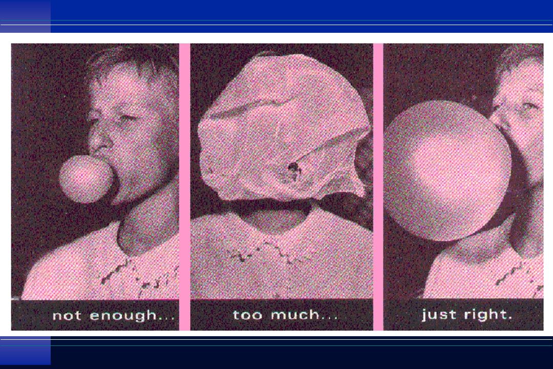
good judgement informed jugement

4
Neo-Puff in the DR manual ventilation of babies <30 weeks gest. Used for all transport ventilation for all babies

5
easy to use, manually operated gas-powered. Neo-Puff Infant Resuscitator

6
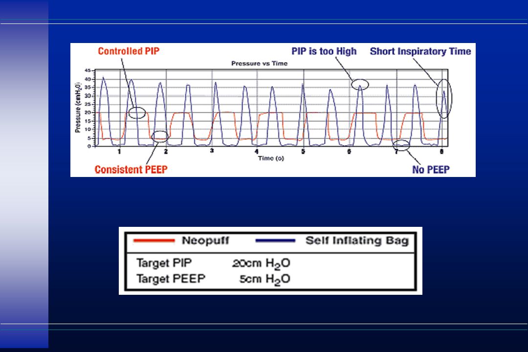
Controlled and Precise Peak Inspiratory Pressure (PIP) The Neopuff™ Infant Resuscitator will inflate the baby’s lungs & provide optimum oxygenation by delivering consistent PIP with each breath, limiting the risks associated with under or over inflation at uncontrolled pressures. Consistent and Precise Positive End Expiratory Pressure (PEEP) The Neopuff™ Infant Resuscitator maintains Functional Residual Capacity (FRC) by providing a consistent PEEP throughout the resuscitation process.
 The Neopuff™ Infant Resuscitator maintains Functional Residual Capacity (FRC) by providing a consistent PEEP throughout the resuscitation process..")
7
The desired PIP is set by turning the inspiratory pressure control. The desired PEEP is set by adjusting the T-piece aperture.

10
Pressure/Volume

11
Over Weaning damages too

12
Ventilator-Associated Lung Injury u Barotrauma (air leak) u Oxygen toxicity u Ventilator associated pneumonia u Over-distention u De-recruitment
 u Oxygen toxicity u Ventilator associated pneumonia u Over-distention u De-recruitment")
13
Slutsky and Tremblay Am J Respir Crit Care Med 1998; 157: 1721-1725 MOSF Death Shear Overdistention Cyclic stretch Inc. intrathoracic pressure Inc alveolar cap permeability Dec cardiac output Dec organ perfusion Tissue injury secondary to Inflamatory mediators/cells Impaired O2 delivery bacteremia Cytokines, prostanoids, Leukotrienes, reactive oxygen species, protease neutrophil Distal Organs Biochemical Injury Biophysical Injury

14
Dreyfuss, Am J Respir Crit Care Med 1998;157:294-323 normal lungs 5 min of 45 cm H 2 O 20 min of 45 cm H 2 O

15
Webb and Tierney, Am Rev Respir Dis 1974; 110:556-565 14/0 45/10 45/0

17
esophageal intubation

18
u Pulmonary u Interstitial u Emphesema u to Pneumo-

20
Assessment Chest x-ray AP 8 rib conventional 9-10 rib Hi-Fi Rise & fall of chest ( slight per NRP ) Listen to breath sounds Vt 5-7 ml/kg (3-5 spont.) follow ABGs
 Listen to breath sounds Vt 5-7 ml/kg (3-5 spont.) follow ABGs")
21
Pressure Wave To Increase Mean Airway Pressure 1. Increase flow 2. Increase peak pressure 3. Lengthen inspiratory time 4. Increase PEEP 5. Increase Rate

23
TYPES OF MECHANICAL VENTILATION o negative pressure ventilation o positive pressure ventilation o high-frequency ventilation o non-invasive positive pressure ventilation

24
Body Box:

25
Outline u Respiratory mechanics and gas exchange u Factors affecting oxygenation and carbon dioxide elimination during mechanical ventilation u Blood gas analysis u Ventilatory management: basics and specifics u High frequency ventilation: the basics

26
Overview u Mechanical ventilation is an integral part of neonatal intensive care, and has led to increased survival of neonates over the last 3 decades u Advances in knowledge of neonatal respiratory physiology have led to optimization of techniques and strategies u Conventional mechanical ventilation (CMV) is most often used, despite the advent of HFV and SIMV
 is most often used, despite the advent of HFV and SIMV")
27
Overview u Respiratory failure in neonates has significant morbidity and mortality (although less than in the past) u Optimal ventilatory management will reduce the risk of chronic lung disease u Optimal ventilatory management should be individualized and be based upon the pathophysiology and certain basic concepts of mechanical ventilation
 u Optimal ventilatory management will reduce the risk of chronic lung disease u Optimal ventilatory management should be individualized and be based upon the pathophysiology and certain basic concepts of mechanical ventilation")
28
Concepts u Goal of mechanical ventilation: to improve gas exchange and to sustain life without inducing lung injury u Factors that should influence ventilator adjustment decisions: l Pulmonary mechanics l Gas exchange l Control of breathing l Lung injury

29
Pulmonary mechanics u Compliance l Property of distensibility of the lungs and chest wall l Change in volume per unit change in pressure C = Volume C = Volume Pressure Pressure l Neonatal lung u Normal 0.003-0.006 L/cm H 2 O u with RDS 0.0005-0.001 L/cm H 2 O

30
Pulmonary mechanics u Resistance: l inherent capacity of the air conducting system (airways and ETT) and tissues to resist airflow l Change in pressure per unit change in flow R = Pressure R = Pressure Flow Flow Total cross-sectional area of airways Total cross-sectional area of airways Resistance Length of the airways Flow rate Flow rate Density and viscosity of gas
 and tissues to resist airflow l Change in pressure per unit change in flow R = Pressure R = Pressure Flow Flow Total cross-sectional area of airways Total cross-sectional area of airways Resistance Length of the airways Flow rate Flow rate Density and viscosity of gas")
31
Pulmonary mechanics u Location of airway resistance: 0 5 10 15 20 0 5 10 15 20 u Distal airways contribute less to resistance due to increased total cross-sectional area u Small ETT and high flow rates can increase resistance markedly Resistance Resistance Airway Generation Distal -->

32
Pulmonary mechanics u Laminar flow (Distal airways) l Driving pressure proportional to flow l R= 8 n l (n = viscosity ; l = length; r = radius) r 4 r 4 u Turbulent flow (Proximal airways) l Driving pressure proportional to square of flow l Reynolds number (Re) = 2 r V d (d = density) n
 l Driving pressure proportional to flow l R= 8 n l (n = viscosity ; l = length; r = radius) r 4 r 4 u Turbulent flow (Proximal airways) l Driving pressure proportional to square of flow l Reynolds number (Re) = 2 r V d (d = density) n")
33
Pulmonary mechanics u A pressure gradient between the upper airway and alveoli is necessary for gas flow during inspiration and expiration u The pressure gradient is required to overcome the elasticity, resistance, and inertance of the respiratory system u Equation of motion: P = 1 V + R V + I V C C Elasticity+Resistance+Inertance

34
Pulmonary mechanics u Time constant l The time taken for the airway pressure (and volume) changes to equilibrate throughout the lung is proportional to the compliance and resistance of the respiratory system l Time constant = Compliance x Resistance
 changes to equilibrate throughout the lung is proportional to the compliance and resistance of the respiratory system l Time constant = Compliance x Resistance")
35
Pulmonary mechanics u % change in pressure in relation to time u Almost full equilibration: 3-5 time constants 100 80 60 40 20 0 1 2 3 4 5 Time constants Change in pressure (%) 63 86 95 98 99
")
36
Pulmonary mechanics u Healthy term neonate: l C = 0.004 L/cm H 2 O; R = 30 cm H 2 O/L/sec l T = 0.004 x 30 = 0.12 sec u Time constants Time (sec) % equilibration 10.1263 20.2486 30.3695 50.6099 u RDS: Shorter time constant
 % equilibration u RDS: Shorter time constant")
37
Pulmonary mechanics u Application of the concept of time constant l Short T I : decreased tidal volume delivery l Inadequate T E : Gas trapping ( FRC, inadvertent PEEP) l Heterogeneous lung disease (BPD): different regions of the lung have different time constants; tendency for atelectasis and hyperexpansion to co-exist
 l Heterogeneous lung disease (BPD): different regions of the lung have different time constants; tendency for atelectasis and hyperexpansion to co-exist")
38
Gas exchange u Total minute ventilation = tidal vol x freq l V E = V T x f u Alveolar ventilation (V A ) = Useful (fresh gas) portion of minute ventilation that reaches gas exchange units; excludes dead space (V D ) l V A = (V T -V D ) x f l Alveolar ventilation equation: V A (L/min) = V CO2 (ml/min) x 0.863 (BTPS P A CO2 (mm Hg) corr.)
 = Useful (fresh gas) portion of minute ventilation that reaches gas exchange units; excludes dead space (V D ) l V A = (V T -V D ) x f l Alveolar ventilation equation: V A (L/min) = V CO2 (ml/min) x (BTPS P A CO2 (mm Hg) corr.)")
39
Gas exchange u Alveolar gas equation: l If R=1, each molecule of O 2 removed from alveoli is replaced by one molecule of CO 2 P A O2 = P I O2 - P A CO2 l Average normal value for R = 0.8 P A O2 = F I O2 x (P B -P H2O ) - P A CO2 x F I O2+ 1- F I O2 R R l Pa CO2 = effective P A CO2 l True P A CO2 = P ET CO2
 - P A CO2 x F I O2+ 1- F I O2 R R l Pa CO2 = effective P A CO2 l True P A CO2 = P ET CO2")
40
Gas exchange u Ventilation-Perfusion matching: matching of gas flow and blood flow required for successful gas exchange l V A = Alveolar ventilation Q Pulmonary blood flow (Fick method: O 2 ) Q Pulmonary blood flow (Fick method: O 2 ) = 0.863 x R x (Ca O2 - C V O2 ) = 0.863 x R x (Ca O2 - C V O2 ) P A CO2 V/Q mismatching usually relevant to effect on alveolar-arterial P O2 difference: (A-a) P O2 V/Q mismatching usually relevant to effect on alveolar-arterial P O2 difference: (A-a) P O2
 Q Pulmonary blood flow (Fick method: O 2 ) = x R x (Ca O2 - C V O2 ) = x R x (Ca O2 - C V O2 ) P A CO2 V/Q mismatching usually relevant to effect on alveolar-arterial P O2 difference: (A-a) P O2 V/Q mismatching usually relevant to effect on alveolar-arterial P O2 difference: (A-a) P O2")
41
Gas exchange u O 2 -CO 2 diagram 406080100120140160 0204060 P CO 2 P O 2 (mm Hg) V/Q = 8 I V/Q = 0 0.2 1.0 1.5 0.5 Ideal V/Q = 0.84 v
 V/Q = 8 I V/Q = Ideal V/Q = 0.84 v")
42
Gas exchange u Causes of hypoxemia l V/Q mismatch l Right to left shunt (venous admixture) l Hypoventilation (e.g. in apnea) l Diffusion abnormalities u Causes of hypercapnia l Hypoventilation l Severe V/Q mismatch
 l Diffusion abnormalities u Causes of hypercapnia l Hypoventilation l Severe V/Q mismatch.")
43
Gas exchange u Factors involved in gas exchange during mechanical ventilation l Oxygenation l Carbon dioxide elimination l Gas transport mechanisms l Patient - ventilator interactions

44
Gas exchange u Factors affecting oxygenation l Mean airway pressure (MAP) : affects V/Q matching. MAP is the average airway pressure during respiratory cycle MAP = K (PIP-PEEP) [T I / (T I +T E )] + PEEP l Oxygen concentration of inspired gas (F I O2 )
 [T I / (T I +T E )] + PEEP l Oxygen concentration of inspired gas (F I O2 ).")
45
Gas exchange u MAP increases with increasing PIP, PEEP, T I to T E ratio, rate, and flow PEEP PIP TITI Rate Flow Pressure Time TITI TETE PEEP PIP

46
Gas exchange u Relation of MAP to Pa O 2 not linear; is like an inverted “U”: l Low MAP: u Atelectasis--> very low Pa O 2 l High MAP: u hyperinflation--> V/Q mismatch; intrapulmonary shunt, hypoventilation due to distended alveoli u decreased cardiac output --> decreased oxygen transport despite adequate Pa O 2

47
Gas exchange u For the same change in MAP, changes in PIP and PEEP improve oxygenation more than changes in I:E ratio u Reversed I:E ratios increase risk of air-trapping u PEEP levels higher than 6 cm H 2 O may not improve oxygenation in neonates u Attainment of optimal MAP may allow weaning of F I O2 u Atelectasis may lead to sudden increase in required F I O2

48
Gas exchange u Carbon dioxide elimination l Proportional to alveolar ventilation (V A ) which depends on tidal volume (V T ) and frequency (rate) l V T changes more effective (but more barotrauma) : dead space constant, so proportion of V T that is alveolar ventilation increases to a greater degree with increases in V T u V T 4 --> 6cc/kg (50% ) with dead space of 2 cc/kg increases V A from 2 (4-2) to 4 (6-2) cc/kg/breath (100% )
 which depends on tidal volume (V T ) and frequency (rate) l V T changes more effective (but more barotrauma) : dead space constant, so proportion of V T that is alveolar ventilation increases to a greater degree with increases in V T u V T 4 --> 6cc/kg (50% ) with dead space of 2 cc/kg increases V A from 2 (4-2) to 4 (6-2) cc/kg/breath (100% )")
49
Gas exchange u Clinical estimation of optimal T I and T E : Short T I Optimal T I Long T I Inadeq V T Short insp. plateau Long plateau Short T E Optimal T E Long T E Air trapping Short exp. plateau Long exp. plateau Chest Wall Motion Time Chest Wall Motion

50
Gas exchange u Synchrony vs. Asynchrony + “fighting” l Synchrony augments ventilation, improves CO 2 elimination, decreases hypoxic episodes l Asynchrony leads to poor tidal volume delivery, and impairs gas exchange l Active exhalation (exhalation during ventilator breath) increases risk of hypoxic episodes
 increases risk of hypoxic episodes.")
51
Blood gas analysis u Arterial blood gas analysis the “gold standard” u Interpretation: l pH: Is it normal, acidotic, or alkalotic? l P CO 2 : Is it normal, (respiratory acidosis), or (respiratory alkalosis)? l HCO 3 : Is it normal, (metabolic acidosis), or (metabolic alkalosis)? l Simple disorder or mixed? Compensated or not? l P O 2 : Normal, hypoxia, or hyperoxia?
, or (respiratory alkalosis). l HCO 3 : Is it normal, (metabolic acidosis), or (metabolic alkalosis). l Simple disorder or mixed. Compensated or not. l P O 2 : Normal, hypoxia, or hyperoxia .")
52
Blood gas analysis u Normal values (1 hr age, not ventilated) l Preterm: pH 7.28-7.32, P CO 2 35-45, P O 2 50-80 l Term: pH 7.30-7.35, P CO 2 35-45, P O 2 80-95 u Target values l RDS: pH > 7.25, P CO 2 45-55, P O 2 50-70 l BPD: pH > 7.25, P CO 2 45-70, P O 2 60-80 l PPHN: pH 7.50-7.60, P CO 2 25-40, P O 2 80-120 u Remember! O 2 content determined mostly by SpO 2 and Hb%.

53
Blood gas analysis u Common errors: l Infrequent ventilator adjustments made only when ABG (q4/q6) is obtained. In acute phase of RDS or PPHN, adjustments should be made with chest rise, SpO 2, TcP O 2 /P CO 2 trends l Room air contamination: P CO 2, P O 2 (if <150 torr ). Amount in butterfly set sufficient ! l Liquid heparin /saline contamination: pH same, but lower P CO 2 (mimics compensated metabolic acidosis)
. Amount in butterfly set sufficient . l Liquid heparin /saline contamination: pH same, but lower P CO 2 (mimics compensated metabolic acidosis).")
54
Ventilatory management u Indications: l Clinical: Absolute: Apnea (intractable), gasping, cyanosis not responsive to O 2 by hood Relative: Severe tachypnea / retractions l Laboratory (while on CPAP or FiO 2 > 0.7): pH 60 pH 60 (or) P O 2 < 45- 50 and / or SpO 2 < 85 (or) P O 2 < 45- 50 and / or SpO 2 < 85 l Other: Surgical procedures, compromised airway
, gasping, cyanosis not responsive to O 2 by hood Relative: Severe tachypnea / retractions l Laboratory (while on CPAP or FiO 2 > 0.7): pH 60 pH 60 (or) P O 2 < and / or SpO 2 < 85 (or) P O 2 < and / or SpO 2 < 85 l Other: Surgical procedures, compromised airway")
55
Ventilator settings u PIP: l affects MAP (P O 2 ) and V T (P CO 2 ) l PIP required depends largely on compliance of respiratory system l Clinical: gentle rise of chest with breath, similar to spontaneous breath l Minimum effective PIP to be used. No relation to weight or airway resistance l Neonate with RDS: 15-30 cm H 2 O. Start low and increase.

56
Ventilator settings u PEEP: l affects MAP (P O 2 ), affects V T (P CO 2 ) depending on position on P-V curve l older infants (e.g. BPD) tolerate higher levels of PEEP (6-8 cm H 2 O) better l RDS: minimum 2-3, maximum 6 cm H 2 O. Pressure Volume PEEP PIP
 tolerate higher levels of PEEP (6-8 cm H 2 O) better l RDS: minimum 2-3, maximum 6 cm H 2 O. Pressure Volume PEEP PIP.")
57
Ventilator settings u Rate: l affects minute ventilation (P CO 2 ) l In general, rate ---> P CO 2 l Rate changes alone do not alter MAP (with constant I:E ratio) or change P O 2, unless PVR changes with changes in pH l However, i f rate --> T E gas trapping--> decreased V T --> P CO 2 l Minute ventilation plateaus, then falls with rate
 l In general, rate ---> P CO 2 l Rate changes alone do not alter MAP (with constant I:E ratio) or change P O 2, unless PVR changes with changes in pH l However, i f rate --> T E gas trapping--> decreased V T --> P CO 2 l Minute ventilation plateaus, then falls with rate")
58
Ventilator settings u T I and T E : l Need to be 3-5 TC for complete inspiration and expiration (Note: TC exp = TC insp) l Usual ranges: T I secT E sec u RDS 0.2-0.45 0.4-0.6 u BPD 0.4-0.80.5-1.5 u PPHN 0.3-0.80.5-1.0 l Chest wall motion / V T may be useful in determining optimal T I and T E
 l Usual ranges: T I secT E sec u RDS u BPD u PPHN l Chest wall motion / V T may be useful in determining optimal T I and T E")
59
Ventilator settings u I : E Ratio l When corrected for the same MAP, changes in I:E ratio do not affect gas exchange as much as changes in PIP or PEEP l Changes in T I or T E do not change V T or P CO 2 unless they are too short (< 3 TC) l Reversed I:E ratio: No change in mortality or morbidity noted in studies. Not often used. May improve V/Q matching and P O 2 at risk of venous return and gas trapping

60
Ventilator settings u Fi O2 l affects oxygenation directly l with Fi O2 <0.6-0.7, risk of oxygen toxicity less than risk of barotrauma l to improve oxygenation, increase Fi O2 to 0.7 before increasing MAP l during weaning, once PIP is low enough, reduce Fi O2 from 0.7 to 0.4. Maintenance of adequate MAP and V/Q matching may permit a reduction in Fi O2

61
Ventilator settings u Flow: l affects pressure waveform l minimal effect on gas exchange as long as sufficient flow used l increased flow--> turbulence l higher flow required if TI short, to maintain TV l flow of 8-10 lpm usually sufficient l change of flow may affect delivery of NO or anesthesia gases

62
Ventilatory management u RDS: l Pathology: decreased compliance, FRC l Once diagnosis established, and if P O2 <50 on 40% oxygen: CPAP (or) early intubation and surfactant. (Prophylactic CPAP for ELBW not useful) l Ventilation if Fi O2 > 0.7 required on CPAP l Surfactant q 6 hrs if intubated and Fi O2 > 0.3- 0.4 (Survanta / Infasurf / Curosurf better than Exosurf). Usually 1-2, rarely 4 doses required.
 l Ventilation if Fi O2 > 0.7 required on CPAP l Surfactant q 6 hrs if intubated and Fi O2 > (Survanta / Infasurf / Curosurf better than Exosurf). Usually 1-2, rarely 4 doses required..")
63
Ventilatory management u RDS (continued): l Use lowest PIP required l moderate PEEP (4-5 cm H 2 O) l permissive hypercarbia (Pa CO2 45-55 mmHg instead of 35-45 is safe, and need for ventilation in first 4 days) l limited use of paralysis, aggressive weaning l chest PT not useful, maybe dangerous in acute phase (increases IVH)
: l Use lowest PIP required l moderate PEEP (4-5 cm H 2 O) l permissive hypercarbia (Pa CO mmHg instead of is safe, and need for ventilation in first 4 days) l limited use of paralysis, aggressive weaning l chest PT not useful, maybe dangerous in acute phase (increases IVH)")
64
Ventilatory management u Chronic lung disease / BPD: l usually heterogeneous lung disease - different areas of lung with different time constants l increased resistance, frequent exacerbations l higher PEEP often helpful (4-7 cm H 2 O) l longer T I and T E, with low rates l hypercarbia and compensated respiratory acidosis often tolerated to avoid increased lung injury
 l longer T I and T E, with low rates l hypercarbia and compensated respiratory acidosis often tolerated to avoid increased lung injury")
65
Ventilatory management u PPHN: l ventilator management controversial l Fi O2 adjusted to maintain Pa O2 80-100 to minimize hypoxia-mediated pulmonary vasoconstriction l ventilatory rates and pressures adjusted to maintain mild alkalosis (pH 7.5-7.6), usually combined with bicarbonate infusion l avoid low Pa CO2 (<20 mm Hg) to prevent cerebral vasoconstriction
, usually combined with bicarbonate infusion l avoid low Pa CO2 (<20 mm Hg) to prevent cerebral vasoconstriction")
66
Volume Guarantee The ventilator automatically adjusts the inspiratory pressure according to changes of compliance, resistance or respiratory drive.

67
Pressure Support Ventilation Working Principle of Breath Termination Erin Browne

68
Flow Sensor Measurement Principle u Two tiny platinum wires are heated to 400°C u Gas flow cools the wire down u From the amount of cooling the amount of gas flowing can be calculated T = 400°C no gas flow with gas flow

69
Endotracheal Tube Leak

70
Lung Function Monitoring Clinical Applications u Identification of Lung Overdistention u Prediction of successful extubation u Prediction of risk of BPD development u Response to Surfactant or Brochodilators u Teaching tool u Titration of optimal PEEP u Trend in development of disease u Check of compliance during HFV u recognition of recovery from suctioning

73
OSCILLATOROSCILLATOR

75
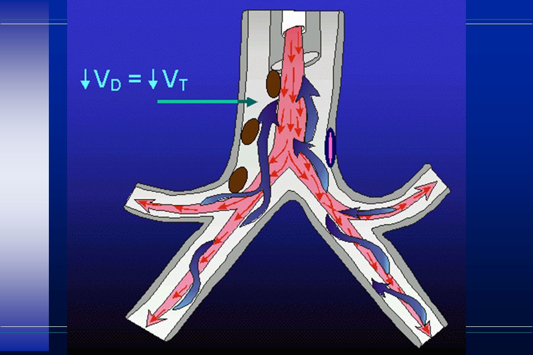
High frequency ventilation u Techniques HFPPV HFJV HFFIHFOV HFPPV HFJV HFFIHFOV V T >dead sp > or or dead sp > or or <ds <ds Exp passive passive passive active Wave- variable triangular triangular sine wave form Entrai- none possible nonenone ment Freq. 60-150 60-600 300-900 300-3000 (/min)
.")
76
High frequency ventilation u HFPPV l conventional ventilators with low- compliance tubing l ventilatory rates of 60-150/min l not very effective: minute ventilation decreases with high frequencies l ventilator and circuit design are not optimal for use at frequencies

77
High frequency ventilation u HFJV (e.g. Bunnell Life Pulse HFJV) l adequate gas exchange with lower MAP l Servo pressure reflects volume ventilated: u increases with improving compliance or resistance or by peri-ET leaks u decreased by worsening compliance, resistance, obstruction, or pneumothorax l Larger babies: 300 bpm; smaller ones: 500 bpm
 l adequate gas exchange with lower MAP l Servo pressure reflects volume ventilated: u increases with improving compliance or resistance or by peri-ET leaks u decreased by worsening compliance, resistance, obstruction, or pneumothorax l Larger babies: 300 bpm; smaller ones: 500 bpm.")
78
High frequency ventilation u HFJV (contd.) MAP controls Pa O2, P (and frequency) control Pa CO2. (MAP controls lung volume. Pa O2 will not respond to increased MAP if FRC normal) smaller TV ( P) with higher PEEP better than larger TV with lower PEEP (--> hypoxia with hypocarbia) l Optimal PEEP: no drop in SpO 2 when CMV off l Parallel conventional ventilation recruits alveoli (use low rate : 1-3 bpm; 0-1 bpm if air leak)
 smaller TV ( P) with higher PEEP better than larger TV with lower PEEP (--> hypoxia with hypocarbia) l Optimal PEEP: no drop in SpO 2 when CMV off l Parallel conventional ventilation recruits alveoli (use low rate : 1-3 bpm; 0-1 bpm if air leak).")
79
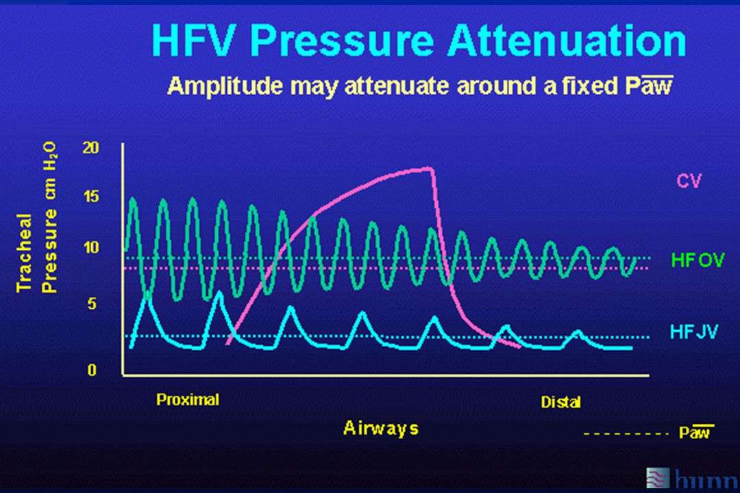
High frequency ventilation u HFOV (e.g. Sensormedics 3100A) l Generally used at more MAP than CMV; optimal MAP difficult to determine as CXR “rib space counting” not very accurate l Frequency: 5-10 Hz better for CO 2 elimination; 10-15 Hz better for improving oxygenation l maybe useful in airleak syndromes l maybe useful in PPHN; may decrease need for ECMO esp. if combined with NO
 l Generally used at more MAP than CMV; optimal MAP difficult to determine as CXR rib space counting not very accurate l Frequency: 5-10 Hz better for CO 2 elimination; Hz better for improving oxygenation l maybe useful in airleak syndromes l maybe useful in PPHN; may decrease need for ECMO esp. if combined with NO.")
80
High frequency ventilation u HFFI (e.g. Infant Star with HFFI module) l active expiration in Infant Star model makes operation more like HFOV l clinical studies have not shown it to be superior to conventional ventilation l more convenient: single ventilator for CMV and HFV makes initiation and weaning easier
 l active expiration in Infant Star model makes operation more like HFOV l clinical studies have not shown it to be superior to conventional ventilation l more convenient: single ventilator for CMV and HFV makes initiation and weaning easier.")
81
High frequency ventilation u Uses of HFOV/ HFJV/ HFFI : l “rescue” for severe RDS l air leak syndromes (pneumothorax, PIE) l PPHN u Primary use controversial: risk of hypocarbia (-->PVL) higher, and reduction of BPD or airleaks seen in some, but not all, studies.
 l PPHN u Primary use controversial: risk of hypocarbia (-->PVL) higher, and reduction of BPD or airleaks seen in some, but not all, studies.")
82
Summary The practice of the art of mechanical ventilation lies in the application of the underlying science and physiologic concepts to the specific clinical situation An individualized flexible approach aimed at maintaining adequate gas exchange with the minimum of ventilatory support, both in magnitude and duration, should optimize the possible outcome

83
Combining IMV and HFV

Similar presentations


>")


