Download presentation
Presentation is loading. Please wait.

1
Human A & P Bone Structure and Function

2
I. Introduction to The Skeletal System A. Background information about the skeletal system: 1. The skeletal system includes the entire framework of ____________ and their _____________. 2. Each bone is considered to be an ___________. Bones Cartilage organ

3
3. Bone tissue is a __________________ tissue. a. The _______________ is: i. _________________________ - to provide hardness ii. ______________ - to provide some flexibility. iii. _____________ connective matrix Crystallized Minerals Collagen fibers Water

4
b. The ______ kinds of cells of bone tissue and their functions: i. ________________ - bone building cells which produce the matrix. (modified fibroblasts). This process is called _________________________. ii. ________________- matured bone cells that develop from osteoblasts, which help to maintain bone _________________. (take in nutrients and release wastes). 3 Osteoblasts Osteocytes metabolism Bone formation
. This process is called _________________________. ii. ________________- matured bone cells that develop from osteoblasts, which help to maintain bone _________________. (take in nutrients and release wastes). 3 Osteoblasts Osteocytes metabolism Bone formation.")
5
iii. ______________________- (modified macrophage) huge cells made from 50+ WBCs that produce lysosomal enzymes & acids to break down bone matrix. This is a process called __________________. Osteoclasts resorption
 huge cells made from 50+ WBCs that produce lysosomal enzymes & acids to break down bone matrix. This is a process called __________________. Osteoclasts resorption.")
6
II. Gross Anatomy of Bone A. Classification of bones based on shape 1. _______________- greater in length than in width. Ex – 2. _______________- nearly equal in length and width Ex - Long Bone Femur, tibia, ulna, humerus, phalanges Short Bone Wrist, & ankle bones

7
3. ________________ - thin and flat Ex – 4. _________________- complex shapes that do not fit other categories. Ex - Flat Bone Ribs, cranium, sternum, shoulder blades Irregular Bone Vertebra, pelvis, some facial bones

9
B. Macroscopic Structure of Bone 1. gross view of outside of bone

10
_____________________- end of bone epiphysis _____________________- end of bone epiphysis

11
_____________________- main middle portion of the bone. Diaphysis

12
_______________ - region in mature bone where diaphysis meets epiphysis metaphysis Metaphysis

13
_____________________- thin layer of cartilage over the epiphysis where the bone connects with another bone. It has two features: 1. ________________ & protects the ends of bone. 2. _________________ability to repair itself. Why???? Articular Cartilage cushions limited Articular Cartilage

14
2. gross view of the inside of the bone

15
_____________________- a layer of cartilage in growing bone where the diaphysis can grow in __________. - when the bone stops growing in length, bone will replace the cartilage and become the _____________________________. Epiphyseal Plate LENGTH Epiphyseal Line

16
Site of _______________________ in babies and adults which is where blood cell production occurs. Red bone marrow

17
_____________________- looks like a network of bone with marrow in between. Spongy Bone

18
_____________________- single layer of bone-forming cells membrane that lines the inside of the medullary cavity. Endosteum

19
_____________________- dense bone that serves to protect and support. Compact Bone

20
_____________________- dense irregular connective tissue that surrounds bone where articular cartilage is absent. Serves the following functions: 1.____________ the bone & assists in fracture repair. 2. ____________ point for ligaments & tendons. 3. _____________ & thickens the bone. BUT DOES NOT LENGTHEN! 4. _______________ bone tissue. ACRONYM HELP – P.A.W.N. Periosteum Protects Attachment Widens Nourishes

21
_____________________- contains __________________ in babies, but as we age, this marrow becomes __________________ as adults which acts as fat storage. Medullary Cavity Red bone marrow Yellow bone marrow

22
_____________________- transports nutrients and waste into & out of bone. (This is how breaking one’s “femur” could be a life- threatening, blood loss situation). Nutrient Artery
. Nutrient Artery.")
24
III. Microanatomy of Compact and Spongy Bone

25
A. Anatomy of both types of bones. 1. Compact bone ___________________- units that compact bone are arranged in. (also called ____________.) Haversian System osteons Circumferential Lamellae Blood vessels
 Haversian System osteons Circumferential Lamellae Blood vessels.")
26
___________________- rings of hard, calcified matrix around the Haversian canal. Concentric Lamellae Circumferential Lamellae Blood vessels

27
Circumferential Lamellae Blood vessels _________________- central canal in the osteon that contains: __________________ _________________. Haversian Canal Nerves, lymph & blood vessels Circumferential Lamellae

28
Blood vessels _________________- Leads to the periosteum Perforating Canal Circumferential Lamellae

29
Concentric Lamellae Compact Bone (continued)
")
30
______________- the bone cell. Osteocyte

31
______________- (“small lake”) – small space that holds the osteocyte. Lacunae
 – small space that holds the osteocyte. Lacunae")
32
__________________- small channels filled with extracellular fluid which connects lacunae w/ each other and Haversian canal Canaliculi

33
Haversian Canal

34
Red Space for ___________ bone marrow. 2. Spongy Bone

35
_______________ - a network of thin columns of bone. Trabeculae

36
Lacuna

37
Concentric Lamellae

38
Canaliculi

39
____________- fiber makers Osteoblasts

40
_______________ - microbe killers Osteoclasts

41
____________- bone “maintainers” Osteocyte

42
B. Differences between compact and spongy bone: 1. anatomical differences of each type: a. __________________ with Haversian canals are unique to compact bone. b. ___________________ are unique to spongy bone. Osteons Trabeculae

43
2. Location of each type in the body: a. COMPACT bone is found in the ________________ of long bones. b. SPONGY bone is found in: i. the _______________ and near the _____________________ of long bone. ii. Makes up most of: ___________ __________________________. Diaphysis epiphysis Medullary cavity flat, short & irregular

45
3. Density differences of each type: a. Spongy bone is _______________ with empty spaces in between for red bone marrow to fill. b. Compact bone is ____________ packed with few spaces in between cells & ___________. lighter tightly matrix

46
III. Physiological Features of Bone Tissue A.Main Functions of the Bones & Skeletal System: (Quick Glance) 1. ___________________or _______________ - occurs in ___________________ only. 2. __________ - provides a framework for muscles to attach to. 3. __________________ (detailed later) 4. _____________________ - works with muscles 5. __________ heart & other internal organs 6. _____________________________ in yellow bone marrow. Acronym help: BS MA P h D Support Protects Assists In Movement Mineral Homeostasis Blood Cell Production Deposits & stores adipose tissue hemopoiesis Red bone marrow
 1. ___________________or _______________ - occurs in ___________________ only. 2. __________ - provides a framework for muscles to attach to. 3. __________________ (detailed later) 4. _____________________ - works with muscles 5. __________ heart & other internal organs 6. _____________________________ in yellow bone marrow. Acronym help: BS MA P h D Support Protects Assists In Movement Mineral Homeostasis Blood Cell Production Deposits & stores adipose tissue hemopoiesis Red bone marrow.")
47
B. Bone formation & ossification 1. Definition of ossification: _____________ _________________________________ 2. When does ossification occur? a. begins about the ________ week of embryonic life and continues into ___________ (ages18-25). the process of bone formation 6th adulthood
. the process of bone formation 6th adulthood.")
48
3. Two methods of ossification: a. _________________________- bone forms directly on or in loose fibrous connective tissue. i. Where does this occur? 1. _____________________ 2. _____________________ Intramembraneous Flat bones of skull Mandible (lower jaw)
.")
49
b. ____________________- bone forms within the cartilage. ____________________ in the body form this way. Endochondral MOST BONES

50
4. Process of endochondral ossification: STEP #1:________________________________ Development of the Cartilage Model a). fetal _______________ cells crowd together in the shape of a future bone. (mesenchymal cells are embryonic tissue cells from which ALL connective tissue arises.) mesenchymal
. fetal _______________ cells crowd together in the shape of a future bone. (mesenchymal cells are embryonic tissue cells from which ALL connective tissue arises.) mesenchymal.")
51
b). mesenchymal cells turn into _________________. chondroblasts
. mesenchymal cells turn into _________________. chondroblasts")
52
c). chondroblasts produce _________________ cartilage and the _________________ - membrane around the cartilage. hyaline perichondrium

53
STEP #2. _________________________________ Growth of Cartilage Model a). Chondroblasts become ________________ and some start to burst, triggering _________________. b). Remaining chondrocytes die b/c they cannot get enough _____________ in the calcifying matrix. c). When they die, _____________ form and merge into small cavities. chondrocytes calcification nutrients lacunae
. Remaining chondrocytes die b/c they cannot get enough _____________ in the calcifying matrix. c). When they die, _____________ form and merge into small cavities. chondrocytes calcification nutrients lacunae.")
54
STEP #3. _________________________________ Development of Primary Ossification Center a). Primary ossification proceeds ____________________________. inward

55
b). A nutrient _________________ penetrates the middle of the cartilage. c) This stimulates _______________ cells to become ___________________ which lay the matrix to form the __________________ of spongy bone artery Osteogenic osteoblasts trabeculae
 This stimulates _______________ cells to become ___________________ which lay the matrix to form the __________________ of spongy bone artery Osteogenic osteoblasts trabeculae.")
56
d). ____________________ dissolves some of the newly formed trabeculae to create the ____________________ which fills with _____________________________. Osteoclasts Medullary cavity Red bone marrow

57
STEP #4. _________________________________ Development of Secondary Ossification Center a). Like primary ossification except the bone remains as _______________. Spongy bone

58
STEP #5. _________________________________ Formation of articular cartilage and epiphyseal plate a). ________________ cartilage is replaced with ___________________ cartilage. hyaline articular
. ________________ cartilage is replaced with ___________________ cartilage. hyaline articular.")
59
b). ______________ remains. Spongy bone
. ______________ remains. Spongy bone")
60
c). ____________________ is the only remaining hyaline cartilage that allows the bone to ______________ until it calcifies. (usually between 18-25). Epiphyseal plate lengthens
. Epiphyseal plate lengthens.")
61
IV. Homeostasis in Bone Tissue & Complications from an Imbalance A. _____________________- the study of bone structure and the treatment of disorders. Osteology

62
B. Bone Tissue “regeneration”: 1. old bone is constantly being ___________ by osteoclasts & new bone tissue is being formed by _______________.(EVEN IN ADULTHOOD.) resorbed osteoblasts
 resorbed osteoblasts.")
63
C. Normal Bone Metabolism depends on: 1. Adequate dietary amounts of: a. minerals- ____________________ _____________________________ b. vitamin - _____________________ 2. Hormone interaction – There are ______ hormones that play a role in bone homeostasis. (Bone resorption hormones – PTH, cortisol; Bone formation – calcitriol, calcitonin, estrogen, thyroid, insulin, growth hormone, insulin growth factors) calcium, phosphorus & magnesium C, A, D 9
 calcium, phosphorus & magnesium C, A, D 9.")
64
D. How bone aids in the homeostasis of Calcium in the blood: 1. if blood-calcium levels are too __________, PTH (___________________), from the parathyroid gland is released which activates the osteoclasts to _______________ calcium into the blood via resoprtion. low Parathyroid hormone release
, from the parathyroid gland is released which activates the osteoclasts to _______________ calcium into the blood via resoprtion. low Parathyroid hormone release.")
65
2. If blood-calcium levels are too _________, then the thyroid gland releases _________________________ which activates the osteoblasts to take up calcium via bone formation. 3. Effects of calcium imbalance: a. Too low ______________________ b. Too high ______________________ high calcitonin Breathing stops Heart stops

66
Calcium Homeostasis Song (Tune: Jingle Bells, start with “Dashing…..”) If calcium gets low Parathyroid goes to work Releases PTH Bones give up calcium Kidneys take in less So it stays in the blood Vitamin D makes intestines take it from grub.
 If calcium gets low Parathyroid goes to work Releases PTH Bones give up calcium Kidneys take in less So it stays in the blood Vitamin D makes intestines take it from grub.")
67
Song continued… If calcium gets too high Stimulates thyroid Thyroid makes calcitonin To bring the level down Calcitonin stimulates deposition in the bones Kidney leave more calcium in urine Now we’re done!

68
E. Impact on Human Growth Hormone (hGH) on Bone Homeostasis 1. hGH stimulates ___________________ to make more _________________ which will cause bone formation. chondrocytes cartilage

69
2. effects of hGH imbalance: a. Undersecretion: i. ______________________ - when too little hGH is secreted and the epiphyseal plate closes too soon. Pituitary dwarfism

71
b. Oversecretion: i. during ________________ could lead to ___________________ which is caused by an abnormal lengthening of bones. childhood gigantism

72
Gigantism slide here

73
ii. During _________________ could lead to ___________________ which causes a thickening of the hand, feet, cheek and jaw bones. (bones won’t lengthen b/c epiphyseal plate is already calcified). adulthood acromegaly
. adulthood acromegaly.")
75
Pic of NFL issue and growth hormone here

76
F. Maintaining Bone Mass 1. How can bone mass be maintained or strengthened? a. ________________________ i. Why? 1. mechanical stress (like the pull of gravity or the pull of skeletal muscles) ____________________ bones. Weight bearing Exercise Strengthens and thickens
 ____________________ bones. Weight bearing Exercise Strengthens and thickens.")
77
2. How can bone mass be lost? a. when bone ________________ occurs faster than bone formation. In healthy individuals, this happens when: i. ___________________________ ii. _______________ - break in bone resorption A limb is unused A bone fractures

78
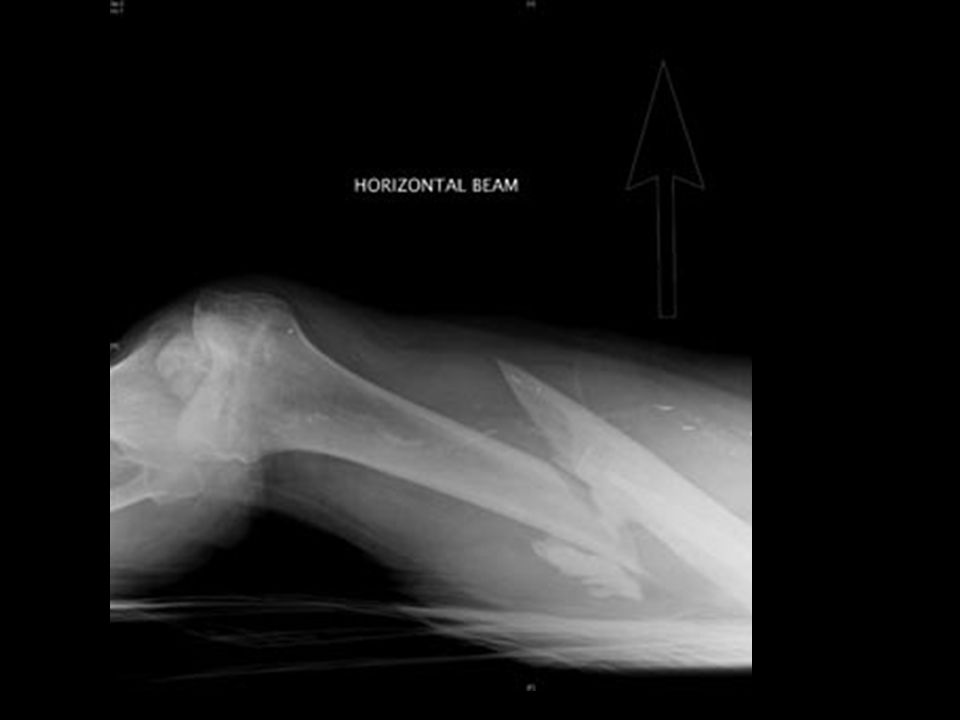
1. four descriptions of fractures: a. _____________________ - an incomplete break across the bone, such as a crack. Partial fracture

80
b. _______________________- a complete break across the bone so that the bone is in two or more pieces. Complete Fracture

82
c. _______________________ - the fractured bone does not break through the skin. (It could be ________________ or ________________) Closed fracture partialcomplete
 Closed fracture partialcomplete.")
84
d. ________________________- the broken end of the bone protrudes through the skin. (Can only be a ________________ fracture.) open fracture compound
 open fracture compound.")
86
G. ____________________- a condition of porous bones caused by a depletion of ____________ in the body. 1. What can it cause? a. ______________________ b. _________________(especially hip fractures) c. ______________________ d. ______________________ Osteoporosis calcium Bone mass loss fractures Shortened height (hunch back) Bone pain
 c. ______________________ d. ______________________ Osteoporosis calcium Bone mass loss fractures Shortened height (hunch back) Bone pain.")
87
2. Who is most likely to get it & why? a. ____________________- estrogen (an osteoblast stimulator) declines dramatically during menopause. 3. What can be done to prevent getting it later in life? a. ________________________ b. ________________________ Middle aged women Adequate calcium in diet Weight bearing exercise in early years
 declines dramatically during menopause. 3. What can be done to prevent getting it later in life. a. ________________________ b. ________________________ Middle aged women Adequate calcium in diet Weight bearing exercise in early years.")
Similar presentations


 Joints ► Cartilages Ligaments ► Divided.>")


 Joints Cartilages Ligaments Divided into two divisions Axial skeleton –>")


