Download presentation
Presentation is loading. Please wait.

1
Muscular System Chapter 8

2
Introduction: A. All movements require muscle which are organs using chemical energy to contract. B. The three types of muscle in the body are skeletal, smooth, and cardiac muscle. C. This chapter focuses on skeletal muscle.

3
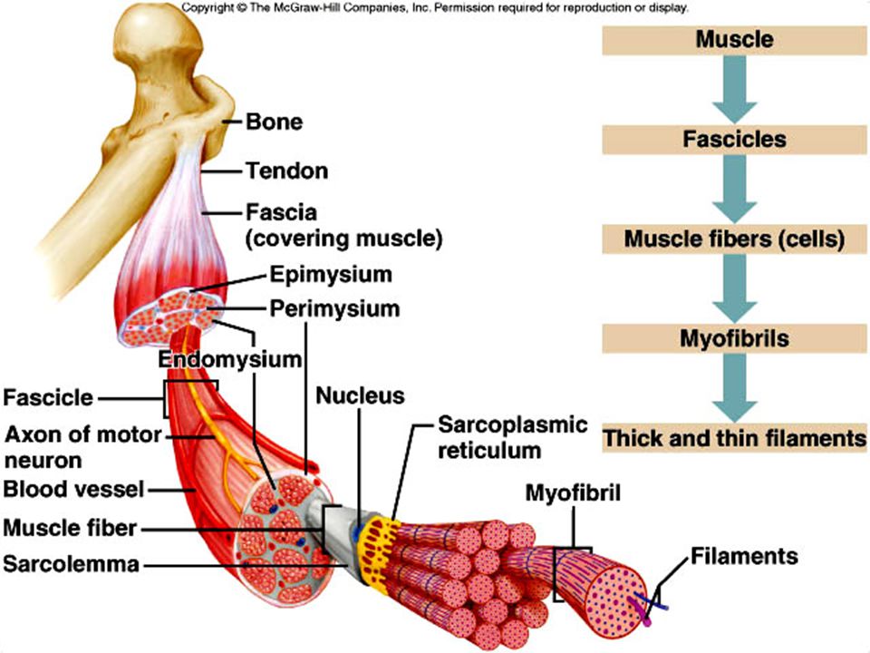
Skeletal Muscle Each skeletal muscle is an organ, comprised of skeletal muscle tissue, connective tissue, nervous tissue, and blood.

4
Connective Tissue Coverings
1. Layers of dense connective tissue called fascia, surround and separate each muscle. 2. This connective tissue extends beyond the ends of the muscle and gives rise to tendons, that are fused together to the periosteum of bones.

5
Connective Tissue Coverings
3. Sometimes muscles are connected to each other by broad sheets of connective tissue called aponeuroses. 4. The layer of connective tissue around each whole muscle is the epimysium; the perimysium surrounds individual bundles (fascicles) within each muscle; and each muscle cell (fiber) is covered by a connective tissue layer called endomysium.
 within each muscle; and each muscle cell (fiber) is covered by a connective tissue layer called endomysium.")
7
Study Analogy Pretend you are going to play a joke on someone and give them 100 pencils. The pencils will represent muscle fibers. First you wrap each individual pencil in tissue paper (dense tissue paper of course!). This would be endomysium. Then you take about 10 pencils in a bundle (a fascicle) and wrap them in paper (perimysium). After that you take all the bundles and wrap them in gift wrap (epimysium). But you are going to mail this joke, so you also have to wrap it in brown paper representing the fascia.
. This would be endomysium. Then you take about 10 pencils in a bundle (a fascicle) and wrap them in paper (perimysium). After that you take all the bundles and wrap them in gift wrap (epimysium). But you are going to mail this joke, so you also have to wrap it in brown paper representing the fascia.")
8
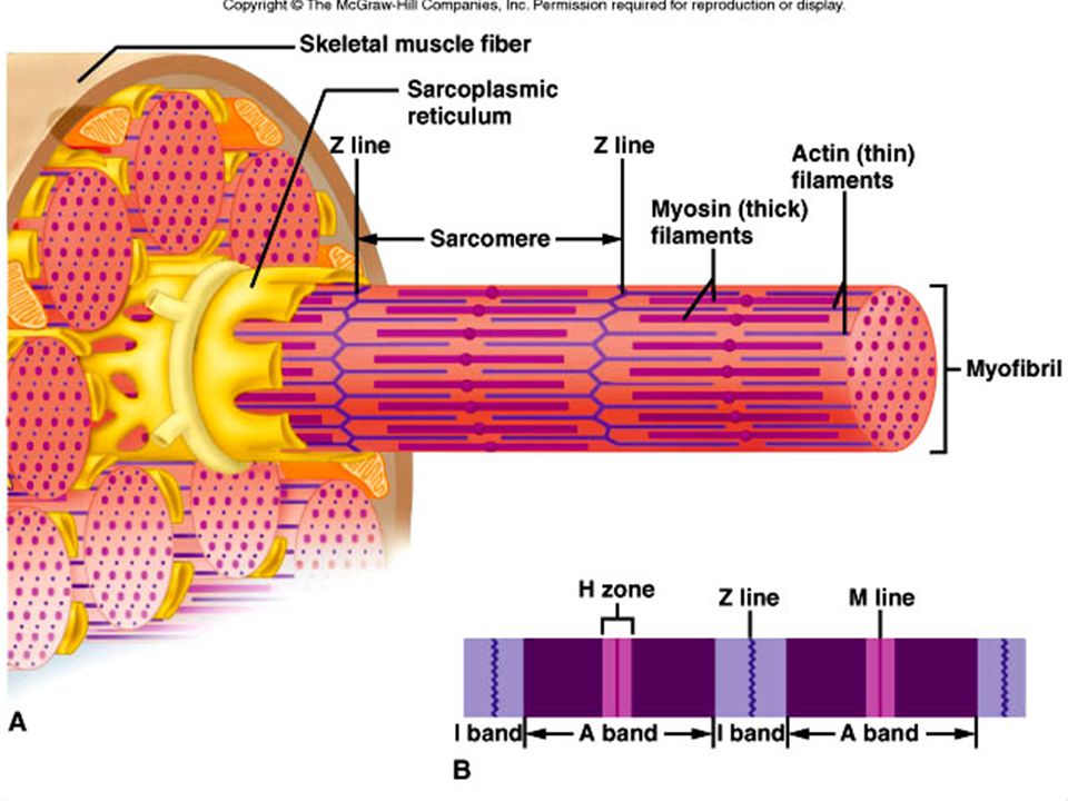
Skeletal Muscle Fibers
1. Each muscle fiber is a single, long, cylindrical muscle cell. 2. Beneath the sarcolemma (cell membrane) lies sarcoplasm (cytoplasm) With many mitochondria and nuclei; the sarcoplasm contains myofibrils. a. Thick filaments of myofibrils are made up of the protein myosin. b. Thin filaments of myofibrils are made up of the protein actin. c. The organization of these filaments produces striations.
 lies sarcoplasm (cytoplasm) With many mitochondria and nuclei; the sarcoplasm contains myofibrils. a. Thick filaments of myofibrils are made up of the protein myosin. b. Thin filaments of myofibrils are made up of the protein actin. c. The organization of these filaments produces striations.")
9
Skeletal Muscle Fibers
A sarcomere extends from Z line to Z line. a. I bands (light bands) made up of actin filaments are anchored to Z lines. b. A bands (dark bands) are made up of overlapping thick and thin filaments. c. In the center of A bands is an H zone, consisting of myosin filaments only.
 made up of actin filaments are anchored to Z lines. b. A bands (dark bands) are made up of overlapping thick and thin filaments. c. In the center of A bands is an H zone, consisting of myosin filaments only.")
11
Skeletal Muscle Fibers
4. Beneath the sarcolemma of a muscle fiber lies the sarcoplasiceticulum (endoplasmic reticulum), which is associated with transverse (T) tubules (invasinations of sacrolemma). a. Each T tubule lies between two cisternae of the sarcoplasmic reticulum and is open to the outside of the muscle fiber. b. The sarcoplasmic reticulum and transverse tubules activate the muscle contraction mechanism when the fiber is stimulated
, which is associated with transverse (T) tubules (invasinations of sacrolemma). a. Each T tubule lies between two cisternae of the sarcoplasmic reticulum and is open to the outside of the muscle fiber. b. The sarcoplasmic reticulum and transverse tubules activate the muscle contraction mechanism when the fiber is stimulated.")
12
Motor Units 1. A motor neuron and the muscle fibers it controls make up a motor unit; when stimulated to do so, the muscle fibers of the motor unit contract all at once.

13
Skeletal Muscle Contractions
A. Muscle contraction involves several components that result in the shortening of sarcomeres, and the pulling of the muscles against its attachments.

14
Skeletal Muscle Contractions
B. Role of Myosin and Actin 1. Myosin consists of two twisted strands with globular cross-bridges projected outward along the strands. 2. Actin is a globular protein with myosin binding sites; tropomysosin and troponin are two proteins associated with the surface of actin filaments.

15
Skeletal Muscle Contractions
3. According to the sliding filament theory of muscle contraction, the myosin crossbridge attaches to the binding site on the actin filament and bends, pulling on the actin filament; it then releases and attaches to the next binding site on the actin, pulling again. 4. Energy from the conversion of ATP to ADP is provided to the cross-bridges from the enzyme ATPase, causing them to be in a “cocked” position.

16
Skeletal Muscle Contractions
C. Stimulus for Contraction 1. The motor neuron must release the neurotransmitter acetylcholine from its synaptic vesicles into the synaptic cleft in order to initiate a muscle contraction. 2. Protein receptors in the motor end plate detect the neurotransmitters, and a muscle impulse spreads over the surface of the sarcolemma and into the T tubules, where it reaches the sarcoplasmic reticulum.

17
Skeletal Muscle Contractions
3. Upon receipt of the muscle impulse, the sarcoplasmic reticulum releases its stored calcium to the sarcoplasm of the muscle fiber. 4. The high concentration of calcium in the sarcoplasm interacts with the troponin and tropomyosin molecules, which move aside, exposing the myosin binding sites on the actin filaments.

18
Skeletal Muscle Contractions
5. Myosin cross-bridges now bind and pull on the actin filaments, causing the sarcomeres to shorten. 6. After the nervous impulse has been received, acetylcholinesterase rapidly decomposes the acetylcholine. 7. Then, calcium is returned to the sarcoplasmic reticulum, and the linkages between myosin and actin are broken.

19
Smooth Muscle Fibers 1. Smooth muscle cells are elongated with tapered ends, lack striations, and have a relatively undeveloped sarcoplasmic reticulum. 2. Multiunit smooth muscle and visceral muscle are two types of smooth muscles. a. In multiunit smooth muscle, such as in the blood vessels and iris of the eye, fibers occur separately rather than as sheets. b. Visceral smooth muscle occurs in sheets and is found in the walls of hollow organs; these fibers can stimulate one another and display rhythmicity, and are thus responsible for peristalsis in hollow organs and tubes.

20
Smooth Muscle Fibers B. Smooth Muscle Contraction
1. The myosin-binding-to-actin mechanism is the mostly same for smooth muscles and skeletal muscles. 2. Both acetylcholine and norepinephrine stimulate and inhibit smooth muscle contraction, depending on the target muscle. 3. Hormones can also stimulate or inhibit contraction. 4. Smooth muscle is slower to contract and relax than is skeletal muscle, but can contract longer using the same amount of ATP.

21
Cardiac Muscle A. The mechanism of contraction in cardiac muscle is essentially the same as that for skeletal and smooth muscle, but with some differences. B. Cardiac muscle has transverse tubules that supply extra calcium, and can thus contract for longer periods.

22
Cardiac Muscle C. Complex membrane junctions, called intercalated disks, join cells and transmit the force of contraction from one cell to the next, as well as aid in the rapid transmission of impulses throughout the heart. D. Cardiac muscle is self-exciting and rhythmic, and the whole structure contracts as a unit.

Similar presentations


, which.>")


 –sheet or band of fibrous C.T. under.>")


