Download presentation
Presentation is loading. Please wait.

1
Histology

2
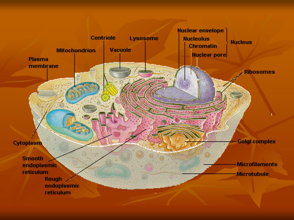
Membranous cell Organelles 1-Cell Membrane 1-Cell Membrane 2-Mitochondria 2-Mitochondria 3-Golgi Apartus 3-Golgi Apartus 4-Peroxisome 4-Peroxisome 5-Endoplasmic reticulum 5-Endoplasmic reticulum 6-Lysosomes 6-Lysosomes 7-coated vesicles 7-coated vesicles 8-Endosomes 8-Endosomes

3
2-Mitochondria Definition:they are membranous cell organelles present in all nucleated cells Definition:they are membranous cell organelles present in all nucleated cells Structures: formed of protein,lipid,DNA,RNA,zinc,calcium&enzyme Structures: formed of protein,lipid,DNA,RNA,zinc,calcium&enzyme L.M.: appear as rods,granules or filaments they are very sensitive to temperature&PH L.M.: appear as rods,granules or filaments they are very sensitive to temperature&PH

4
Number: of mitochondria: it varies from one to another Number: of mitochondria: it varies from one to another About 1000 mitochondria present in liver cell About 1000 mitochondria present in liver cell No mitochondria are present in blood corpuscle No mitochondria are present in blood corpuscle

5
Mitochondria Loction:Mitochondria occur in nearly all eukaryotic cells&most are dispersed throughout the cytoplasma Loction:Mitochondria occur in nearly all eukaryotic cells&most are dispersed throughout the cytoplasma -Accumulate in cell types&intracellular regions -Accumulate in cell types&intracellular regions -Cardiac muscle cell are notable for abundant mitochondria -Cardiac muscle cell are notable for abundant mitochondria -Epithelial cells lining kidney tubules -Epithelial cells lining kidney tubules

6
Function: Function: -responsible for cell respiration -responsible for cell respiration -power house of the cell -power house of the cell -they supply energy to all cellar activities -they supply energy to all cellar activities -mitochondria regulate the concentration of calcium&magnesium ions -mitochondria regulate the concentration of calcium&magnesium ions Theelecron transport systemof the mitochondria can produce and store energy Theelecron transport systemof the mitochondria can produce and store energy Formation ATP from ADP Formation ATP from ADP

8
Mitochondria have a double-membrane: outer membrane & highly convoluted inner membrane inner membrane has folds or shelf-like structures called cristae that contain elementary particles; these particles contain enzymes important in ATP production primary function is production of adenosine triphosphate (ATP)
")
9
Energy production & storage Cellular respiration In the form of ATP molecules : mainly from glucose & fatty acid Glucose to pyruvic acid degraded to Co2 & Ho2 a large quantity of ATP occurs in Mitochondria Fatty acids directly to mitochondria Mitochondria are the principal organelles involved in cellular respiration.

10
3-Golgi Complex Participates in many activities Participates in many activities Structures: 1-stack slightly curved,flattened cisternae Structures: 1-stack slightly curved,flattened cisternae 2-numerous small vesicles peripheral to the stack 2-numerous small vesicles peripheral to the stack 3-few large condensing vacuoles 3-few large condensing vacuoles

11
Function: 1-polsaccharde synthesis 1-polsaccharde synthesis 2-modification of secretory products 2-modification of secretory products 3-sorting of secretory products 3-sorting of secretory products 4-packaging of secretory products 4-packaging of secretory products 5-concentration&storge of secretory product 5-concentration&storge of secretory product Location:golgi comlex typically is near the nucleus best developed in neurons&glandular cells Location:golgi comlex typically is near the nucleus best developed in neurons&glandular cells

12
Excretion of secretion Excretion of secretion By four principal mechanisms, By four principal mechanisms, Packaged within membrane (packaging process by golgi apparatus) Packaged within membrane (packaging process by golgi apparatus) Golgi apparatus : consists of a series of flattened sacs (or cisternae) functions include: synthesis (of substances likes phospholipids), packaging of materials for transport (in vesicles), and production of lysosomes
 Packaged within membrane (packaging process by golgi apparatus) Golgi apparatus : consists of a series of flattened sacs (or cisternae) functions include: synthesis (of substances likes phospholipids), packaging of materials for transport (in vesicles), and production of lysosomes")
13
4-Peroxisomes Contain 40 enzymes as enzymes of oxidation of long chain fatty acids&urate oxidase enzyme Contain 40 enzymes as enzymes of oxidation of long chain fatty acids&urate oxidase enzyme In some animals peroxisome many contain crystalline In some animals peroxisome many contain crystalline They can be found in liver cells,proximal&distal convoluated tubules&small intestin They can be found in liver cells,proximal&distal convoluated tubules&small intestin They are originated from rEr They are originated from rEr They can renew them self by division They can renew them self by division

14
Function Function Oxidation energy is produced but this energy can not be stored a ATP as in mitochondria Oxidation energy is produced but this energy can not be stored a ATP as in mitochondria

15
5-Endoplasmic Reticulum(ER) 1---Rough endoplasmic reticulum 1---Rough endoplasmic reticulum 2---Smooth endoplasmic 2---Smooth endoplasmic
 1---Rough endoplasmic reticulum 1---Rough endoplasmic reticulum 2---Smooth endoplasmic 2---Smooth endoplasmic")
16
Rough endoplasmic reticulum: also called granular ER also called granular ER Structures: Structures: composed of cisternae are typically parallel.flattened and elongated especially in cells specialized in proteins secreting (pancreatic acinar cells,plasma cells) composed of cisternae are typically parallel.flattened and elongated especially in cells specialized in proteins secreting (pancreatic acinar cells,plasma cells)
 composed of cisternae are typically parallel.flattened and elongated especially in cells specialized in proteins secreting (pancreatic acinar cells,plasma cells)")
17
Function: synthesize proteins for sequestration from the cytoplasm,including secretory proteins Function: synthesize proteins for sequestration from the cytoplasm,including secretory proteins Location: is suspended in the cytoplasm&in proteins secreting epithelial cells--------basal cytoplasm between the plasma membrane&the nucleus Location: is suspended in the cytoplasm&in proteins secreting epithelial cells--------basal cytoplasm between the plasma membrane&the nucleus

18
Smooth endoplasma reticulum Structures cisternae are more tubular or vesicular Structures cisternae are more tubular or vesicular Function:SER cannot synthesize proteins Function:SER cannot synthesize proteins Location:abundant in cells synthesizing steroid hormones (adrenal gland,gonads)it is abundant in liver cells(hepatocytes) Location:abundant in cells synthesizing steroid hormones (adrenal gland,gonads)it is abundant in liver cells(hepatocytes) Specialized SER termed sarcoplasmic reticulum is found in striated muscle cells,where it regulates muscle contraction releasing calcium ions Specialized SER termed sarcoplasmic reticulum is found in striated muscle cells,where it regulates muscle contraction releasing calcium ions
it is abundant in liver cells(hepatocytes) Location:abundant in cells synthesizing steroid hormones (adrenal gland,gonads)it is abundant in liver cells(hepatocytes) Specialized SER termed sarcoplasmic reticulum is found in striated muscle cells,where it regulates muscle contraction releasing calcium ions Specialized SER termed sarcoplasmic reticulum is found in striated muscle cells,where it regulates muscle contraction releasing calcium ions")
19
6-Lysosomes Lysosomes: spherical membrane limited vesicles Lysosomes: spherical membrane limited vesicles 1-primary lysosomes 1-primary lysosomes 2-secondary lysosomes 2-secondary lysosomes 3-residual bodies ……..pigments,crystals,lipofuscin pigment 3-residual bodies ……..pigments,crystals,lipofuscin pigment Containing 40 digestive hydrolytic enzymes called hydrolases ex. Acid phosphatase,nuclease&lipases Containing 40 digestive hydrolytic enzymes called hydrolases ex. Acid phosphatase,nuclease&lipases The hydrolytic enzymes are found&packed in vesicles in R.endoplasmic reticulum&transferred to golgi that packed them as primary lysosomes The hydrolytic enzymes are found&packed in vesicles in R.endoplasmic reticulum&transferred to golgi that packed them as primary lysosomes They are numerous in macrophages,neutrophils&osteoclast They are numerous in macrophages,neutrophils&osteoclast

20
Function: 1-They are responsible for intracytoplasm digestion of nutritive matenals 1-They are responsible for intracytoplasm digestion of nutritive matenals 2-phagocytosis of bacteria&viruses 2-phagocytosis of bacteria&viruses 3-digestive of old organelles 3-digestive of old organelles

21
7-Coated vesicles Cell membrane contains 25 types of specific receptors that help in transport of macromolecles into the cytoplasma&lipoprotein&protein of somes hormones&antibodies Cell membrane contains 25 types of specific receptors that help in transport of macromolecles into the cytoplasma&lipoprotein&protein of somes hormones&antibodies Coated vesicles fused with endosomes and resulting is dissociation of receptors Coated vesicles fused with endosomes and resulting is dissociation of receptors

22
Function Help imacromolecules to be internalized in to the cytoplasm Help imacromolecules to be internalized in to the cytoplasm

23
8-Endosome Contain electron lucent material&arevesicular or tubular in shape Contain electron lucent material&arevesicular or tubular in shape They are present between golgi&surface of cells They are present between golgi&surface of cells They are similar to lysosomes but without hydrolytic enzymes They are similar to lysosomes but without hydrolytic enzymes

24
Function Carry receptors to cell membrane again Carry receptors to cell membrane again They transport to lysosomes or to golgi complex They transport to lysosomes or to golgi complex

25
Non-Membranous Cytoplasmic org. A-Ribosomes A-Ribosomes B-Centrioles B-Centrioles C-microtubules C-microtubules D-Microfilaments D-Microfilaments E-Cilia E-Cilia F-Flagella F-Flagella

26
Ribosomes -composed of rRNA (ribosomal RNA) & protein -may be dispersed randomly throughout the cytoplasm or attached to surface of rough endoplasmic reticulum -often linked together in chains called polyribosomes or polysomes -Primary function to produce proteins
 & protein -may be dispersed randomly throughout the cytoplasm or attached to surface of rough endoplasmic reticulum -often linked together in chains called polyribosomes or polysomes -Primary function to produce proteins")
27
Function: Function: They are the site for protien synthesis They are the site for protien synthesis They are considered as factories for protien formation They are considered as factories for protien formation

28
B-Cenrioles Is cilinder of microtubules Is cilinder of microtubules Location: Near the nucleus often surrounded by golgi complex Location: Near the nucleus often surrounded by golgi complex Function:Cells structures organizers during mitoses Function:Cells structures organizers during mitoses

29
Microfilaments Mainly fine strands of a protein called Actin Arranged with another type called Myosin

30
Intermediate filaments A stable fibrous structure, specific to particular cell types Cytokeratins = tonofibrils epithelial cells Vimentin mesoderrmal cells Desmin muscle Neurofilament nerve cells

31
Microtubules *to provide for alterations in cell shape & position of organelles -Alpha & beta tubulin -Centrosome -dynein & kinesin

32
Actin and tubulin components of the cytoskeleton

33
Linear polymers of tubulin (globular protein) Organized in a linear row called a “ protofilament ” What is function? Tracks for organelles Move vesicles, granules, organelles Also serve a cytoskeletal role

34
The cytoskeleton & cell movement Three main structural elements : 1)Microfilaments 1)Microfilaments 2)Microtubules 2)Microtubules 3)Intermediate filaments 3)Intermediate filaments
Microfilaments 1)Microfilaments 2)Microtubules 2)Microtubules 3)Intermediate filaments 3)Intermediate filaments")
35
Glycolysis : involves the breakdown of glucose. Cells obtain glucose from the blood. Blood glucose levels are maintained by the interaction of two processes: glycogenesis and glycogenolysis. Glycogenesis is the production of glycogen from glucose and occurs (primarily in the liver and skeletal muscles) when blood glucose levels are too high (for example, after a meal )
 when blood glucose levels are too high (for example, after a meal ).")
36
Cytoplasmic Inclusion a-stored food (glucogen&lipid) a-stored food (glucogen&lipid) b-glycogen is stored specially in liver&muscle b-glycogen is stored specially in liver&muscle glycogen can be stained with periodic acid Schiff(PAS)by LM glycogen can be stained with periodic acid Schiff(PAS)by LM c-lipid is stored in fat cells can be stained by suddan black give black color c-lipid is stored in fat cells can be stained by suddan black give black color large globules in fat cells of adipos connective tissue large globules in fat cells of adipos connective tissue
 a-stored food (glucogen&lipid) b-glycogen is stored specially in liver&muscle b-glycogen is stored specially in liver&muscle glycogen can be stained with periodic acid Schiff(PAS)by LM glycogen can be stained with periodic acid Schiff(PAS)by LM c-lipid is stored in fat cells can be stained by suddan black give black color c-lipid is stored in fat cells can be stained by suddan black give black color large globules in fat cells of adipos connective tissue large globules in fat cells of adipos connective tissue")
37
Pigment(Exgenous&endogenous) A-Exogenous pigments A-Exogenous pigments 1-carotene pigment present under the skin 1-carotene pigment present under the skin When used in huge amount deposition When used in huge amount deposition 2-Dust which may be inhaled can be seen in lung as black pigments 2-Dust which may be inhaled can be seen in lung as black pigments 3-Tattoo which may be introduced by needles for drawing on skin 3-Tattoo which may be introduced by needles for drawing on skin
 A-Exogenous pigments A-Exogenous pigments 1-carotene pigment present under the skin 1-carotene pigment present under the skin When used in huge amount deposition When used in huge amount deposition 2-Dust which may be inhaled can be seen in lung as black pigments 2-Dust which may be inhaled can be seen in lung as black pigments 3-Tattoo which may be introduced by needles for drawing on skin 3-Tattoo which may be introduced by needles for drawing on skin")
38
B-Endogenous pigments A-Hemoglobin is present RBCs&responsible for carrying O2 to tissue and CO2 to lung ……….alveola ………..hemosidren A-Hemoglobin is present RBCs&responsible for carrying O2 to tissue and CO2 to lung ……….alveola ………..hemosidren B-melanin is dark abrown or black pigment,this pigment is formed by melanocyteseye,sik epidermis B-melanin is dark abrown or black pigment,this pigment is formed by melanocyteseye,sik epidermis C-lipofuscin is brown pigments is present in liver&cardiac muscle,increased with age C-lipofuscin is brown pigments is present in liver&cardiac muscle,increased with age D-crystals as calcium oxalate,phosphate&carbonte D-crystals as calcium oxalate,phosphate&carbonte

Similar presentations


 The McGraw-Hill Companies, Inc.>")


 – constructed a crude microscope to analyze “animalcules” in pond water &>")

