Download presentation
Presentation is loading. Please wait.

1
The Neutral Zone Concept In Complete Denture

2
Table of content Introduction Definition
Anatomy : Muscles of mastication Muscles of facial expression Tongue Age changes

3
Neutral zone concept Neutral zone and denture surface Steps in complete denture fabrication based on neutral zone principle Summary Conclusion Reference

4
Introduction

5
Incorrect tooth placement and arbitrary shaping of the polished surfaces may have an adverse effect on the success of the prosthesis. This is particularly true for patients with reduced mandibular residual ridges, yielding flat or concave foundations due to severe bone resorption. When patient gives a history of numerous unstable denture Neutral Zone concept is a viable alternative technique.

6
The concept considers the actions of the tongue, lips, cheeks, and floor of the mouth during a specific oral function, to push the soft material into a position where buccolingual forces are neutralized. A number of techniques, relying on function to develop the shape of the neutral zone and polished surface of dentures, have been described.

7
Definition neutral zone: the potential space between the lips and cheeks on one side and the tongue on the other; that area or position where the forces between the tongue and cheeks or lips are equal. (GPT 7) Beresin & Schisser :The neutral zone is that areain the mouth where, during function, the forces of the tongue pressing outward are neutralized by the forces of the cheeks and lips pressing inward
 Beresin & Schisser :The neutral zone is that areain the mouth where, during function, the forces of the tongue pressing outward are neutralized by the forces of the cheeks and lips pressing inward.")
8
This zone is referred to by various names
Dead space (fish) zone of minimal conflict (Matthew) Stable zone
 zone of minimal conflict (Matthew) Stable zone.")
9
Anatomy

10
Anatomy Muscles of mastication Masseter Temporalis Medial pterygoid
Lateral pterygoid

11
Masseter Most powerful of the three closing muscles
Origin: It originates in three layers from zygomatic arch Insertion: Moves downward and backward to get inserted into the lateral surface of mandible

13
The dynamic nature of lower denture space :
Significance: The posterior extension of the inferior buccal part of the denture space is determined by the action of masseter muscle. If masseter is relaxed while recording the impression, the denture will tend to displace when muscle contracts as the tissues covering the masseter muscle are displaced anteriorly. The dynamic nature of lower denture space : Brill et al : JPD 1965

15
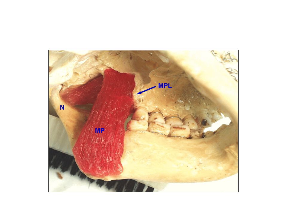
Internal pterygoid Elevator muscle
Origin: It has a superficial and a deeper head. The former arises from the tuberosity of maxilla & later from the medial surface of lateral pterygoid plate. Insertion: Together they run downward, backward & laterally to get inserted into the medial surface of angle of ramus.

16
Significance Just as buccinator decides the lower buccal posterior extension, internal pterygoid muscle determines the extension of a denture in the lower posterior lingual part of the denture space

18
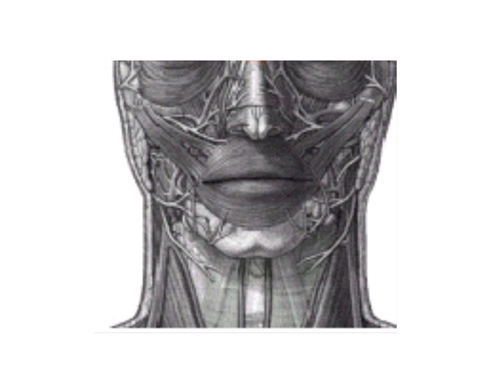
Muscles of facial expression

19
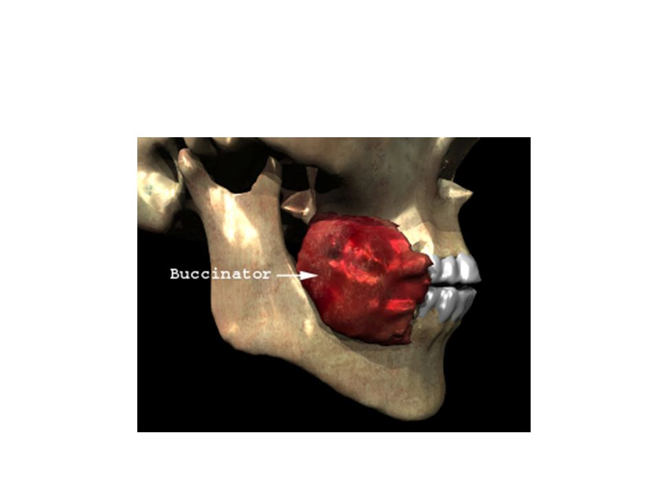
Buccinator Principle muscle of the cheek, which has horseshoe shaped origin Originates in the molar region at the base of the alveolar process and runs posteriorly and inferiorly past the maxillary tuberosity to continue into the pterygomandibular raphae. From there it runs to external oblique line and ends in 2nd molar region

21
Insertion: The upper, middle and lower fibres of the buccinator run horizontally to decussate and insert in upper and lower lip and modiolus.

22
Mandibular Rearmament :Merkeley : JPD 1959
Significance: Cheeks are pressed against the dental arches when buccinator contracts. During chewing and swallowing the muscle rhythmically contracts with muscles of mastication. It assists in placing the food between the teeth and returning the food to occlusal table which has escaped into the vestibule. Mandibular Rearmament :Merkeley : JPD 1959

24
The buccinator muscle gains attachment to the mandible via the external oblique ridge and the adjacent bone. Overextensions of a lower denture are common in this region, either because the impression material is viscous enough to excessively displace the soft tissues, or because the buccinator attachment is more medially than expected. Any such overextension will interfere with the buccinator in function and result in either displacement of the denture or pain from the traumatized mucosa.

25
Mentalis Origin: frontal surface of mandible between the lateral incisor and canine eminence, alveolar process Insertion: Muscles of both the side fuse and get inserted into the skin of the chin

27
Significance Origin of the mentalis is located closer to the crest of the residual ridge then the mucosal reflection in the alveololabial sulcus. The bottom of the sulcus is lifted when the muscle contracts thereby reducing the depth and the space of the oral vestibule.

28
Anteriorly, when resorption has been particularly severe, the mentalis muscle insertions can become prominent as two elevations on either side of the Mental foveae midline. The denture must be relieved over, and contoured around them. Extensions beyond their crest will interfere with the mentalis muscle movement and lead to denture instability.

29
Incisive labii inferioris
Origin: Arises from oblique ridge of the mandible below the canine Insertion: passes upward and medially to get inserted into the modiolus become fused with the fibres of orbicularis oris

31
Significance: It has the same characteristic course as the mentalis i
Significance: It has the same characteristic course as the mentalis i.e it originates near to the crest of the ridge and extends down and below the alveololabial sulcus. It’s contraction can reduce the denture space. In action it pulls the modioli forward and tenses the buccinator thereby applying pressure on the polished surface.

32
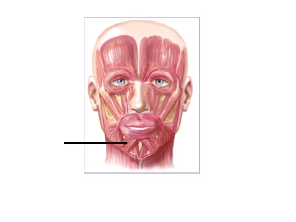
Orbicularis oris Origin: It is attached to maxilla by incisive labii superior and attached to the mandible by incisive labii inferior. Insertion: does not insert in to bone rather runs around the mouth Significance : It is active when the lips are pressed against the teeth. Like buccinator, orbicularis oris rhythmically contracts during chewing, sucking and swallowing

34
Modioli Orbicularis oris Zygomaticus major Zygomaticus minor
Levator labii superioris Levator aguli oris Buccinator Triangularis Risorius

35
Triangularis

36
Modiolus Hub of muscles which forms a knot of considerable strength with a wide versatility of movement ; up, down, forward and backward. Situated at the corner of the mouth it is in a strategic position to unseat the lower denture and sometimes the upper denture too. This may occur if the arch form is too wide and restricts the movement of the modiolus.

38
Modiolus can mold a soft material on the occlusal rim to correctly establish the shape and the anteroposterior position of the arch form of the anterior part thus establishing the buccal limit to which the bicuspids must be restricted. They help stabilize upper denture by placing premolar in a position above them. Thus during functional activities involving elevation of the lips there are fixing and elevation of the modioli and uplifting of the denture. At trial denture stage it restores the natural appearance of the mouth by moulding the material

39
Quadratus labii superioris
Levator anguli oris Zygomaticus major Risorius Triangularis

40
Other muscles

41
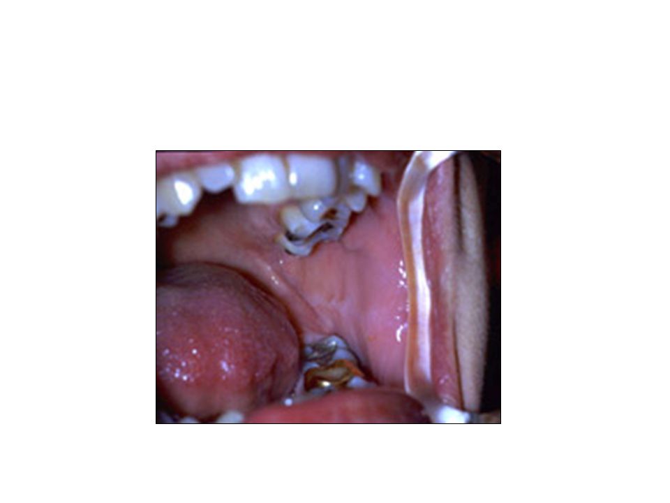
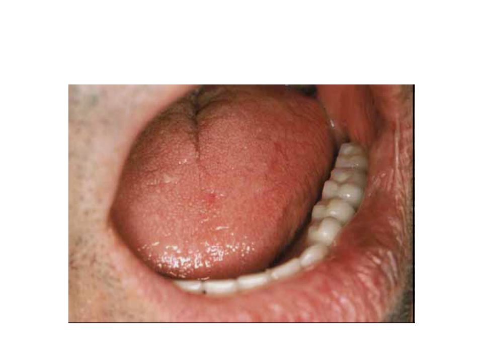
Tongue Powerful and extremely adaptable
It has two set of muscles: intrinsic and extrinsic Participates in speech swallowing sucking etc Normal tongue fills the floor of mouth and maintains the seal of mandibular denture Must be observed with the patients mouth half closed

43
A retracted tongue exposes the floor and compromises denture retention by losing the border seal.
A narrow dental arch encroaches upon the tongue, which can no longer occupy it’s rest position and tends to push the lower denture out. Occlusal plane placed at the level of tongue helps stabilize the denture and guide the food on to it.

44
Genioglossus On the lingual side of the mandible, also in the midline, the insertion of the genioglossus into the superior genial tubercle can appear surprisingly large especially if resorption . Further, the Large superior genial tubercle absence of an adequate alveolus means that antero/posterior movement of the denture is unrestrained and trauma commonly results.

45
Mylohyoid On the medial side of the mandible, extensions over the attachment of the mylohyoid can be made, but with care. It is essential that any extension integrates with the direction of insertion of the mylohyoid muscle and is inclined downwards and medially at an angle of approximately 45° to the sagittal plane occupying the cleft between mylohyoid and hyoglossus muscles.

46
Pterygomandibular raphae

47
Freni

48
Their importance lies in the fact that the denture periphery must be relieved around them otherwise pain and ulceration follow. In carrying this out the operator must be aware that the ‘notching’ of the base that results can cause structural weakness.

49
changes in edentulous mouth

50
Aging changes and the complete denture
: Lammie: JPD 1956

51
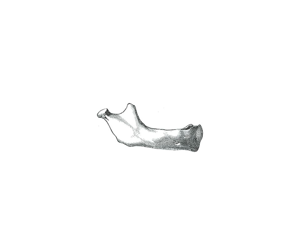
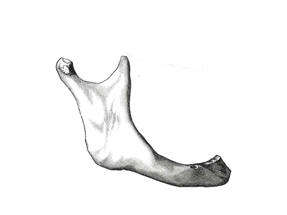
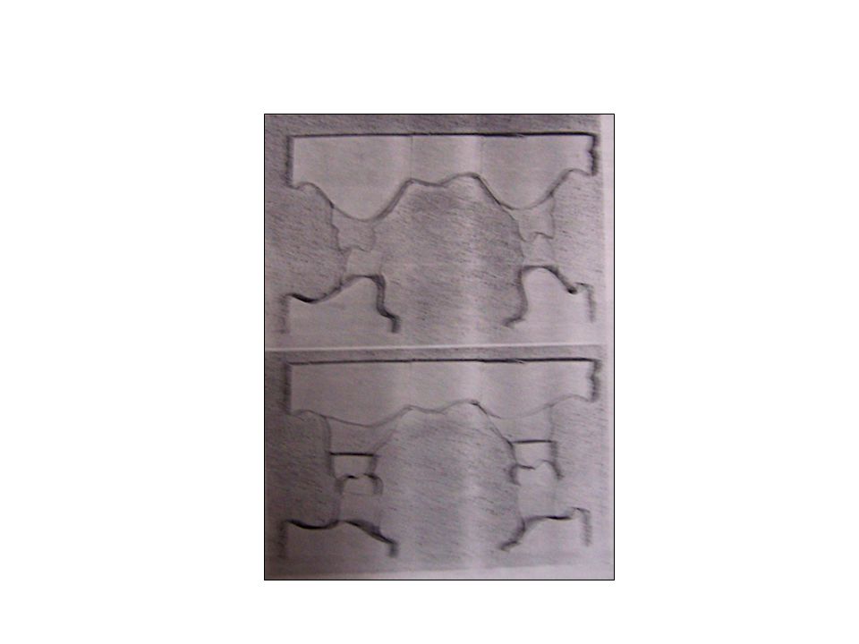
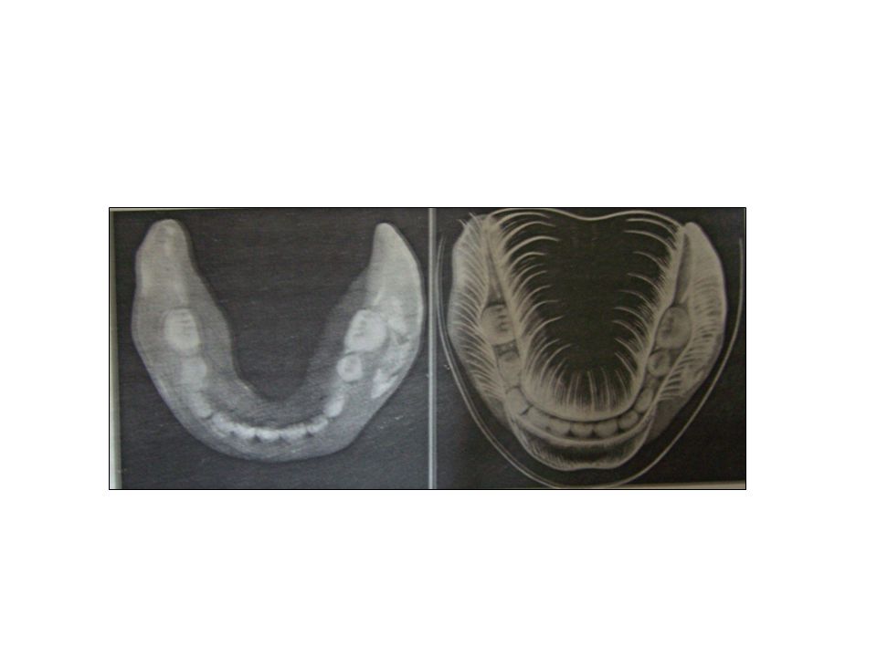
Maxilla and mandible Neither alveolar ridge resorbs uniformly. Mandibular residual alveolar ridges tend to resorb more from the lingual while maxillary residual alveolar ridges resorb more from the buccal. Usually, the longer a patient is edentulous, the greater is this interridge facial/lingual and facial/palatal dimensional disparity.

56
Changes over a period of 11 yrs

57
Lips and modioli Collapse of upper lip
Reduced prominence of philtrum and vermilion border Drooping of corners of mouth Modioli becomes sagging, less active, shape changes

58
Clinical applications of concepts of functional anatomy
Muscle attachment As the mandibular ridge resorbs the crest falls below the level of the mentalis. As a result mentalis tends to fold over and rests on the ridge. It pushes the neutral zone posteriorly. The freni occupy a more superior position on the ridge Clinical applications of concepts of functional anatomy : Martone : JPD 1962

59
Analysis of tongue factor : Kessler
In addition, it is estimated that tongue size increases by approximately 10% in the edentulous patient (Wright et al, 1961). This lingual increase contributes further to the confusion about optimum tooth placement, under the dislodging forces. Analysis of tongue factor : Kessler : JPD 1955
. This lingual increase contributes further to the confusion about optimum tooth placement, under the dislodging forces. Analysis of tongue factor : Kessler. : JPD")
60
The longer a patient remains edentulous the more facially (buccally/labially) the neutral zone will develop in relation to the mandibular residual alveolar ridge (Fahmi, 1992). Since residual alveolar ridges are spatially changing in a tight spaced functioning stomatognathic apparatus, it appears prudent to build prostheses that fit into current functional spaces and use local forces to enhance prosthetic function, stabilization and retention.

61
Lost-fine arts in fallacy of ridges:
Pound: JPD 1954

63
Denture surfaces

64
Impression surface Polished surface Occlusal surface

65
Denture surfaces Sir wilfred fish described (1948) three surfaces of a denture Impression surface Occlusal surface Polished surface

66
Impression surface That part of the denture in contact with the tissues and on which the denture rests. The retention of the denture depends on physical forces developed by adhesion and atmospheric pressure.

67
Occlusal surface It is that area in contact with the teeth, either natural or artificial of the opposite side. The forces develop by the muscles of mastication are received and directed by the occlusal surface. The stability of the denture in occlusion is determined by the fit of the impression surface against the tissues and occlusal surface against each other.

68
Polished surface This surface is constantly in contact with the cheek, tongue and lips. With RRR the impression surface decreases in size and the polished surface becomes more critical for stability and retention

69
Since most of the time jaws are at rest stability is more dependent on forces on external surface as transmitted to impression surface . The only time teeth make contact is during mastication and swallowing. In order to construct dentures that function properly not only in chewing but also in speaking and swallowing , one must develop the fit and contour of the external surface as accurately as that of impression surface and occlusal surface

70
Neutral Zone concept

71
Razek et al, 1981; McDonald et al, 1984; Searl, 1987;
Fish, 1933; 1947; Pound, 1954; Russell, 1959; Wright et al, 1961; 1966; Martone, 1962; Raybin, 1963; Heartwell, 1968; Strain, 1969; Boucher, 1970; Beresin et al, 1973; 1976; 1978; 1980; Winkler, 1979; Razek et al, 1981; McDonald et al, 1984; Searl, 1987; Dawson, 1989; Fahmi, 1992; Massad et al, 1993; 1997

72
Neutral zone is that area in the mouth where, during function, the forces of tongue pressing outward are neutralized by the forces of the cheeks and lips pressing inwards. Since these forces are developed through muscular contraction during chewing, speaking, swallowing etc they vary in magnitude and direction in different individuals and in different periods of life

73
The way these forces are directed against the denture will either stabilize or dislodge them. Our objective is to utilize this information to so position the teeth and the external surface that the force the musculature exerts will have a seating effect. This can be only accomplished by a knowledge of neutral zone and by positioning the teeth and developing the external surface so that all the forces exerted are neutralized.

74
Perhaps the greatest controversy lies in the arrangement of teeth
Perhaps the greatest controversy lies in the arrangement of teeth. This concept does not advocate placement of teeth on the ridge. Rather it is most of the time buccal or labial to it. According to pound “tooth over the ridge concept is a fallacy”.

75
Boucher (1975) “ Formerly all teeth were placed over the ridge. This was done for mechanical reasons when leverage was the big concern. Now however teeth are being successfully placed in the neutral zone which is in fact the zone previously occupied by the natural teeth. Leverage is not ignored but a lack of favourable leverage is counterbalanced by the controlling action of cheek, lips and tongue that confine the dentures. Thus the same factors that helped to position the natural teeth in the dental arches can help to maintain the artificial teeth in their places”.

77
Steps in fabrication of a complete denture based on neutral zone concept

78
Primary impression The primary and secondary impression are based on Tench”s Neuromuscular concept wherein functions of Sucking and Swallowing are used for recording an impression.

79
Physiologic complete denture impressions
: Barone : JPD 1963

80
Select the appropriate stock impression tray
Load the tray with irreversible hydrocolloid place it in the mouth To mold the labial and buccal segment either ask the patient to close the mouth and suck on the tray handle as hard as possible or ask the patient to say “Proo-wiss” with exaggerated movements of the mouth Ask the patient to move the mandible from side to side

81
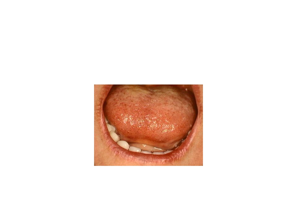
For mandibular impression ask the patient to push his tongue out
For mandibular impression ask the patient to push his tongue out . According to shanahan this forms the lingual handle. Lingual handle is an extension of the lingual flanges of the lower denture three spaces : sublingual crescent shaped space in the anterior part of the mouth Sublingual fossa over the mylohyoid muscle Retromylohyoid fossa which is below and behind the retromolar pad

82
Secondary impression

83
Prepare the custom tray with cold cure acrylic and the wax spacer.
Tray is adjusted in the mouth to ensure there is no interference with the free movement of lips, cheeks, freni , tongue. Tray must be 2mm short of the borders. Now border moulding is done with low fusing compound with movement similar to primary impression. Final impression is recorded after removing the spacer

84
The working impression of the denture-bearing surface is taken in such a way as to avoid the denture periphery encroaching onto muscle insertions. The most significant of these are the insertions of the buccinator, mylohyoid, and genioglossus, and the muscles of the lip (especially the mentalis).
..")
85
Functional trimming is often carried out when impressions are taken, during which the muscles surrounding the impression are stretched either physiologically or via traction by the clinician. Trimming in this way will ensure the periphery of the final denture does not cover muscle insertions and displacing forces from this source are minimized.

86
Many materials have been suggested for
shaping the neutral zone: modeling plastic impression compound,5 soft wax,7 a polymer of dimethyl siloxanefilled with calcium silicate,8 silicone,9 and tissue conditioners and resilient lining materials.

87
The special tray is a plate of acrylic adapted to the lower ridge, with spurs or fins projecting upwards towards the upper arch. These help with retention of the impression material. The upper base plate is made without spurs

88
A lower acrylic special tray with metal spurs to aid retention of the impression material

89
The upper wax rim is adjusted as in normal registration for a complete denture. The lower special tray is placed in the mouth. Two occlusal pillars are then built up in green stick compound on opposite sides of the lower arch. These pillars are moulded and adjusted to the correct height so as to give the usual 3mm freeway space.

90
Occlusal pillars have been built up in green stick to the correct occlusal height

91
Establishing the correct occlusal height

92
A thick mix of viscogel is then placed around the rest of the lower special tray, distally and mesially to the occlusal pillars. The patient is then asked to talk, swallow, drink some water etc. After 5-10 minutes the set impression is removed from the mouth and examined. The viscogel material will have been moulded by the patient's musculature into a position of balance.

93
The viscogel rim being moulded within the mouth

94
Viscogel Tissue conditioner Powder: polyethyl methacrylate
Liquid: ethyl alcohol+aromatic ester Mixed in high powder to liquid ratio to get high viscosity Visco-gel; Dentsply/DeTrey, Surrey, United Kingdom)
")
95
A completed viscogel impression

96
Russel (1959) Resin base is covered with a layer of sticky wax
then a portion of soft wax is added and built into occlusal rim Soft wax: kerr tissue seal wax + 4 spoon of mineral oil The height of rim is adjusted to corner of mouth and width is kept at 3-4mm. The rim is placed in water F for 3 min to soften it. Functional molding: for labial side patient is asked to make pro-wiss movement

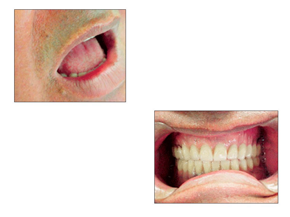
97
For lingual side patient is asked to touch the upper lip and move it from side to side
Reflect the lips and observe the molding The orbicularis oris will make a horizontal trough from one premolar area o another..In premolar area , modiolus will form a dish shaped depression. The lingual portion will be hollowed out Later the wax is chilled out.

98
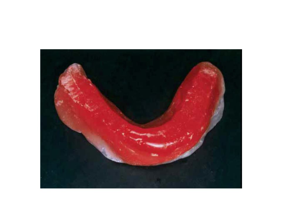
Mandibular record base with modeling plastic impression compound formed to patient’s neutral zone.

99
Imprint of maxillary occlusal rim into mandibular occlusal rim

100
Final maxillomandibular registration.

101
Flange technique(lott and levin)
")
103
Scheisser technique

105
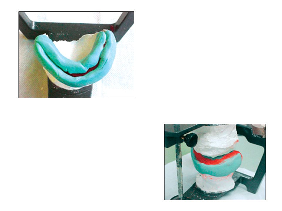
The record base with the molded rim was placed
on the mandibular cast, and buccal and lingual indices were carefully fabricated with silicone putty impression material. The TCM then was removed from the base. Indices were returned and sealed into place with sticky wax, and the empty space, representing the neutral zone, was filled with molten pink denture wax slightly below the level of the occlusal plane. The tentatively completed wax neutral zone rim then was ready for the final maxillomandibular registration

109
Separated index

111
Silicone lab putty to make indexes for neutral zone

118
Concave labial surface of lower denture
If the polished surfaces of a denture are generally concave, the action of the surrounding musculature is to stabilise the dentures. This is of special importance in the mandibular labial region , and if this surface is convex, the lower denture will tend to displace backwards and upwards by the powerful labial muscles.

Similar presentations