Download presentation
Presentation is loading. Please wait.

2
Dynamics of Dental Pulp & Periapical Pathologies
Deepti Awasthi P G student

3
CONTENTS Introduction Development Anatomy Structural features
Blood vessels Lymph vessels Nerves Functions Differences in primary & permanent pulp organ

4
Age changes Classification of pulpal pathologies Reversible pulpitis Irreversible pulpitis Acute apical periodontitis Phoenix abscess Periapical granuloma Radicular cyst External resorption Internal resorption Pulp necrosis Conclusion References

5
Definition Dental pulp, a small mass of connective tissue, blood vessels, and nerves located in a chamber within the dentin layer of a tooth. The pulp chamber is found in the crown and the root of a tooth. Mosby's Medical Dictionary, 8th edition. © 2009, Elsevier

6
:// Introduction

7
Regeneration of tooth pulp dentin
Development Regeneration of tooth pulp dentin

8
ANATOMY General features volume

9
Coronal pulp 6 surfaces pulp horns

10
Radicular pulp single , multiple tubular shape growth –more dentin – pulp gets narrower

11
location & shape may undergo changes
Apical foramen avg. size in adults location & shape may undergo changes frequently , there are 2 or more foramina seperated by a portion of dentin & cementum or by cementum only. orbans

12
Textbook of Endodontics – Nisha Garg
Accessory canals from radicular pulp laterally through the root dentin to the pdl tissue apical third clinically significant in spread of infection Mechanism of accessory canal formation : Textbook of Endodontics – Nisha Garg

13
STRUCTURAL FEATURES Histologically, Human anatomy II, DDS 09

14
Principal cells : odontoblasts fibroblasts undifferentiated mesenchymal macrophages lymphocytes dendritic cells Intercellular substances :

15
Intercellular substances
Dense & gel like Acid muco polysaccharide & protein polysaccharide During early development, chondroitin A,B & hyaluronic acid. Lends support to the cells of the pulp Transport of nutrients & metabolites Gag –hydrophilic,forms a gel – high tissue fluid pressure. orbans

16
Fibroblasts Most numerous Function –
Stellate shape & extensive processes Electron micrograph – RER , mitochondria etc Young pulp – divide & are active in protein synthesis Older pulp – spindle shaped , fibrocytes Mature & immature pulp- orbans

18
Inflammation & healing Fibres – type I & III length – 10 – 100nm
Dual function Inflammation & healing Fibres – type I & III length – 10 – 100nm cross striation at 64 nm After root completion, pulp matures & bundles of collagen fibres increase in number. Apical region orbans

19
Textbook of endodontics
Fibres are more numerous in radicular pulp than coronal & greatest concentration of collagen occurs in the most apical portion of the pulp. This is of practical significance as engaging the pulp with a barbed broach in the apex region affords a better oppurtunity to remove the tissue intact. Textbook of endodontics

20
Undifferentiated mesenchymal cells
Totiopotent cell Light microscope – large polyhedral cell ,large lightly stained centrally placed nucleus. Decrease in old age – reduces the regenerative potential of the pulp orbans

21
Odontoblasts 2nd most prominent
Adjacent to the predentin with cell bodies in the pulp & processes in the dentinal tubules. Diameter um Length – um Cell bodies – large oval nuclei , fills the basal part of the cell Adjacent to the nucleus basally, RER & golgi app Pulpal predentin junction – no organelles orbans

22
Odontoblasts mainly synthesize type I collagen ,proteoglycans & also secretes dentin sialoprotein, alkaline phosphatase , phosphophoryn involved in extracellular mineralization. Pathways of pulp

25
More cylindrical & longer – crown More cuboid – middle of root
Cell junctions : macula adherens tight junctions gap junctions More cylindrical & longer – crown More cuboid – middle of root Ovoid & spindle shaped – close to apex Releases IL-8 , chemotactic for neutrophils Nitric oxide synthetase – vasodilation & blood pressure regulation Berkowitz

26
DEFENCE CELLS Histiocyte / macrophage –
Inactive –difficult to distinguish from fibroblasts Pulpal inflammation- exhibit granules & vacuoles in their cytoplasm , nuclei increases Small blood vessels & capillaries Aggregate of vesicles or phagosomes orbans

27
Similar to langerhan’s In deciduous teeth – odontoblasts
Dendritic cells Similar to langerhan’s In deciduous teeth – odontoblasts Increase in areas affected by caries, attrition or restorative procedures Immunocompetent cells in deciduous teeth increased in number during shedding. Tencates

28
Mast cells – vessels in the inflamed pulp. Plasma cells – antibodies
Lymphocytes Mast cells – vessels in the inflamed pulp. Plasma cells – antibodies light microscope – nucleus appears small & concentric cart wheel appearance Orbans

29
lymphocyte Mast cell

30
BLOOD VESSELS The blood vessels – arise from inferior or superior alveolar artery & drain by same veins Small arteries & arterioles – apical canal & pursue a direct route to the coronal pulp. Along their course they give off numerous branches in the radicular pulp that passes peripherally to form plexus in the odontogenic region . Pulpal pressure – among the highest Tencates

31
Regeneration of tooth pulp & dentin

32
LYMPH VESSELS Endothelium lined tubes that join thin walled lymph venules or veins in the central pulp. Capillaries – thin walls Central part of the pulp. Lymph vessel draining the pulp & pdl – common outlet. Anterior teeth – submental Posterior teeth – submandibular & deep cervical nodes

33
NERVES non-myelinated 150-1200
Larger fibre range 5 & 13 um, majority are smaller than 4 um Perineurium & epineurium – thin Parietel layer, plexus of rashkow pulp horns berkovitz

34
The sensory nerves of the pulp arise from the trigeminal nerve.
The mature deciduous teeth-well innervated, esp coronal pulp , has many nerve endings terminating in or near odontoblast layer , with a few penetrating into dentin. No clear evidence that any sensation other than pain can be experienced fron pulp. The sensory nerves of the pulp arise from the trigeminal nerve. Pathways of pulp

35
Textbook of endodontics
A$ - myelinated faster pulp- dentin junction sharp,pricking, localized C – sympathetic efferents non myelinated throughout the pulp dull & more diffuse pain Textbook of endodontics

36
EPT stimulates A delta fibres first because of their lower threshold
EPT stimulates A delta fibres first because of their lower threshold. As intensity of stimulus is increased, some of the C fibres also get stimulated. The relatively late appearance of A fibres in the pulp may explain why EPT tends to be unreliable in young teeth since A fibres are more easily stimulated than C fibres. Pathways of pulp

37
FUNCTIONS Tencates INDUCTIVE FORMATIVE NUTRITIVE PROTECTIVE REPARATIVE
DEFENSIVE/ Tencates

38
Differences in primary & permanent pulp tissues
PRIMARY PULP Function for only about 8yrs & 3 mths duration 3 time periods : Period of growth of pulp organ : in about 1yr time , the crown & roots of the teeth develop orbans

39
2. Period of maturation of pulp:
root is completed & resorption of root begins at about 3yrs to 9 mths of age 3. Regression of pulp : period of regression of deciduous radicular pulp depends on the time from the completion of the permanent crown till the time of permanent tooth eruption. Max. life of primary pulp organ : 9yrs & 6mths. orbans

40
Primary teeth – 4yrs, 2mths
PERMANENT PULP ORGAN The pulp undergoes development for about 12 yrs ,4mths.( crown calcification to root completion ) Primary teeth – 4yrs, 2mths Period of pulp aging is much accelerated in primary teeth. Maxillary arches require slightly longer to complete each process of development. orbans
 Primary teeth – 4yrs, 2mths. Period of pulp aging is much accelerated in primary teeth. Maxillary arches require slightly longer to complete each process of development. orbans.")
41
AGE CHANGES Most conspicuous change – decreasing volume of the pulp chamber & root canal In old teeth , RC is often a thin channel. sometimes almost completely obliterated. Continued restriction in pulp volume –decrease in vascular supply & initiates other age changes. orbans

42
Reduction in size & no. of cytoplasmic organelles.
Cell changes Reduction in size & no. of cytoplasmic organelles. 20 yrs of age , cells gradually reduce in no. until age 70 ,cell density has decreased by about half. orbans

43
Fibrosis Radicular pulp – arranged longitudnally in bundles
Coronal pulp – random/diffuse Any external trauma – localized fibrosis or scarring effect. Collagen increase is noted in media & adventitia layer of blood vessel

44
Vascular changes Reduced vascularity Blood flow is decreased.
Calcification in the walls of blood vessels – near apical foramen Atherosclerotic plaques may appear Tencates

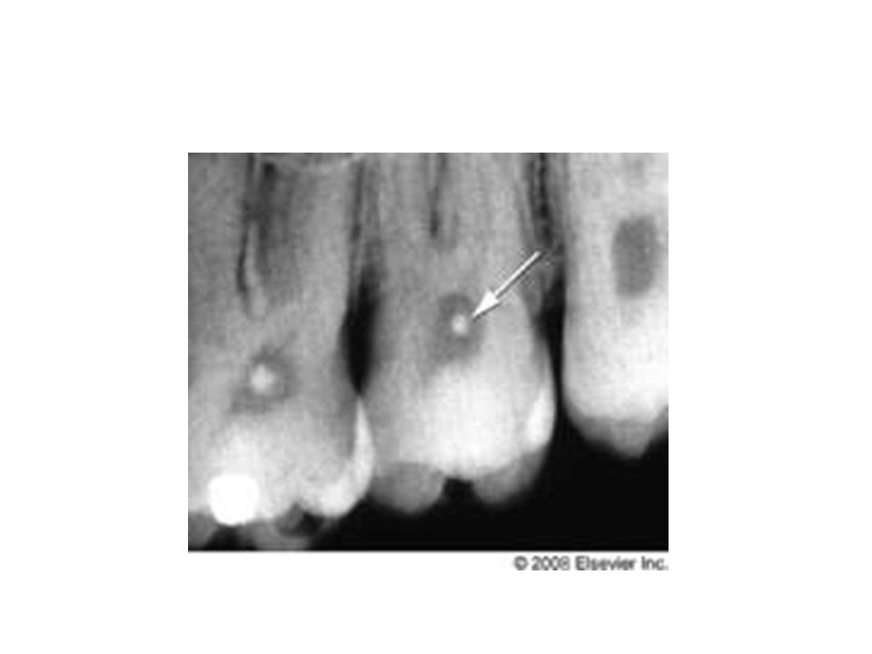
45
Pulp stones Denticles Calcified masses appearing in either or both the coronal & root portions of the pulp organ Usually are asymptomatic, unless impinge on nerve or blood vessel.

47
Textbook of endodontics
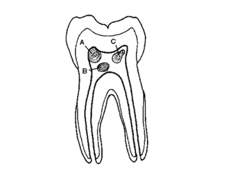
CLASSIFICATION Acc to structure : true false Acc to relation with Dentin : free attached embedded Textbook of endodontics

49
True Similar to dentin Close to the apical foramen
Development – inclusion of remnants of HERS within the pulp. they induce the cells of the pulp to differentiate into odontoblasts which form the dentin masses. Tencates

50
False Appear as concentric layers of calcified tissues
They appear within a bundle of collagen fibres. Some arise around vessels. In the centre – remnants of necrotic & calcified cells. Phlebolith – may serve as nidi for false denticles. Tencates

51
Free – Attached- Embedded- 90% - > 50yrs of age
Tencates

52
Diffuse calcification
Irregular calcific deposits in the pulp tissue usually following collagenous fibre bundles or blood vessels. usually found in the root canal , whereas denticles are more in coronal pulp. orbans

53
Textbook of endodontics
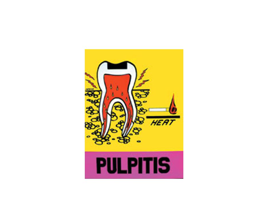
CLASSIFICATION Acc to Grossman: Pulpitis Reversible symptomatic asymptomatic Irreversible acute responsive to cold responsive to heat Textbook of endodontics

54
chronic Asymptomatic hyperplastic pulpitis internal resorption Pulp degeneration calcific Others necrosis

55
WHO Classification K04.4 – AAP K04.5 – CAP K04.6 – PA with sinus
K04.60 – PA with sinus to max antrum K PA with sinus to nasal cavity K PA with sinus to oral cavity K PA with sinus to skin K PA without sinus K PC K apical & lateral cyst K Residual cyst K inflammatory paradental cyst Pathways of pulp

56
Ingle’s Classification
Painful Pulpoperiapical Pathologies 1. AAP 2. Advanced AP a. Acute Apical Abscess b. phoenix abscess c. suppurative apical periodontitis

57
Non – Painful 1. Condensing osteitis 2. CAP - incipient stage 3. CAP - Periapical granuloma - periapical cyst - suppurative apical periodontitis

59
Acute Apical Periodontitis

60
Acute Apical Abscess

61
Phoenix Abscess An acute inflammatory reaction superimposed on an existing chronic lesion, such as cyst or granuloma.

62
Periapical Granuloma

63
Radicular Cyst

64
External Root Resorption

65
Internal Resorption

66
Pulp Necrosis

67
Textbook of endodontics
Effect of posture on pulpal flow In normal upright posture , there is less pressure effect in the structure of head. Lying down increases blood flow to pulp by removal of both gravitational & baroreceptor effect. Textbook of endodontics

68
Regeneration of tooth pulp & dentin
Regeneration of pulp dentin Regeneration of tooth pulp & dentin

69
conclusion

70
References Orban’s Oral Histology &Embryology- 12th ed
Pathways of the pulp –stephen Cohen 9th ed Textbook of endodontics – Nisha garg & Amit garg Oral histology –Tencates Berkovitz Regeneration of tooth pulp and dentin : trends and advances. Sarang Sharma, Vimal Sikri.Annals of Neurosciences, Volume 17, Number 1, January 2010

Similar presentations


>")


 - epithelial rest cells -immune system cells - cells associated with neurovascular elements.>")
