Download presentation
Presentation is loading. Please wait.

1
FEI NovaNano FEG-SEM 630

2
Technical Objective To train you to use the FEI FEG HR-SEM electron microscope High vacuum operation for conductive samples Low vacuum operation for non-conductive samples and avoidance of “crud deposition” This machine requires your full attention –Very easy to damage –Very expensive to repair –Damage is not covered under the very expensive service contract

3
Vacuum system layout Chamber Ion Getter pumps Turbo pump Scroll pump Capacitance gage Cold cathode gage (Penning) Chamber
 Chamber")
4
Scroll pump (Oil-free)
")
5
IR CCD camera Ion getter pumps

6
Ion getter pumps Everhart – Thornley detector

7
Specimen current monitor Plasma cleaner Cold cathode ion gage Plasma head Low vac detector power supply Door! (Linear Bearings) Aperture adjustment Outer ring: size Inner ring: position Height adjusting “elephant” “600 A”
 Aperture adjustment Outer ring: size Inner ring: position Height adjusting elephant 600 A .")
9
Stage Height gage “elephant” “600 A” Sample holder

10
Sample mounting fixture

11
IR CCD Pole piece (No detectors attached Everhart- Thornley Detector Low Vac detector jack

12
Console Accounting system User interface

13
Knob panel Brightness Contrast Stigmation FocusImage Shift Magnification Coarse Fine

14
Sample Mounting FEI uses mounting stubs with 1/8” shafts Carbon tape (double sticky) is simplest Carbon dots have lower vapor pressure Multiple samples can be mounted on “turret” Make a drawing in your research notebook of the locations of your samples on the “turret” Sample locations are 19 mm apart and 19 mm off center Single mount is easiest for tilting; tilting the “turret” will be extremely dangerous Vertical mount is also an option Tighten gently with Allen wrench; ditto for sample holder on stage
 is simplest Carbon dots have lower vapor pressure Multiple samples can be mounted on turret Make a drawing in your research notebook of the locations of your samples on the turret Sample locations are 19 mm apart and 19 mm off center Single mount is easiest for tilting; tilting the turret will be extremely dangerous Vertical mount is also an option Tighten gently with Allen wrench; ditto for sample holder on stage")
15
Notes on the computer Left computer is “support computer”: left monitor –Connected to the internet –No internet use whatsoever except data transfer –Use USB ports on front to extract your data –Never install software Right computer runs microscope: right monitor –Never insert a memory stick –Never change anything –Never, ever, ever install software! Move between computers by moving mouse left or right

16
Administrative matters Nothing in life is free Log onto the SEM with the sheet on the desktop Enter your name Enter the name and contact information of whoever will be paying Record how many times you coated samples Enter the time of day when you start. Enter the time of day when you finish. If we learn that your times are inaccurate, you will be required to pay an operator to document your time

17
Getting started with the FEI NovaNano HR-SEM Typical conditions when you walk in: –All pumps are running –High Voltage is off –FEG is hot (FEG is always hot!)
")
18
Status module

19
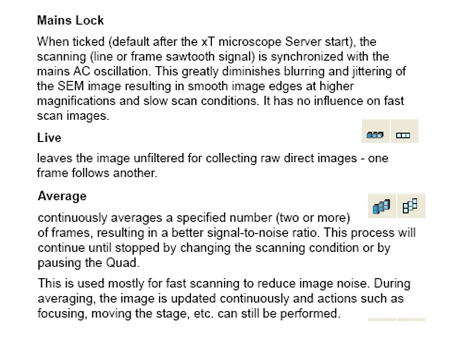
Random stuff F5 cycles between full screen and 4-quad mode. Activate one of the four quadrants by clicking anywhere in it. The databar at the bottom will become blue. Double green bars in the upper right-hand corner of an image mean that it has been paused and is not active Un-pause a window by clicking on the pause button on the button bar

20
How the software works

23
Right click to access these options

24
Starting and stopping the control software On desktop

26
Tool bar and button bar (Many SEM functions are mouse-controlled)
")
27
Log off user: log off when you are done! User Use “Open” to Import an image Use “Save as” the first time you save an image

29
Extremely useful when you have no image! “Horizontal line”: current raster

31
..and e-beam lithography!

37
This must be done every time The software Is rebooted. n.b.! Stage will tilt; your sample will fall off if not fastened!

39
Magnification control options

40
Conductive or non-conducting sample? –High vacuum for conductive –Low vacuum for non-conductive (coat if possible) Install low-vac detector Choose initial parameters –High voltage (20 kV) for maximum resolution –Low voltage (1-3 kV) for maximum topographic detail –Low current (spot size 1-2) for max resolution –Higher current for better signal-to-noise
 Install low-vac detector Choose initial parameters –High voltage (20 kV) for maximum resolution –Low voltage (1-3 kV) for maximum topographic detail –Low current (spot size 1-2) for max resolution –Higher current for better signal-to-noise.")
41
To insert your sample Check that high voltage (HV) is off (no yellow background) Click on Vent Insert sample. Wear gloves or use tools. Check sample height with “elephant nose”. Bottom of nose is where highest point on sample should be. Click on quadrant showing CCD image Make sure image is live, not paused! (No double green bars in upper right corner.) Slowly close door while watching monitor to assure that sample/holder is clear of pole piece and detector Click on “Pump” with left hand while holding door shut with right hand. Check that door is shut!
 Slowly close door while watching monitor to assure that sample/holder is clear of pole piece and detector Click on Pump with left hand while holding door shut with right hand. Check that door is shut!.")
42
Getting started toward an image When adequate vacuum has been reached, chamber icon becomes green and HV button changes from grey to black. Click on HV!

43
Get an image! Black screen: –Check that H(igh) V(oltage) is on: yellow background? HV automatically shuts off when switching between High Vac and Low Vac! –Go to minimum magnification Can you see the aperture? If not, you are in big trouble. Check HV! Try spot size = 4, 15 kV –Turn on waveform feature Increase contrast and brightness so waveform fills region between lines

44
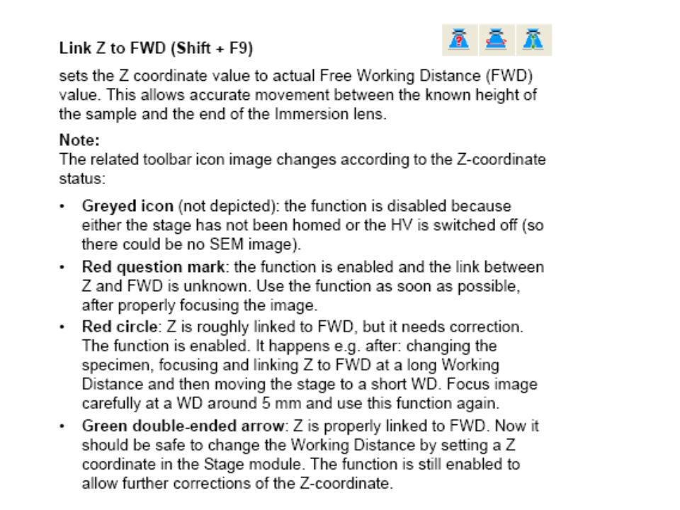
Link Initially your stage should be about 13 mm down..but could be anywhere: it has no way of knowing where you mounted the top of your sample! Focus Increase magnification Focus Link Z-coordinate is now correct Less crucial if using only samples of minimal thickness Absolutely crucial for samples of widely varying thickness Extremely crucial if sample is taller than elephant nose!

45
Image optimization With Everhart-Thornley or Low Vac Detector, raise stage to about 5 mm –Typically during pump-down with roller ball –With cursor low on screen, press roller ball and roll mouse up –Watch active (not paused!) image of sample mount on CCD –Translation speed is proportional to distance cursor is from starting point
 image of sample mount on CCD –Translation speed is proportional to distance cursor is from starting point")
46
Increase magnification Re-adjust focus Repeat process At about 5kX, link Raise sample to 5 mm Switch to TLD (Through Lens Detector) Re-focus Go to immersion mode Optimize image
 Re-focus Go to immersion mode Optimize image")
47
Ian’s Recipe for Image Optimization Set reduced raster to a tall, narrow box Scan as rapidly as allowed by contrast Try to focus on a vertical feature Adjust stigmation for optimal image Move box across screen if object degrades Blank beam Shift stage or beam to virgin sample (arrow keys move state 80% of screen dimension) Return to full screen scanning Reduce scan speed for high image quality CTRL R restarts scan Immediately un-blank beam Click “Pause”
 Return to full screen scanning Reduce scan speed for high image quality CTRL R restarts scan Immediately un-blank beam Click Pause")
48
Labeling your Image

50
(At the bottom of the Scan menu)
")
52
To save your image Pause symbol will blink as image scans Pause symbol will stop blinking when scanning has stopped Insert memory stick into Support (left) pc File/Save as… Choose “Network places” Choose e:USB…on Support pc Choose an appropriate filename for your sample, preferably ending in …_001.tif For black and white images, choose tif8 For images with color markings (labels), choose tif24
 pc File/Save as… Choose Network places Choose e:USB…on Support pc Choose an appropriate filename for your sample, preferably ending in …_001.tif For black and white images, choose tif8 For images with color markings (labels), choose tif24")
53
Beam Detectors Navigation Processing Control Double click to enter arbitrary number Right menu common to “all” “pages” “Pages”

54
Beam control

56
Focus wobble Probably better to start via the “crosshair target” button on the button bar.

58
Immersion mode

59
Apertures Available sizes and uses –2@30 μm: highest resolution –40 μm: low kV, low vac –50 μm: low kV, EDX –100 μm: EDX –1 mm: for service engineer for alignment Apertures are changed mechanically by rotating the knob on the side of the column…the one on the right After changing apertures it is necessary to align it with the screws on the sides (coarse beam align) Also “manually” change the value stored for “aperture” under the “beam” menu
 Also manually change the value stored for aperture under the beam menu")
60
Detectors page Proper grid voltage is very important in Low vac mode Choose detector Choose operating mode: SEBSE Charge neutralization Down-hole High vacuum Everhart Thornley TLD Low vac LVD Helix STEM

62
Secondary electrons give best topographic Information BSE give elemental contrast

63
TLD has four operating modes THROUGH THE LENS DETECTOR (TLD) Secondary electron Backscatter electron Charge Neutralization Down-hole visibility ETD and TLD both feed the same output, so you cannot view them simultaneously. You can view them sequentially in Quad 1 and Quad 2, then mix the images in Quad 3.

64
Magnetic hazards Ferromagnetic particles are potentially the most dangerous thing you can image If they get sucked into the column, the rebuild will cost $60-100k! Trying to image loose ferromagnetic particles in immersion mode will be fatal!!! They can be imaged in EDX mode They can be imaged in immersion mode if firmly embedded in plastic. Do not use tape!!

65
Secondary Electron Detector Backscattered Electron Detector Fractured Aluminum

66
More stuff on running the NovaNano The generic working distance for the NovaNano is 5 mm. –ETD –TLD –LVD –Also the eucentric working distance –Focus to at least 5000X, then Link. Do this early in the process. Do it repeatedly…especially if using the Helix detector

67
Furthermore If you are using the 2-sample holder, the pins are 16 mm apart If you are using the 8-sample holder, the pins are 19 mm apart. –Always make a drawing of the locations of your samples on the holder –Be extremely careful of orientation: get the sides of the holder on the translation axes –Remember that the CCD camera is mounted in the rear of the chamber

68
Further still more You can move the stage in the x- and y- directions by typing coordinates into the boxes in the “Navigation” page You can also move the stage from the image (not CCD) by holding down the roller ball and moving the mouse –The arrow points in the direction you want to look –The arrow points in the direction opposite to the one in which the stage moves
 by holding down the roller ball and moving the mouse –The arrow points in the direction you want to look –The arrow points in the direction opposite to the one in which the stage moves")
69
Still further more yet If you raise the stage by typing a value into the box on the “Navigation” page –Activate by clicking the GoTo button –“GoTo” will immediately change to “Stop” –Watch the rising stage on the CCD monitor –If the stage is in danger of hitting the detector or pole piece, immediately click “Stop” –“Escape” is an alternative “Panic button”

70
“"When I Use a Word, It Means Precisely What I Want It To Mean....“ Humpty Dumpty In the lexicon of Leo, “Beam current” is the current leaving the filament and “Probe current” is an approximation to the current incident on the sample, usually high by about a factor of two. From FEI, “Beam current” is an excellent approximation to the current actually striking the sample.

71
Low vacuum Why? For non-conducting samples or when you want to avoid rapid deposition of crud on your sample. ETD does not work New detector must be installed…while wearing gloves –LVD is relatively cheap and adequate for images up to about 50 kX –Helix is very expensive but gives killer images on non-conducting samples to 500 kX or more! PLA must be installed sufficiently tightly that it does not leak and kill the column

72
Pressure limiting Aperture (PLA)
")
Similar presentations