Download presentation
Presentation is loading. Please wait.

1
Cutaneous Vascular Diseases, Part 1 Rick Lin, DO MPH KCOM Dermatology Residency Program

3
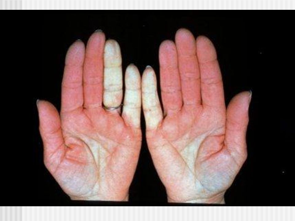
Raynaud’s Phenomenon Intermittent constriction of the small digital arteries and arterioles Persistently cyanotic and painful Aggrevated by cold weather Young middle aged women Assoc c scleroderma, dermatomyositis, LE, Mixed connective tissue diseases, Sjogren’s RA, and paroxysmal hemoglobinuria.

4
Raynaud’s Phenomenon Scleroderma is the underlying condition for more than half of the patients Maybe caused by medications, ie bleomycin

8
The LDI images below graphically illustrate the vasospasm of Raynaud’s phenomenon following a cold provocation. Baseline Cold Exposure 7.5 min recovery

9
Raynaud’s Disease Primary disorders Pallor, cyanosis, hyperemia, and numbness of the finger Precipitated by cold. Present for 2 years with out associated disease finding Good prognosis

10
Raynaud’s Disease Multifactorial. Increase alpha-2 sympathetic receptor activity on vessels. Endothelia dysfunction Deficiency in calcitonin gene related protein Central thermoregulatory defects

11
Raynaud’s Disease Treatment include avoidance of aggravating factor, ie cold. Vasodilating drugs, nifedipine, 10-20 mg tid; prazosin 1-3 mg tid Nitroglycerin 2% local application

12
Erythromelalgia Aka erythermalgia and acromelalgia May be secondary to myeloproliferative disease such as polycythemia vera, TTP Responds to treatment of primary disorders Cold water immersion

14
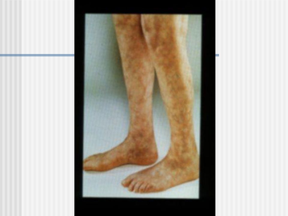
Livedo Reticularis Mottled or reticulated pink/reddish/blue discoloration Assoc c LE, DM, scleroderma, RA Side effect of amantadine

18
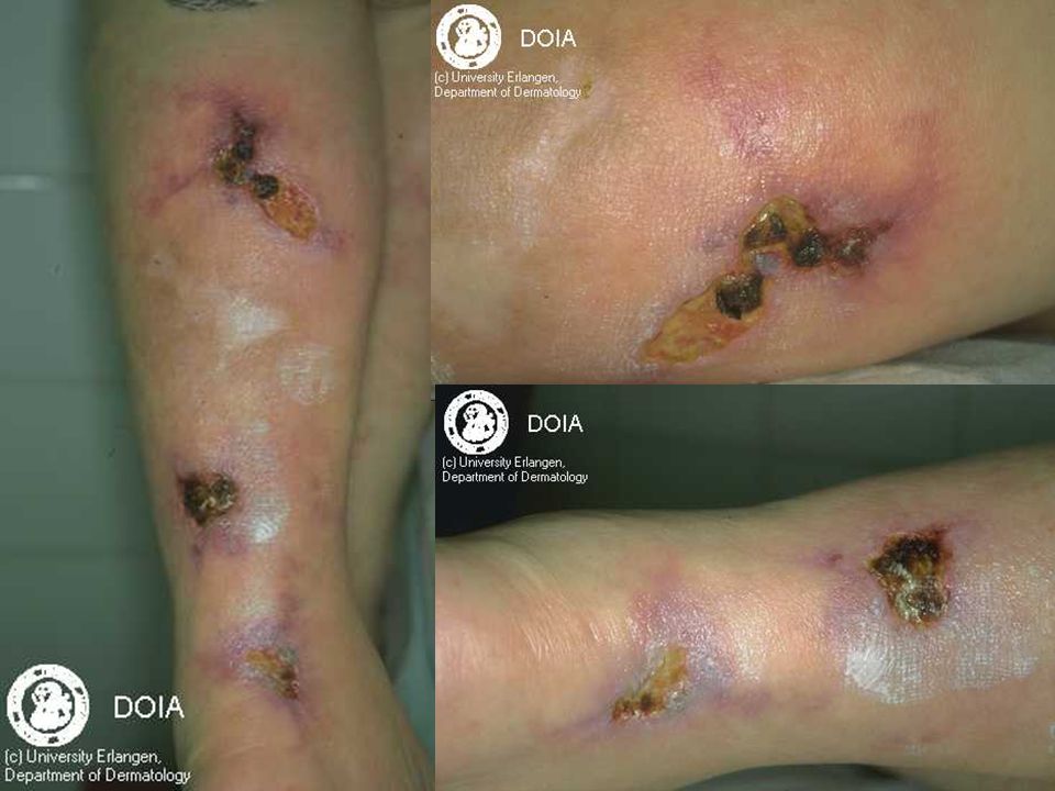
Necrotizing livedo reticularis Assoc c nodules and ulcerations Result from sever atherosclerotic disease Sneddon’s syndrome

19
Livedoid Vasculitis atrophie blanche White stellate scars of ulcers PURPLE (Painful purpuric ulcer with reticular pattern of the lower extremity) Histologically shows chronic perivascular hemorrhage.
 Histologically shows chronic perivascular hemorrhage.")
20
Treatment Low Dosage of Aspirin 325mg qd Nifedipine 10mg TID Pentoxifylline 400mg BID-TID

23
Marshall-White Syndrome Bier’s Spot: White spot appear on hand with blood pressure cuff Consist of Bier’s spot and is associated with insomnia and tachycardia White middle age men

25
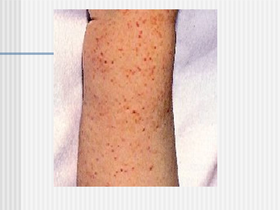
Purpura Multifocal extravasation of blood into the skin Petechiae <3mm Ecchymosis Vobices (vibex) – Linear Hematoma – pool-like collection
 – Linear Hematoma – pool-like collection")
26
Purpura Complete blood count PT and PTT

27
Thrombocytopenic Purpura Three Large Categories: Accelerated platelet destruction Deficient platelet production Unknown pathogenesis

28
Idiopathic Thrombocytopenic Purpura Aka autoimmune thrombocytopenic purpura Aka Werlhof’s disease Bleeding occurs when platelet count drops below 50,000 Risk greatly increased for serious hemorrhage when count goes below 10,000

29
Idiopathic Thrombocytopenic Purpura Acute variety occurs in children following season viral illness in 50% of the patient. Lag between illness and onset of purpura is 2 weeks Resolve spontaneously with minimal therapy Chronic case may result in death.

30
Idiopathic Thrombocytopenic Purpura Chronic form most often occur in adult Evaluate patient with Tc99M radionuclide scan to look for accessory spleen Result of platelet injury by antibodies of IgG class Treatment include Splenectomy, systemic corticosteroid, IVIg

32
These are the kidneys from a case of idiopathic thrombocytopenic purpura. Petechiae are found throughout the renal parenchyma.

35
Drug-Induced Thrombocytopenia Drug induced antiplatelet antibodies May be caused by sulfonamides, digoxin, quinine, quinidine, PCN, furosemide, Lidocaine, methyldopa

36
Thrombotic Thrombocytopenic Purpura Aka Moschcowitz syndrome Pentad of thrombocytopenia, hemolytic anemia, renal abnormalities, fever, CNS disturbance. Delay in diagnosis may lead to a mortality rate as high as 90%

37
Thrombotic Thrombocytopenic Purpura Positive histologic diagnosis require gingival biopsies looking for subendothelial hyaline deposits Exchange plasmapheresis is required for treatment. 80% patient survive if treatment is instituted.

38
Dysproteinemic Purpura Aka Nonthrombocytopenic purpura Aka purpura cryoglobulinemica Aka cryofibrinogenemia Occur most frequently in multiple myeloma and macroglobulinemia of monoclonal IgM, IgG, or Bence Jones cryoglobulin. Tx with plasmaphoresis, systemic steroid, and immunosuppressors.

39
Purpura Hyperglobulinemica Aka Waldenstrom’s hyperglobulinemic purpura Consist of episodic showers of petechiae occuring on all parts of body Diffuse peppery distribution, resembling Schamberg’s Most useful labtest is protein electrophoresis

40
Purpura Hyperglobulinemica Hyperglobulinemic purpura occurs most commonly in women. Frequently seen with Hepatitis C and Sjogren’s syndrome, keratoconjunctivitis sicca, RA Histologically: derma vessels with perivascular infiltrate of mononuclear cells. Benign and chronic course. Assoc c various of connective tissue diseases.

41
Waldenstrom’s Macroglobulinemia Bleeding from mucous membrane of the mouth and nose, lymphadenopathy, hepatomegaly, retina hemorrhage, and RARELY the purpura Perivasular infiltrate containing lymphocytes and neutrophils and eosinophils

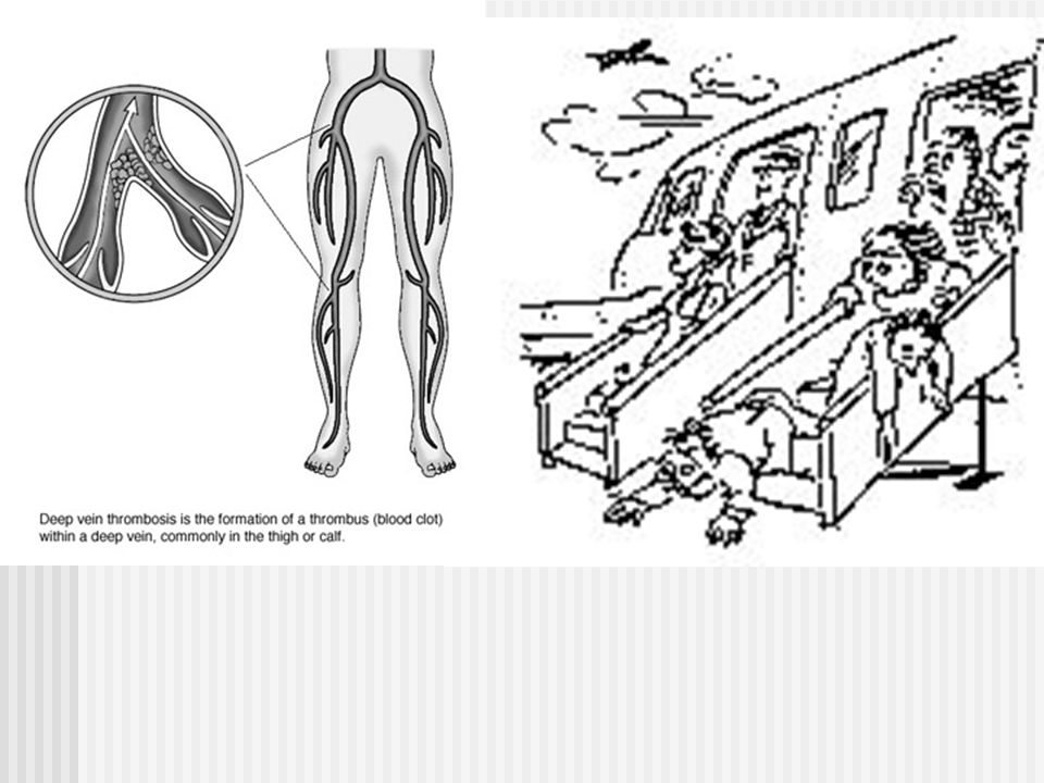
42
Waldenstrom’s Macroglobulinemia Plasmaphoresis until adequate dose of chlorambucil is administered. Cyclophosphamide and corticosteriods are treatment options as well

43
A Skull X Ray showed a single, small, left-sided lytic Defect.

44
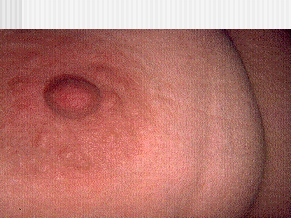
Drug- and Food Induced Purpura Drug induced purpura may occurs without platelet destruction. Cocaine induced thrombosis with infarct skin assoc c skin popping. Rumpel-Leede sign: distal shower of petechiae that occurs immediately after the release of pressure from a tourniqut release. Associated with capillary fragility. Topical EMLA can induce purpura in 30m.

45
Solar Purpura Large, sharply outlined 1-5 cm dark purplish red ecchymoses on dormsum of the forearm Less frequently, back of the hand

47
Purpura Fulminans Aka purpura gangrenosa Severe, rapidly fatal reaction occurring most commonly in children after infectious illness May follows scarlet fever, strep pharyngitis, and meningococcal meningitis. Assoc c Protein C or S deficiency in Neonates

48
Purpura Fulminans Management is supportive Protein C replacement if protein C deficiency is present Fresh frozen plasma maybe useful

52
Disseminated Intravascular Coagulation Up to 2/3 of DIC patients have skin lesions Minute, widspread petechiae, ecchymoses, ischemic necrosis of the skin and hemorrhage bullae. Elevated PT and PTT, fibrin degradation products Decrease platelets, decreased fibrinogen

53
Disseminated Intravascular Coagulation All patient needs to receive vitamin K replacement to exclude vitamin K deficiency.

54
Fibrinolysis Syndrome An acute hemorrhagic state brought by inability of the blood to clot Massive hemorrhages into the skin produce blackish, purplish swelling. Can be a complication of pregnancy in cases of placental previa, eclampsia, and fatal death Excessive fibrinolysis

55
Blue Muffin Baby Purpuric lesions observed in newborns with congenital rubella Assoc c disease that produce extramedullary erythropoesis Generalized dark blue to magenta nonblanchable, indurated, round, oval hemispheric papules 1-7mm Evaluation c biopsy, TORCH serology, CBC, viral culture.

56
Fig. 1. Blueberry muffin baby. A, Extensive lesions of dermal erythropoiesis in infant with erythroblastosis fetalis. B, Facial lesions in infant with congenital cytomegalovirus infection.

57
Itching Purpura Aka diseminated pruriginous angiodermatitis Orange-purpleish-red petechiae evolve completely and may become confluent in 2 weeks Runs its course in 3-6 months. May become chronic Unknown etiology

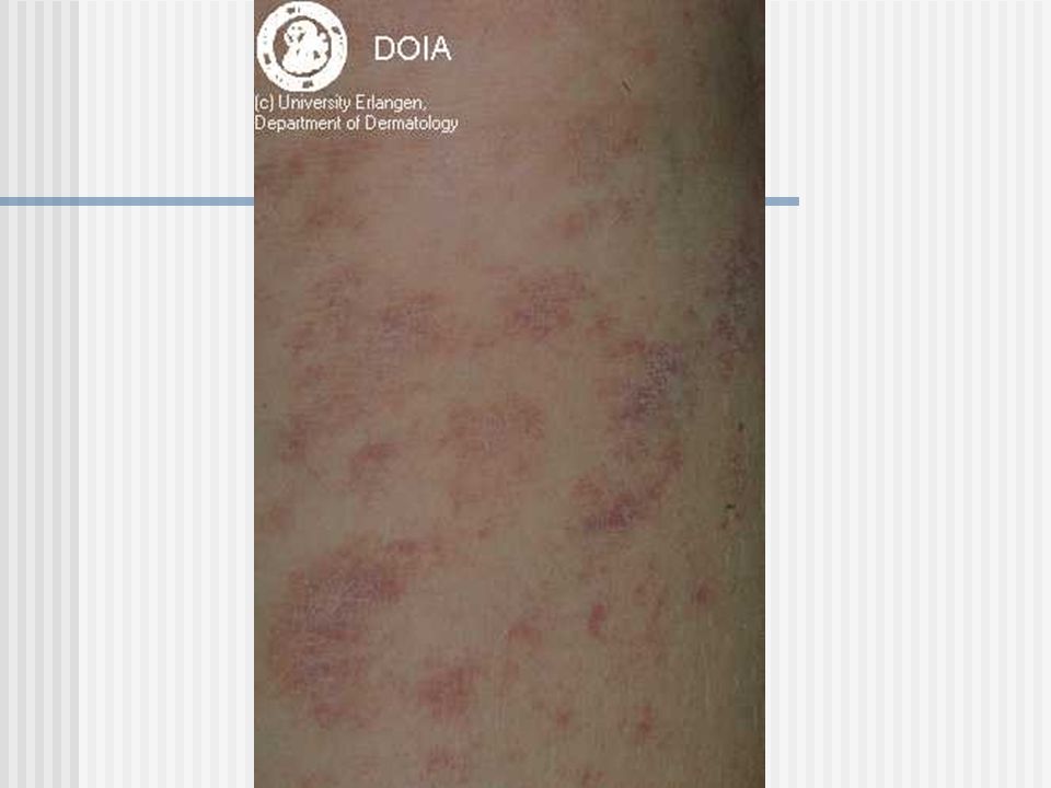
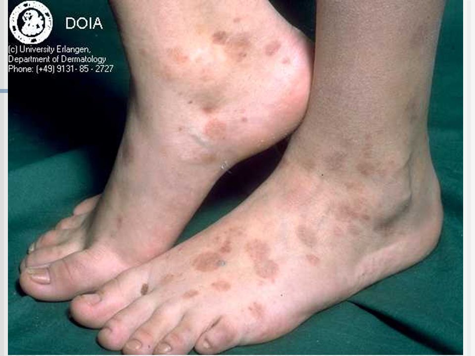
58
Deep Vein Thrombosis Almost always affect femoral vein Leads to Reversible ischemia or frank gangrene Significant superficial vein thrombosis is a risk factor for DVT Pulmonary Embolism is a major concern Malignant Neoplasms are the most common underlying condition

59
Deep Vein Thrombosis Pulmonary Embolism has 40% mortality DVT is assoc c 35% of cancer associated causes as the first sign Patient younger than 40 with DVT prompt for search for cancer

62
Superficial Thrombophlebitis Painful induration with erythema Linear or branching configuration forming cords Assoc c hypercoagulable state Need to be evaluated for possibility of deep veous disease

63
Mondor’s Disease 3:1 = women:men Age range 30-60 Sudden appearance of a cord like thrombosed vein along the anterior- lateral chest wall First red and tender and subsequently change into a painless tough, fibrous band.

64
Mondor’s Disease No systemic symptom associated Treatment of the symptom: hot moist dressing with NSAID Runs its course for 3-6 months.

68
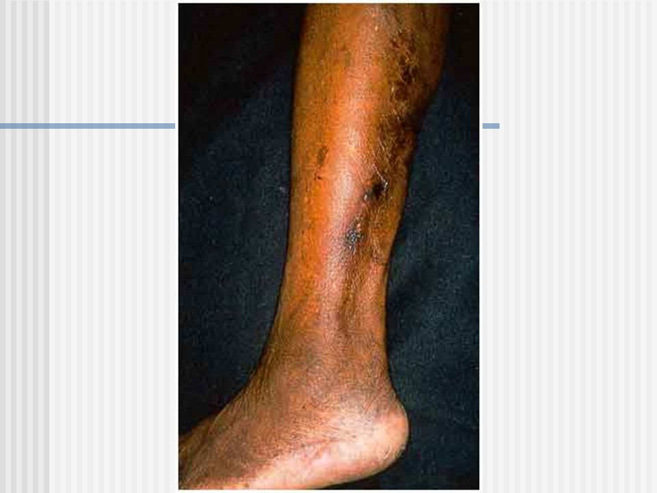
Calciphylaxis End-stage renal disease patients with metastatic calcification are most exclusively affected by this disease. Reticulated violaceous, mottled patches Progress into ecchymosis, central necrosis, and ulceration

69
Calciphylaxis 50%+ morbidity and mortality Death is usually caused by staphylococcal sepsis after infection Hyperbaric oxygen has used with some success

73
On low magnification, basophilic alteration of a fibrous septum can be seen Fibrin thrombi are also present within many of the blood vessels of the subcutaneous adipose tissue (Figure 3). Higher magnification identifies calcium deposition within the fibrous septum, primarily on elastic fibers (Figure 4, Figure 5). Higher magnification of the same area with Verhoeff-van Gieson stain confirms the presence of fragmented elastic fibers (Figure 6). Special stain for calcium (von- Kossa) identifies calcium deposition both within the septum of the fat lobule (Figure 7) and within the walls of blood vessels (Figure 8).Figure 3Figure 4Figure 5Figure 6Figure 7Figure 8
. Higher magnification of the same area with Verhoeff-van Gieson stain confirms the presence of fragmented elastic fibers (Figure 6). Special stain for calcium (von- Kossa) identifies calcium deposition both within the septum of the fat lobule (Figure 7) and within the walls of blood vessels (Figure 8).Figure 3Figure 4Figure 5Figure 6Figure 7Figure 8.")
74
Fibrin thrombi are also present within many of the blood vessels of the subcutaneous adipose tissue

75
Higher magnification of the same area with Verhoeff-van Gieson stain confirms the presence of fragmented elastic fibers

76
Special stain for calcium (von- Kossa) identifies calcium deposition both within the septum of the fat lobule
 identifies calcium deposition both within the septum of the fat lobule")
77
Scorbutic Purpura Bleeding gums Deficiency in Vitamin C

78
Achenbach’s Syndrome Aka Paroxysmal Hand Hematoma Spontaneous focal hemrhage into palm or volar suface Transitory localized pain followed by rapid swelling and bluish discoloration Acute nature with rapid resolution

79
Painful Bruising Syndrome Aka Autoerythrocyte Sensitization Aka Gardner-Diamond Syndrome Distinctive localized purpuric reaction Young and middle-aged women with some emotional disturbance

80
Painful Bruising Syndrome Emotional upset is the precipitating factor Intracutaneous injections of erythrocytes stroma evoke lesions Some believe the symptome to be artifactual.

81
Psychogenic Purpura Similar purpura as Painful bruising syndrome Absence of erythrocytes sensitivity Secretan’s syndrome: factitial lymphedema of the hand L’oedeme bleu: factitial lymphadema of the arm

82
Pigmentary Purpuric Eruption Pigmented purpuric eruptions of the lower extremities Similar histologic finding Shamberg’s Majocci’s Gougerot-Blum

83
Shamberg Diseases Aka progressive pigmentary dermatosis Grains of cayenne pepper Lesions seldom itch Favors lower shins and ankles

86
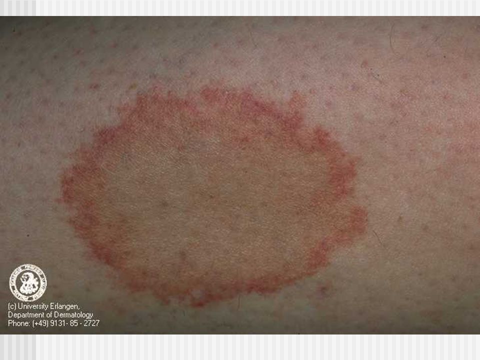
Majocchi’s Disease Aka purpura annularis telangiectodes Bluish annular macules 1-3cm in diameter with telangiectatic puncta Begins symmetrically at lower extremities Involution requires as long as a year, and may prolong indefinitely Asymptomatic.

92
Gougerot-Blum Pigmented purpuric lichenoid dermatitis Minute, rust-colored lichenoid papules that fuse into plaques of various hues Legs, thighs, and lower trunk Differentiate from Schamberg based on distribution and lichenoid lesions. Lichen Aureus

93
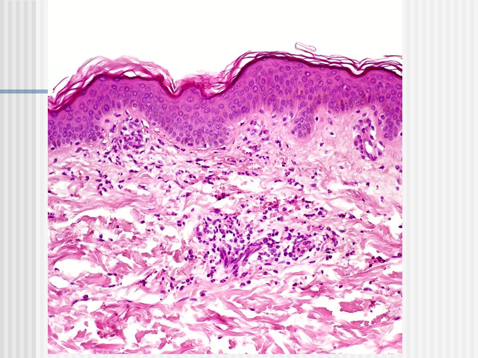
Ducas Kapetanakis pigmented purpura Histologically present with distinguished from others by presence of spongiosis Must be distinguished from mycosis fungoides

95
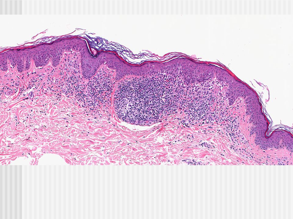
Histology Purupra Majocchi-Schamberg is characterized by slight alteration of superficial capillaries with hemorrhage and perivascular lymphocytic infiltrate.

100
Luckily, Rick's computer was equipped with an airbag and he was able to walk away from this system crash See you next time…

Similar presentations


 Nicola Davis.>")