Download presentation
Presentation is loading. Please wait.

1
Diseases of Blood Cells

2
Back to Basics Blood is a liquid tissue A mixture of cells and water
The water contains Protein, glucose, cholesterol, calcium, hormones, metabolic waste and hundreds of other substances Plasma is the liquid portion of the blood containing the blood clotting protein Fibrinogen Serum is the fluid remaining after the blood clots Does not contain Fibrinogen

3
plasma (55%) red blood cells (5-6-million /ml) white blood cells (5000/ml) platelets
 red blood cells (5-6-million /ml) white blood cells (5000/ml) platelets")
4
Plasma liquid part of blood plasma transports:- soluble food molecules waste products hormones antibodies

5
Platelets if you get cut:- platelets produce tiny fibrin threads these form a web-like mesh that traps blood cells. these harden forming a clot, or "scab." 150,000 to 400,000 per mm3

6
Physical Characteristics of Blood
Average volume of blood: 5–6 L for males; 4–5 L for females (Normovolemia) Hypovolemia - low blood volume Hypervolemia - high blood volume Viscosity (thickness) (where water = 1) The pH of blood is 7.35–7.45; x = 7.4 Osmolarity = 300 mOsm or 0.3 Osm This value reflects the concentration of solutes in the plasma Salinity = 0.85% Reflects the concentration of NaCl in the blood Temperature is 38C, slightly higher than “normal” body temperature Blood accounts for approximately 8% of body weight
 Hypovolemia - low blood volume. Hypervolemia - high blood volume. Viscosity (thickness) (where water = 1) The pH of blood is 7.35–7.45; x = 7.4. Osmolarity = 300 mOsm or 0.3 Osm. This value reflects the concentration of solutes in the plasma. Salinity = 0.85% Reflects the concentration of NaCl in the blood. Temperature is 38C, slightly higher than normal body temperature. Blood accounts for approximately 8% of body weight.")
7
Composition of Blood 2 major components Liquid = plasma (55%)
Formed elements (45%) Erythrocytes, or red blood cells (RBCs) Leukocytes, or white blood cells (WBCs) Platelets - fragments of megakaryocytes in marrow
 Erythrocytes, or red blood cells (RBCs) Leukocytes, or white blood cells (WBCs) Platelets - fragments of megakaryocytes in marrow.")
9
Blood Plasma Blood plasma components: Water = 90-92% Proteins = 6-8%
Albumins; maintain osmotic pressure of the blood Globulins Alpha and beta globulins are used for transport purposes Gamma globulins are the immunoglobulins (IgG, IgA, etc) Fibrinogen; a clotting protein Organic nutrients – glucose, carbohydrates, amino acids Electrolytes – sodium, potassium, calcium, chloride, bicarbonate Nonprotein nitrogenous substances – lactic acid, urea, creatinine Respiratory gases – oxygen and carbon dioxide
 Fibrinogen; a clotting protein. Organic nutrients – glucose, carbohydrates, amino acids. Electrolytes – sodium, potassium, calcium, chloride, bicarbonate. Nonprotein nitrogenous substances – lactic acid, urea, creatinine. Respiratory gases – oxygen and carbon dioxide.")
10
Laboratory Assessment of Blood Cells
Complete Blood Count (CBC) includes White Blood Cell Count (WBC) Red Blood Cell Count (RBC) Percentage of white cells that are neutrophils, eosinophis or basophils (white cell differential count) Amount of hemoglobin Hematocrit Percent of blood volume occupied by red blood cells
 includes. White Blood Cell Count (WBC) Red Blood Cell Count (RBC) Percentage of white cells that are neutrophils, eosinophis or basophils (white cell differential count) Amount of hemoglobin. Hematocrit. Percent of blood volume occupied by red blood cells.")
11
Red Cell Indices Mean Cell Volume (MCV) Average size of a RBC
Mean Cell Hemoglobin (MCH) Average amount of hemoglobin per RBC Mean Corpuscular Hemoglobin Concentration (MCHC) Average concentration of hemoglobin in all RBCs
 Average amount of hemoglobin per RBC. Mean Corpuscular Hemoglobin Concentration (MCHC) Average concentration of hemoglobin in all RBCs.")
12
Red Cell Indices Used to Diagnose Disease
Macrocytic Red Blood Cells may be too large Microcytic Red Blood Cells may be too small Normocytic Red Blood Cells are normal size Hypochromic Too little hemoglobin Normochromic Normal amount of hemoglobin Normochromic Normal amount of hemoglobin

13
Normochromic

14
Macrocytic Red Blood Cells may be too large

15
Macrocytic

16
The RBC's here are smaller than normal and have an increased zone of central pallor. This is indicative of a hypochromic (less hemoglobin in each RBC) microcytic (smaller size of each RBC) anemia. There is also increased anisocytosis (variation in size) and poikilocytosis (variation in shape).
 microcytic (smaller size of each RBC) anemia. There is also increased anisocytosis (variation in size) and poikilocytosis (variation in shape)..")
17
Microcytic

18
The most common cause for a hypochromic microcytic anemia is iron deficiency. The most common nutritional deficiency is lack of dietary iron. Thus, iron deficiency anemia is common. Persons most at risk are children and women in reproductive years (from menstrual blood loss and from pregnancy).
..")
19
the RBC size remains normal, whereas the overall volume of the haemoglobin is found below the healthy minimum of 11.

20
Not enough hemoglobin - hypochromic

21
Erythrocytes (RBCs) Biconcave disc
Folding increases surface area (30% more surface area) Plasma membrane contains spectrin Give erythrocytes their flexibility Anucleate, no centrioles, no organelles End result - no cell division No mitochondria means they generate ATP anaerobically Prevents consumption of O2 being transported Filled with hemoglobin (Hb) - 97% of cell contents Hb functions in gas transport Hb + O HbO2 (oxyhemoglobin) Most numerous of the formed elements Females: 4.3–5.2 million cells/cubic millimeter Males: 5.2–5.8 million cells/cubic millimeter
 Plasma membrane contains spectrin. Give erythrocytes their flexibility. Anucleate, no centrioles, no organelles. End result - no cell division. No mitochondria means they generate ATP anaerobically. Prevents consumption of O2 being transported. Filled with hemoglobin (Hb) - 97% of cell contents. Hb functions in gas transport. Hb + O2 HbO2 (oxyhemoglobin) Most numerous of the formed elements. Females: 4.3–5.2 million cells/cubic millimeter. Males: 5.2–5.8 million cells/cubic millimeter.")
22
Erythrocytes (RBCs) Figure 17.3
 Figure 17.3")
23
Erythrocyte Function Erythrocytes are dedicated to respiratory gas transport Hemoglobin reversibly binds with oxygen and most oxygen in the blood is bound to hemoglobin Composition of hemoglobin A protein called globin made up of two alpha and two beta chains A heme molecule Each heme group bears an atom of iron, which can bind to one oxygen molecule Each hemoglobin molecule thus can transport four molecules of oxygen

24
Structure of Hemoglobin
Figure 17.4

25
Hemoglobin Oxyhemoglobin – hemoglobin bound to oxygen
Oxygen loading takes place in the lungs Deoxyhemoglobin – hemoglobin after oxygen diffuses into tissues (reduced Hb) Carbaminohemoglobin – hemoglobin bound to carbon dioxide Carbon dioxide loading takes place in the tissues
 Carbaminohemoglobin – hemoglobin bound to carbon dioxide. Carbon dioxide loading takes place in the tissues.")
26
Life Cycle of Red Blood Cells

27
Fate and Destruction of Erythrocytes
The life span of an erythrocyte is 100–120 days Travels about 750 miles in that time (LA to Albuquerque) Old erythrocytes become rigid and fragile, and their hemoglobin begins to degenerate Dying erythrocytes are engulfed by macrophages Heme and globin are separated Iron is removed from the heme and salvaged for reuse Stored as hemosiderin or ferritin in tissues Transported in plasma by beta-globulins as transferrin
 Old erythrocytes become rigid and fragile, and their hemoglobin begins to degenerate. Dying erythrocytes are engulfed by macrophages. Heme and globin are separated. Iron is removed from the heme and salvaged for reuse. Stored as hemosiderin or ferritin in tissues. Transported in plasma by beta-globulins as transferrin.")
28
Fate and Destruction of Erythrocytes
Heme is degraded to a yellow pigment called bilirubin Liver secretes bilirubin into the intestines as bile Intestines metabolize bilirubin into urobilinogen Urobilinogen leaves the body in feces, in a pigment called stercobilin Globin is metabolized into amino acids which are then released into the circulation

29
Figure 17.9

30
Production of Erythrocytes
Hematopoiesis – blood cell formation Occurs in the red bone marrow (myeloid tissue) Axial skeleton and girdles Epiphyses of the humerus and femur Marrow contains immature erythrocytes Composed of reticular connective tissue Hemocytoblasts give rise to ALL formed elements Lymphoid stem cells - give rise to lymphocytes Myeloid stem cells - give rise to all other blood cells
 Axial skeleton and girdles. Epiphyses of the humerus and femur. Marrow contains immature erythrocytes. Composed of reticular connective tissue. Hemocytoblasts give rise to ALL formed elements. Lymphoid stem cells - give rise to lymphocytes. Myeloid stem cells - give rise to all other blood cells.")
31
Production of Erythrocytes: Erythropoiesis
A hemocytoblast is transformed into a committed cell called the proerythroblast Proerythroblasts develop into early erythroblasts The developmental pathway consists of three phases Phase 1 – ribosome synthesis in early erythroblasts Phase 2 – hemoglobin accumulation in late erythroblasts and normoblasts Phase 3 – ejection of the nucleus from normoblasts and formation of reticulocytes Reticulocytes then become mature erythrocytes Reticulocytes make up about 1 -2 % of all circulating erythrocytes

32
Production of Erythrocytes: Erythropoiesis

33
Regulation and Requirements for Erythropoiesis
Circulating erythrocytes – the number remains constant and reflects a balance between RBC production and destruction Too few red blood cells leads to tissue hypoxia Too many red blood cells causes an undesirable increase in blood viscosity Erythropoiesis is hormonally controlled and depends on adequate supplies of iron, amino acids, and B vitamins

34
Hormonal Control of Erythropoiesis
Erythropoietin (EPO) released by the kidneys is triggered by: Hypoxia due to decreased RBCs Decreased oxygen availability Increased tissue demand for oxygen Enhanced erythropoiesis increases the: RBC count in circulating blood Oxygen carrying ability of the blood
 released by the kidneys is triggered by: Hypoxia due to decreased RBCs. Decreased oxygen availability. Increased tissue demand for oxygen. Enhanced erythropoiesis increases the: RBC count in circulating blood. Oxygen carrying ability of the blood.")
35
Erythropoietin Mechanism
Imbalance Start Normal blood oxygen levels Stimulus: Hypoxia due to decreased RBC count, decreased availability of O2 to blood, or increased tissue demands for O2 Imbalance Increases O2-carrying ability of blood Reduces O2 levels in blood Erythropoietin stimulates red bone marrow Kidney (and liver to a smaller extent) releases erythropoietin Enhanced erythropoiesis increases RBC count
 releases erythropoietin. Enhanced erythropoiesis increases RBC count.")
36
An Electronmicrograph of a Platelet

37
Dietary Requirements of Erythropoiesis
Erythropoiesis requires: Proteins, lipids, and carbohydrates Iron, vitamin B12, and folic acid The body stores iron in Hb (65%), the liver, spleen, and bone marrow Intracellular iron is stored in protein-iron complexes such as ferritin and hemosiderin Circulating iron is loosely bound to the transport protein transferrin
, the liver, spleen, and bone marrow. Intracellular iron is stored in protein-iron complexes such as ferritin and hemosiderin. Circulating iron is loosely bound to the transport protein transferrin.")
38
Anemia ? Production? Survival/Destruction?

39
Causes of Anemia Decreased erythrocyte production
Decreased erythropoietin production Inadequate marrow response to erythropoietin Erythrocyte loss Hemorrhage Hemolysis

40
Erythrocyte Disorders
Polycythemia Abnormal excess of erythrocytes Increases viscosity, decreases flow rate of blood Anemia Abnormally low hemoglobin in blood Caused by decreased numbers of RBC’s, decreased amount of hemoglobin in RBC’s, or both

41
Anemia Anemia – blood has abnormally low oxygen-carrying capacity
It is a symptom rather than a disease itself Due to some underlying condition Blood oxygen levels cannot support normal metabolism Signs/symptoms include fatigue, paleness, shortness of breath, and chills

42
Morphological Approach (big versus little)
First, measure the size of the RBCs: Use of volume-sensitive automated blood cell counters, such as the Coulter counter. The red cells pass through a small aperture and generate a signal directly proportional to their volume. Other automated counters measure red blood cell volume by means of techniques that measure refracted, diffracted, or scattered light By calculation from an independently-measured red blood cell count and hematocrit: MCV (femtoliters) = 10 x HCT(percent) ÷ RBC (millions/µL)
 = 10 x HCT(percent) ÷ RBC (millions/µL)")
43
Diagnosis of Anemia CBC and Determination of Red Blood Cell Indices
Different types of Anemia are generally characterized by red blood cells of a certain size For Example, small (microcytic, low MCV) RBCs occur with iron deficiency RBCs contain less hemoglobin and are pale (hypochromic, low MCHC)
 RBCs occur with iron deficiency. RBCs contain less hemoglobin and are pale (hypochromic, low MCHC)")
44
Underproduction (morphological approach)
MCV>115 B12, Folate Drugs that impair DNA synthesis (AZT (Zidovudine, chemo) MDS (myelodysplastic syndromes) Ineffective production (or dysplasia) of the myeloid class of blood cells MCV = Ditto Endocrinopathy (hypothyroidism) Reticulocytosis Increased number of immature RBCs
 MDS (myelodysplastic syndromes) Ineffective production (or dysplasia) of the myeloid class of blood cells. MCV = Ditto. Endocrinopathy (hypothyroidism) Reticulocytosis. Increased number of immature RBCs.")
45
Underproduction Microcytic Iron deficiency Thalassemia trait
abnormal form of hemoglobin Anemia due to chronic disease (30-40%) Sideroblastic anemia bone marrow produces ringed sideroblasts rather than healthy RBCs Normocytic Anemia from a chronic disease Renal failure
 Sideroblastic anemia. bone marrow produces ringed sideroblasts rather than healthy RBCs. Normocytic. Anemia from a chronic disease. Renal failure.")
47
Petechial Hemorrhages on the Heart found when a coagulopathy is due to a low platelet count. They can also appear following sudden hypoxia.

48
Ecchymoses are larger than petechiae
Ecchymoses are larger than petechiae. In between in size are hemorrhages called purpura.

49
A localized collection of blood outside the vascular system within tissues is known as a hematoma

50
Review red blood cell disorders
Marrow production Thalassemias Myelodysplasia Myelophthisic Aplastic anemia Nutritional deficiencies Red cell destruction Hemoglobinopathies Enzymopathies Membrane disorders Autoimmune

51
Thalassemia Genetic defect in hemoglobin synthesis
synthesis of one of the 2 globin chains ( or ) Imbalance of globin chain synthesis leads to depression of hemoglobin production and precipitation of excess globin (toxic) “Ineffective erythropoiesis” Ranges in severity from asymptomatic to incompatible with life (hydrops fetalis) Found in people of African, Asian, and Mediterranean heritage
 Imbalance of globin chain synthesis leads to depression of hemoglobin production and precipitation of excess globin (toxic) Ineffective erythropoiesis Ranges in severity from asymptomatic to incompatible with life (hydrops fetalis) Found in people of African, Asian, and Mediterranean heritage.")
52
Alpha Thalassemia This is alpha thalassemia major. There have been two major variations of alpha thalassemia arise in human history. One variation, most prevalent in Southeast Asia, is known as alpha thalassemia 1. In this variant, two alpha globin genes are deleted on one chromosome 16. The other variant, known as alpha thalassemia 2, is most common in Africa and the Mediterranean region, and differs in that a single alpha globin gene is missing from one chromosome 16. Alpha thalassemia 1, with alpha globin gene deletions on a single chromosome 16, can give rise to alpha thalassemia major in the homozygous state, when both chromosomes are affected. Affected persons become anemic in utero, because even fetal hemoglobin cannot be produced, and severe hydrops fetalis results, which leads to stillbirth, or death soon after birth from pulmonary hypoplasia or cardiac failure. Hemoglobin electrophoresis will reveal affected fetuses or neonates to have about 80% hemoglobin Barts (a tetramer of gamma chains) and about 20% hemoglobin Portland (or sometimes hemoglobin Gower 1) normally present only in embryonic life in the first trimester. RBCs that contain mostly hemoglobin Barts have marked anisocytosis and poikilocytosis, and there is expansion of erythropoiesis with many immature RBCs in the peripheral blood, as evidenced by polychromasia, nucleated RBCs and even erythroblasts as shown here.
 and about 20% hemoglobin Portland (or sometimes hemoglobin Gower 1) normally present only in embryonic life in the first trimester. RBCs that contain mostly hemoglobin Barts have marked anisocytosis and poikilocytosis, and there is expansion of erythropoiesis with many immature RBCs in the peripheral blood, as evidenced by polychromasia, nucleated RBCs and even erythroblasts as shown here.")
53
Thalassemias Dx: Smear: microcytic/hypochromic, misshapen RBCs
-thalassemia will have an abnormal Hgb electrophoresis (HbA2, HbF) The more severe -thalassemia syndrome can have HbH inclusions in RBCs Fe stores are usually elevated
 The more severe -thalassemia syndrome can have HbH inclusions in RBCs. Fe stores are usually elevated.")
54
Thalassemia The oxygen depletion in the body becomes apparent within the first 6 months of life. If left untreated, death usually results within a few years. Note the small, pale (hypochromic), abnormally-shaped red blood cells. The darker cells likely represent normal RBCs from a blood transfusion.
, abnormally-shaped red blood cells. The darker cells likely represent normal RBCs from a blood transfusion.")
55
Thalassemia The only treatments are stem cell transplant and simple transfusion. Chelation therapy to avoid iron overload has to be started early.

57
Marrow Production - Myelodysplasia
Used to be referred to as “Preleukemia” Most commonly in the elderly. Occurs when something goes wrong in your bone marrow

58
Signs and Symptoms of Myelodysplasia
Fatigue Shortness of breath Unusual paleness (pallor) due to anemia Easy or unusual bruising or bleeding Pinpoint-sized red spots just beneath your skin caused by bleeding (petechiae) Frequent infections
 due to anemia. Easy or unusual bruising or bleeding. Pinpoint-sized red spots just beneath your skin caused by bleeding (petechiae) Frequent infections.")
59
Causes Caused by poorly formed or dysfunctional blood cells due to either Unknown causes Chemical exposure

60
Myelodysplasia

61
Marrow Production - Myelophthisic
Myelophthisic anemia is a normocytic-normochromic anemia that occurs when normal marrow space is infiltrated and replaced by nonhematopoietic or abnormal cells. Causes: Most often due to replacement of the bone marrow by metastatic cancers such as breast or prostate; less often, kidney, lung, adrenal, or thyroid. Marrow fibrosis often occurs. Splenomegaly may develop.

62
Myelophthisic

63
Marrow Production - Aplastic Anemia
The body stops producing enough new blood cells. Signs and symptoms may include: Fatigue Shortness of breath with exertion Rapid or irregular heart rate Pale skin Frequent or prolonged infections Unexplained or easy bruising Nosebleeds and bleeding gums Prolonged bleeding from cuts Skin rash Dizziness Headache

64
NORMAL BONE MARROW

65
Here we see a sample of bone marrow in a patient with Aplastic Anaemia
Here we see a sample of bone marrow in a patient with Aplastic Anaemia. Notice there are very few cells except for the fat cells

66
Factors that can temporarily or permanently injure bone marrow and affect blood cell production include: Acquired Radiation and chemotherapy treatments Exposure to toxic chemicals. Exposure to benzene Use of certain drugs even some antibiotics. Autoimmune disorders Viral infections Epstein Barr, CMV, Parvovirus B19, HIV Pregnancy. Unknown factors. This is called idiopathic aplastic anemia.

67
Marrow Production - Aplastic Anemia
Hereditary Fanconi a rare, inherited blood disorder that leads to bone marrow failure. Diamond-Shwachman a rare autosomal recessive disorder characterized by exocrine pancreatic insufficiency, bone marrow dysfunction

68
Marrow Production - Aplastic Anemia
Treatment Most patients require red cell transfusions. Bone MarrowTransplant when possible. Stem Cell Transplant Medication: Bone Marrow Stimulants Sargramostim (Leukine) Filgrastim (Neupogen) Pegfilgrastim (Neulasta) Epoetin alfa (Epogen, Procrit)
 Filgrastim (Neupogen) Pegfilgrastim (Neulasta) Epoetin alfa (Epogen, Procrit)")
69
Hemolytic Anemia – RBC Destruction
Hemolytic anemias are either acquired or congenital. Hemolytic anemia is a condition in which there are not enough RBCs in the blood Due to premature RBC destruction Hemolytic anemia can result from: infection certain drugs autoimmune disorders in which the body attacks and destroys its own red blood cells inherited disorders such as sickle cell anemia or thalassemia.

70
Hemolytic anemias

71
Symptoms of Hemolytic Anemia
Dark Urine Enlarged spleen Fatigue Fever Pale skins color Rapid heart rate Shortness of breath Yellow skin color (jaundice) Chills
 Chills.")
72
Sickle Cell Anemia Single base pair mutation results in a single amino acid change. Under low oxygen, Hgb becomes insoluble forming long polymers This leads to membrane changes (“sickling”) and vasoocclusion
 and vasoocclusion.")
76
Red Blood Cells from Sickle Cell Anemia
Deoxygenation of SS erythrocytes leads to intracellular hemoglobin polymerization, loss of deformability and changes in cell morphology. OXY-STATE DEOXY-STATE

77
Transfusion in Sickle Cell (Controversy!)
Used correctly, transfusion can prevent organ damage and save the lives of sickle cell disease patients. Used unwisely, transfusion therapy can result in serious complications.

78
Transfusion in Sickle Cell (Controversy!)
Simple transfusion – give blood Partial exchange transfusion - remove blood and give blood Erythrocytapheresis – use apheresis to maximize blood exchange When to use each method?

79
Transfusion in Sickle Cell
In general, patients should be transfused if there is sufficient physiological derangement to result in heart failure, dyspnea, hypotension, or marked fatigue. Tends to occur during an acute illness or when hemoglobin falls under 5 g/dL.

80
Transfusion in Sickle Cell (exchange transfusion)
Bleed one unit (500 ml), infuse 500 ml of saline Bleed a second unit and infuse two units. Repeat. If the patient has a large blood mass, do it again.
, infuse 500 ml of saline. Bleed a second unit and infuse two units. Repeat. If the patient has a large blood mass, do it again.")
81
Transfusion in Sickle Cell (chronic transfusion therapy)
Stroke Chronic debilitating pain Pulmonary hypertension Setting of renal failure and heart failure

82
Transfusion in Sickle Cell (chronic transfusion therapy)
Controversial uses: Prior to contast media exposure Sub-clinical neurological damage Priapism Leg Ulcers Pregnancy

83
Pernicious Anemia Pernicious anemia is a decrease in red blood cells that occurs when the body cannot properly absorb vitamin B12 from the GI Tract Common causes include: Weakened stomach lining (atrophic gastritis) The body's immune system attacking the cells that make intrinsic factor (autoimmunity against gastric parietal cells) or intrinsic factor itself
 The body s immune system attacking the cells that make intrinsic factor (autoimmunity against gastric parietal cells) or intrinsic factor itself.")
84
Symptoms Diarrhea or constipation Fatigue Loss of appetite Pale skin
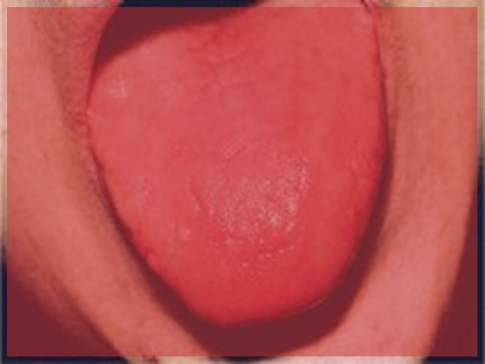
Shortness of breath, mostly during exercise Swollen, red tongue or bleeding gums Nerve damage

85
Red cell destruction Elevated reticulocyte count Mechanical Autoimmune
Drug Congenital

89
Red cell destruction – membrane disorders
Hereditary spherocytosis Hereditary elliptocytosis Hereditary pyropoikilocytosis Southeast Asian ovalocytosis

90
Review red blood cell disorders Red cell destruction – membrane disorders

91
Review red blood cell disorders Red cell destruction – enzymopathies
G6PD deficiency Pyruvate kinase deficiency Other very rare deficiencies

92
Deoxyhemoglobin S Polymer Structure
A) Deoxyhemoglobin S 14-stranded polymer (electron micrograph) B) Paired strands of deoxyhemoglobin S (crystal structure) C) Hydrophobic pocket for 6b Val D) Charge and size prevent 6b Glu from binding. Dykes, Nature 1978; JMB 1979 Crepeau, PNAS 1981 Wishner, JMB 1975
 Deoxyhemoglobin S. 14-stranded polymer. (electron micrograph) B) Paired strands of. deoxyhemoglobin S. (crystal structure) C) Hydrophobic pocket. for 6b Val. D) Charge and size prevent. 6b Glu from binding. Dykes, Nature 1978; JMB Crepeau, PNAS Wishner, JMB")
93
Transfusion in Sickle Cell
In severely anemic patients, simple transfusions should be used. Common causes of acute anemia: acute splenic sequestration transient red cell aplasia Hyperhemolysis (infection, acute chest syndrome, malaria). If the patient is stable and the reticulocyte count high, transfusions can (and should) be deferred.
. If the patient is stable and the reticulocyte count high, transfusions can (and should) be deferred.")
94
Transfusion in Sickle Cell (exchange transfusion)
A comprehensive transfusion protocol should include accurate records of the patient’s red cell phenotype, alloimmunization history, number of units received, serial Hb S percentages, and results of monitoring for infectious diseases and iron overload. Transfusions are used to raise the oxygen-carrying capacity of blood and decrease the proportion of sickle red cells.

95
Transfusion in Sickle Cell (exchange transfusion)
Transfusions usually fall into two categories: episodic, acute transfusions to stabilize or reverse complications. long-term, prophylactic transfusions to prevent future complications.

96
Transfusion in Sickle Cell (exchange transfusion)
episodic, acute transfusions to stabilize or reverse complications. Limited studies have shown that aggressive transfusion (get Hgb S < 30%) may help in sudden severe illness. May be useful before general anesthesia. Vichinsky et al., NEJM 1995
 may help in sudden severe illness. May be useful before general anesthesia. Vichinsky et al., NEJM")
97
Transfusion in Sickle Cell
Inappropriate uses of transfusion: Chronic steady-state anemia Uncomplicated pain episodes Infection Minor surgery Uncomplicated pregnancies Aseptoic necrosis

98
Transfusion in Sickle Cell (exchange transfusion)
Except in severe anemia, exchange transfusion offers many benefits and is our first choice Phenotypically matched, leukodepleted packed cells are the blood product of choice. A posttransfusion hematocrit of 36 percent or less is recommended. Avoid hyperviscosity, which is dangerous to sickle cell patients.

99
Iron overload and chelation
Can occur in any patient requiring chronic transfusion therapy or in hemochromatosis. Liver biopsy is the most accurate test though MRI is being investigated. Ferritin is a good starting test. 120 cc of red cells/kg of body weight is an approximate point at which to think about iron overload

100
Iron overload and chelation
Chelator, deferoxamine 25 mg/kg sq per day over 8 hours. Supplementation with vitamin C may aid excretion. Otooxicity, eye toxicity, allergic reactions. Discontinue during an infection. Oral chelators are in development.

101
Conclusions Transfuse for any severe anemia with physiologic compromise. Decide early whether transfusion will be rare or part of therapy. Avoid long-term complications by working with your blood bank and using chelation theraoy.

102
Anemia: Insufficient Erythrocytes
Hemorrhagic anemia – result of acute or chronic loss of blood Hemolytic anemia – prematurely ruptured erythrocytes Aplastic anemia – destruction or inhibition of red bone marrow

Similar presentations


Transportation -Gases (O 2 and CO 2 ) -Nutrients -Heat and waste -Hormones 2)Regulation.>")


