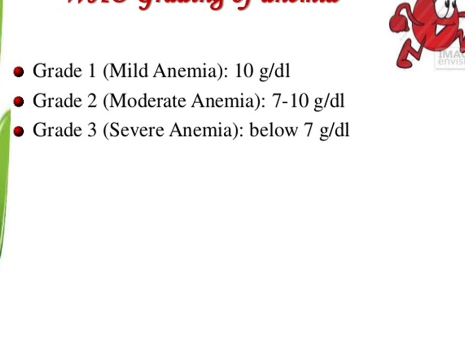
Download presentation
Presentation is loading. Please wait.

1
Anemia Premed 3 Pathophysiology

2
Anemia is a sign, not a disease. Anemia is a sign, not a disease. Anemias are a dynamic process. Anemias are a dynamic process. Decrease in whole red cell mass Decrease in whole red cell mass Causes: Causes: 1. decreased in RBC production 1. decreased in RBC production 2. increased RBC loss 2. increased RBC loss Increased RBC destruction. Increased RBC destruction.

3
Normal blood smear

6
Iron metabolism Only 5-10 % or about 1.0 mg of dietary iron is absorbed as ferrous iron (Fe++), mainly in the duodenum and upper jejunum. Only 5-10 % or about 1.0 mg of dietary iron is absorbed as ferrous iron (Fe++), mainly in the duodenum and upper jejunum. The mucosal cells oxidize the ferrous iron to ferric iron The mucosal cells oxidize the ferrous iron to ferric iron complexed with apoferritin to form ferritin; complexed with apoferritin to form ferritin; Apoferritin a protein present in the intestinal mucosa that binds and stores iron by combining with a ferric hydroxide phosphate composed to form ferritin. Apoferritin a protein present in the intestinal mucosa that binds and stores iron by combining with a ferric hydroxide phosphate composed to form ferritin.
, mainly in the duodenum and upper jejunum. The mucosal cells oxidize the ferrous iron to ferric iron The mucosal cells oxidize the ferrous iron to ferric iron complexed with apoferritin to form ferritin; complexed with apoferritin to form ferritin; Apoferritin a protein present in the intestinal mucosa that binds and stores iron by combining with a ferric hydroxide phosphate composed to form ferritin. Apoferritin a protein present in the intestinal mucosa that binds and stores iron by combining with a ferric hydroxide phosphate composed to form ferritin..")
7
some of the ferritin is transported out of the mucosal cell into the plasma bound to transferrin some of the ferritin is transported out of the mucosal cell into the plasma bound to transferrin

8
Transferrin Are iron binding blood plasma glycoprotein that control the level of free iron in biological fluids. Are iron binding blood plasma glycoprotein that control the level of free iron in biological fluids. The major function of transferrin is to transport iron to cells. The major function of transferrin is to transport iron to cells. iron has to be bound to transferrin because unbound iron is toxic. iron has to be bound to transferrin because unbound iron is toxic. Serum iron has a normal concentration of 60- 150 ug/dL and largely reflects iron bound to transferrin. Serum iron has a normal concentration of 60- 150 ug/dL and largely reflects iron bound to transferrin.

9
TIBC Is a medical lab. test that measures the blood’s capacity to bind iron with transferrin. Is a medical lab. test that measures the blood’s capacity to bind iron with transferrin. Total iron binding capacity (TIBC) is the sum of the unbound apotransferrin plus the iron bound transferrin Total iron binding capacity (TIBC) is the sum of the unbound apotransferrin plus the iron bound transferrin represents the maximum capacity for iron binding represents the maximum capacity for iron binding 250- 360 ug/dL 250- 360 ug/dL Serum iron in males: 65- 177ug/dL Serum iron in males: 65- 177ug/dL Females: 50- 170ug/dL Females: 50- 170ug/dL
 is the sum of the unbound apotransferrin plus the iron bound transferrin Total iron binding capacity (TIBC) is the sum of the unbound apotransferrin plus the iron bound transferrin represents the maximum capacity for iron binding represents the maximum capacity for iron binding ug/dL ug/dL Serum iron in males: ug/dL Serum iron in males: ug/dL Females: ug/dL Females: ug/dL.")
10
When not bound to iron, it is known as apotransferrin. When not bound to iron, it is known as apotransferrin. bounded iron can be transported to the bone marrow or iron storage sites. bounded iron can be transported to the bone marrow or iron storage sites.

11
stored as either ferritin or hemosiderin stored as either ferritin or hemosiderin The main storage site is liver. The main storage site is liver. The receptor-transferrin-iron complex is then incorporated into the cytosol by endocytosis. The receptor-transferrin-iron complex is then incorporated into the cytosol by endocytosis. at an acid pH the iron (Fe++) is released from transferrin at an acid pH the iron (Fe++) is released from transferrin transported to mitochondria where it is incorporated into heme. transported to mitochondria where it is incorporated into heme.
 is released from transferrin at an acid pH the iron (Fe++) is released from transferrin transported to mitochondria where it is incorporated into heme. transported to mitochondria where it is incorporated into heme..")
12
the bulk of body iron is found in erythrocytes with lesser amounts in myoglobin. the bulk of body iron is found in erythrocytes with lesser amounts in myoglobin. Large amounts of iron are required during growth periods in infant, childhood and teenage years. Large amounts of iron are required during growth periods in infant, childhood and teenage years.

13
Globin chains Synthesis controlled by Chr 16 an 11 Synthesis controlled by Chr 16 an 11 location and rate of synthesis varies from embryonic to fetal to neonatal to adult life. location and rate of synthesis varies from embryonic to fetal to neonatal to adult life.

18
Iron deficiency anemia Iron deficiency is a common form of malnutrition that affects more than 2 billion people globally. Iron deficiency is a common form of malnutrition that affects more than 2 billion people globally.

19
Iron-deficiency anemia Major cause in adults: chronic blood loss, insufficient absorption of iron, parasitic inf. Major cause in adults: chronic blood loss, insufficient absorption of iron, parasitic inf. menorrhagia ( heavy menses) bleeding from GIT Dietary deficiency is common after 6 months of age in infants Dietary deficiency is common after 6 months of age in infants Pallor, fatigue, dyspnea on exertion, anxiety,hair loss,KOILONYCHIA, mouth ulcers. Pallor, fatigue, dyspnea on exertion, anxiety,hair loss,KOILONYCHIA, mouth ulcers.
 bleeding from GIT Dietary deficiency is common after 6 months of age in infants Dietary deficiency is common after 6 months of age in infants Pallor, fatigue, dyspnea on exertion, anxiety,hair loss,KOILONYCHIA, mouth ulcers. Pallor, fatigue, dyspnea on exertion, anxiety,hair loss,KOILONYCHIA, mouth ulcers..")
21
Iron-deficiency anemia Low hemoglobin Low hemoglobin Low hematocrit Low hematocrit Low RBC count Low RBC count Hypochromic, microcytic RBC on smear Hypochromic, microcytic RBC on smear Low serum iron Low serum iron High total iron binding capacity(TIBC) High total iron binding capacity(TIBC) Low iron store; low ferritin Low iron store; low ferritin
 High total iron binding capacity(TIBC) Low iron store; low ferritin Low iron store; low ferritin")
22
Iron deficiency anemia

25
Megaloblastic anemia Presence of large, abnormal-looking erythroid precursor cells or MEGALOBLASTS in the bone marrow Presence of large, abnormal-looking erythroid precursor cells or MEGALOBLASTS in the bone marrow Decrease DNA synthesis decrease DNA replication and nuclear division Decrease DNA synthesis decrease DNA replication and nuclear division Impaired RBC production Impaired RBC production Inhibition of DNA synthesis in red blood cells results in the formation of large, fragile megaloblastic erythrocytes Inhibition of DNA synthesis in red blood cells results in the formation of large, fragile megaloblastic erythrocytes

26
Megaloblastic anemia Pancytopenia Pancytopenia low RBC low platelets low WBC Oval macrocytosis (“ big RBC”) Oval macrocytosis (“ big RBC”) Hypersegmented neutrophils (more than 5 lobes) Hypersegmented neutrophils (more than 5 lobes) Megaloblastic hyperplasia of the bone marrow Megaloblastic hyperplasia of the bone marrow
 Oval macrocytosis ( big RBC ) Hypersegmented neutrophils (more than 5 lobes) Hypersegmented neutrophils (more than 5 lobes) Megaloblastic hyperplasia of the bone marrow Megaloblastic hyperplasia of the bone marrow")
27
Hypersegmented neutrophils

28
Megaloblast

29
Megaloblastic anemia Vitamin B12 deficiency Vitamin B12 deficiency Pernicious Anemia: autoimmune parietal cell destruction results in insufficient intrinsic factor production Pernicious Anemia: autoimmune parietal cell destruction results in insufficient intrinsic factor production Lemon-yellow skin Lemon-yellow skin Stomatitis Stomatitis Glossitis Glossitis Demyelination of the posterior and lateral columns of the spinal cord Demyelination of the posterior and lateral columns of the spinal cord Ataxic gait, hyperreflexia, impaired position and vibration reflexes Ataxic gait, hyperreflexia, impaired position and vibration reflexes Labs: Antibodies against Intrinsic factor Abnormal schilling test: to evaluate absorption of vit B12 and to evaluate pernicious anemia.

30
Megaloblastic anemia Folate deficiency Folate deficiency Seen in alcoholics Seen in alcoholics Pregnancy Pregnancy infancy infancy Fad dieters Fad dieters Contraceptive pills Contraceptive pills Dilantin, anticonvulsant drugs Dilantin, anticonvulsant drugs Chemotherapy agents Chemotherapy agents Same presentation as B12 deficiency Women with folate deficiency who become pregnant are more likely to give birth to low birthweight, premature infants with neural tube defects.

31
Pernicious anemia is one of many types of the larger family of megaloblastic anemias. It is caused by loss of gastric parietal cells which are responsible, in part, for the secretion of intrinsic factor, a protein essential for subsequent absorption of vitamin B 12 in the ileum. Pernicious anemia is one of many types of the larger family of megaloblastic anemias. It is caused by loss of gastric parietal cells which are responsible, in part, for the secretion of intrinsic factor, a protein essential for subsequent absorption of vitamin B 12 in the ileum.megaloblastic anemiasintrinsic factormegaloblastic anemiasintrinsic factor

32
Anemia of Chronic disease Impaired red cell production associated` with chronic diseases Second most common form Impaired red cell production associated` with chronic diseases Second most common form May be due to rheumatoid arthritis,regional enteritis, chronic infection, neoplasms. May be due to rheumatoid arthritis,regional enteritis, chronic infection, neoplasms. Normochromic, normocytic Normochromic, normocytic In chronic disease: it may be similar to IDA, but the TIBC is low In chronic disease: it may be similar to IDA, but the TIBC is low

33
Aplastic anemia Aplastic anemia is a condition where bone marrow does not produce sufficient new cells to replenish blood cells. The condition, as the name indicates, involves both aplasia and anemia. Typically, anemia refers to low red blood cell counts, but aplastic anemia patients have lower counts of all three blood cell types: red blood cells, white blood cells, and platelets, termed pancytopenia. Aplastic anemia is a condition where bone marrow does not produce sufficient new cells to replenish blood cells. The condition, as the name indicates, involves both aplasia and anemia. Typically, anemia refers to low red blood cell counts, but aplastic anemia patients have lower counts of all three blood cell types: red blood cells, white blood cells, and platelets, termed pancytopenia.cellsblood cellsaplasia anemia red blood cellswhite blood cellsplateletspancytopeniacellsblood cellsaplasia anemia red blood cellswhite blood cellsplateletspancytopenia

34
Anemia with malaise, pallor and associated symptoms such as palpitations Anemia with malaise, pallor and associated symptoms such as palpitations Anemiamalaisepallor Anemiamalaisepallor Thrombocytopenia (low platelet counts), leading to increased risk of hemorrhage, bruising and petechiae Thrombocytopenia (low platelet counts), leading to increased risk of hemorrhage, bruising and petechiae Thrombocytopeniahemorrhagebruising petechiae Thrombocytopeniahemorrhagebruising petechiae Leukopenia (low white blood cell count), leading to increased risk of infection Leukopenia (low white blood cell count), leading to increased risk of infection Leukopeniainfection Leukopeniainfection Reticulocytopenia (low reticulocyte counts) Reticulocytopenia (low reticulocyte counts) Reticulocytopeniareticulocyte Reticulocytopeniareticulocyte
, leading to increased risk of hemorrhage, bruising and petechiae Thrombocytopenia (low platelet counts), leading to increased risk of hemorrhage, bruising and petechiae Thrombocytopeniahemorrhagebruising petechiae Thrombocytopeniahemorrhagebruising petechiae Leukopenia (low white blood cell count), leading to increased risk of infection Leukopenia (low white blood cell count), leading to increased risk of infection Leukopeniainfection Leukopeniainfection Reticulocytopenia (low reticulocyte counts) Reticulocytopenia (low reticulocyte counts) Reticulocytopeniareticulocyte Reticulocytopeniareticulocyte")
35
Aplastic anemia Most common cause: toxic exposure Most common cause: toxic exposureradiation chemicals – benzene antibiotics – chloramphenicol cancer drugs viruses – hepatitis C and Parvovirus

36
Aplastic anemia ***Hypocellular bone marrow ***Hypocellular bone marrow Loss of hematopoietic cells Loss of hematopoietic cells Peripheral pancytopenia Peripheral pancytopenia

37
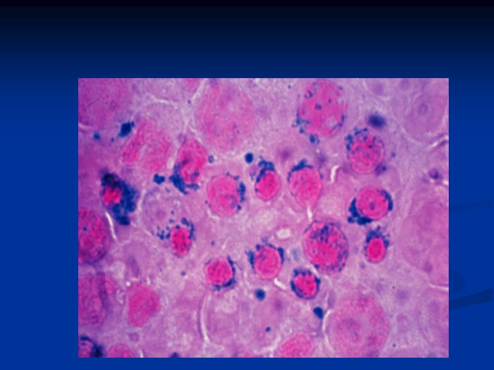
SIDEROBLASTIC ANEMIA a sideroblastic anemia caused by a deficiency of pyridoxine (vitamin B 6 ). The most common cause of pyridoxine deficiency is isoniazid therapy for tuberculosis. Sideroblastic anemias are due to defects in heme synthesis in the mitochondria of developing RBC precursors in the bone marrow. a sideroblastic anemia caused by a deficiency of pyridoxine (vitamin B 6 ). The most common cause of pyridoxine deficiency is isoniazid therapy for tuberculosis. Sideroblastic anemias are due to defects in heme synthesis in the mitochondria of developing RBC precursors in the bone marrow.
. The most common cause of pyridoxine deficiency is isoniazid therapy for tuberculosis. Sideroblastic anemias are due to defects in heme synthesis in the mitochondria of developing RBC precursors in the bone marrow..")
38
Most acquired types of sideroblastic anemia are microcytic. Iron accumulates in the mitochondria producing ringed sideroblasts, which are identified with a Prussian blue stain. Most acquired types of sideroblastic anemia are microcytic. Iron accumulates in the mitochondria producing ringed sideroblasts, which are identified with a Prussian blue stain.

40
The photograph shows a rim of blue staining iron granules around the nucleus of a nucleated RBC precursor. Iron overload occurs, which causes an increase in serum ferritin. The photograph shows a rim of blue staining iron granules around the nucleus of a nucleated RBC precursor. Iron overload occurs, which causes an increase in serum ferritin.

41
Hemolytic anemia Hemolytic anemia is a form of anemia due to hemolysis, the abnormal breakdown of red blood cells, either in the blood vessels (intravascular hemolysis) or elsewhere in the human body (extravascular). Hemolytic anemia is a form of anemia due to hemolysis, the abnormal breakdown of red blood cells, either in the blood vessels (intravascular hemolysis) or elsewhere in the human body (extravascular).anemia hemolysisred blood cellsblood vesselsanemia hemolysisred blood cellsblood vessels
 or elsewhere in the human body (extravascular).anemia hemolysisred blood cellsblood vesselsanemia hemolysisred blood cellsblood vessels.")
42
Hemolytic anemias Immune hemolytic anemias Immune hemolytic anemias cold agglutinin disease Hemolytic disease of the newborn Membrane skeletal protein abnormalities Membrane skeletal protein abnormalities Hereditary spherocytosis Enzyme deficiency HA Enzyme deficiency HA G6PD deficiency Pyruvate kinase deficiency Hemoglobinopathies Hemoglobinopathies Hemoglobin S disorder : Sickle cell anemia Thalassemias: alpha and beta

43
Hemolytic anemias Shortened life span of RBC Shortened life span of RBC Increased destruction Increased destruction Hemoglobinemia, hemoglobinuria Hemoglobinemia, hemoglobinuria Hemosiderosis Hemosiderosis

44
Immune hemolytic anemia immune hemolytic anemia is a condition in which there is a reduced blood cell count due to the premature destruction of red blood cells by the immune system. immune hemolytic anemia is a condition in which there is a reduced blood cell count due to the premature destruction of red blood cells by the immune system.

45
Causes Causes Immune hemolytic anemia occurs when antibodies form against the body's own red blood cells. The antibodies destroy the blood cells because the immune system mistakenly recognizes these blood cells as foreign material within the body. Immune hemolytic anemia occurs when antibodies form against the body's own red blood cells. The antibodies destroy the blood cells because the immune system mistakenly recognizes these blood cells as foreign material within the body. antibodies

46
The antibodies may be caused by: The antibodies may be caused by: Complication of another disease Complication of another disease Past blood transfusions Past blood transfusions Pregnancy (if the baby's blood type is different from the mother's) Pregnancy (if the baby's blood type is different from the mother's) Reaction to certain medications Reaction to certain medications Reaction to certain infections Reaction to certain infections
 Pregnancy (if the baby s blood type is different from the mother s) Reaction to certain medications Reaction to certain medications Reaction to certain infections Reaction to certain infections")
48
IHA: Hemolytic disease of the newborn Erythroblastosis fetalis Erythroblastosis fetalis Maternal antibodies attack the D antigen of the Rh blood group Maternal antibodies attack the D antigen of the Rh blood group Mom: Rh (-) or “ d” Baby: Rh (+) or “D” Also seen in ABO incompatibility Also seen in ABO incompatibility Mom O, Baby A or B Mom A, Baby B or AB Mom B, Baby A or AB
 or d Baby: Rh (+) or D Also seen in ABO incompatibility Also seen in ABO incompatibility Mom O, Baby A or B Mom A, Baby B or AB Mom B, Baby A or AB")
50
Hemolytic disease of the newborn Kernicterus is a very rare type of brain damage that occurs in a newbornwith severe jaundice. It happens when a substance in the blood, called bilirubin, builds up to very high levels and spreads into the brain tissues. This causes permanent brain damage. Kernicterus is a very rare type of brain damage that occurs in a newbornwith severe jaundice. It happens when a substance in the blood, called bilirubin, builds up to very high levels and spreads into the brain tissues. This causes permanent brain damage.

51
Kernicterus: unconjugated bilirubin accumulates in the basal ganglia and the CNS Kernicterus: unconjugated bilirubin accumulates in the basal ganglia and the CNS Hydrops fetalis Hydrops fetalis infant may be stillborn or die shortly after birth. infant may be stillborn or die shortly after birth. stillborn

52
Enzyme deficiency HA Glucose 6 phosphate dehydrogenase defeciency Glucose 6 phosphate dehydrogenase defeciency Most common form Most common form X-linked recessive hereditary disease X-linked recessive hereditary disease Common in 10% of African Americans; Mediterrenean Common in 10% of African Americans; Mediterrenean Acute, self-limited Acute, self-limited Hemoglobinemia, hemoglobinuria Hemoglobinemia, hemoglobinuria Triggering factors: Triggering factors:infectionsprimaquinesulfonamides

53
Heinz bodies are formed by damage to the hemoglobin component molecules, usually through oxidant damage, or from an inherited mutation. Heinz bodies are formed by damage to the hemoglobin component molecules, usually through oxidant damage, or from an inherited mutation.hemoglobin

55
Heinz bodies

56
Enzyme deficiency HA Pyruvate kinase (PK) deficiency is an autosomal recessive hemolytic disease with extravascular hemolysis. PK normally converts phosphoenolpyruvate to pyruvate leading to a net gain of 2 ATP. In PK deficiency, lack of ATP damages the membrane causing a loss of K + and dehydration of the RBC (echinocytes with thorny projections, which are present in the photograph). Pyruvate kinase (PK) deficiency is an autosomal recessive hemolytic disease with extravascular hemolysis. PK normally converts phosphoenolpyruvate to pyruvate leading to a net gain of 2 ATP. In PK deficiency, lack of ATP damages the membrane causing a loss of K + and dehydration of the RBC (echinocytes with thorny projections, which are present in the photograph).
. Pyruvate kinase (PK) deficiency is an autosomal recessive hemolytic disease with extravascular hemolysis. PK normally converts phosphoenolpyruvate to pyruvate leading to a net gain of 2 ATP. In PK deficiency, lack of ATP damages the membrane causing a loss of K + and dehydration of the RBC (echinocytes with thorny projections, which are present in the photograph)..")
58
SUMMARY OF PK DEFECIENCY pyruvate kinase which affects the survival of red blood cells and causes them to deform into echinocytes on peripheral blood smears. pyruvate kinase which affects the survival of red blood cells and causes them to deform into echinocytes on peripheral blood smears. pyruvate kinasered blood cells pyruvate kinasered blood cells commonly, the inheritance is autosomal recessive. commonly, the inheritance is autosomal recessive. Pyruvate kinase deficiency is the second most common cause of enzyme-deficient hemolytic anemia, following G6PD deficiency. Pyruvate kinase deficiency is the second most common cause of enzyme-deficient hemolytic anemia, following G6PD deficiency.hemolytic anemiaG6PD deficiencyhemolytic anemiaG6PD deficiency

59
causes A variety of mutations can lead to lowered production, activity, or stability of pyruvate kinase, an enzyme essential to glycolysis. A total lack of this enzyme's activity will be lethal.enzyme glycolysis

60
Hemoglobinopathies Hemoglobin is produced by genes that control the expression of the hemoglobin protein. Defects in these genes can produce abnormal hemoglobins and anemia, which are conditions termed "hemoglobinopathies". Hemoglobin is produced by genes that control the expression of the hemoglobin protein. Defects in these genes can produce abnormal hemoglobins and anemia, which are conditions termed "hemoglobinopathies". Most commonly involved: Hemoglobin S Most commonly involved: Hemoglobin S Point mutation in codon 6 of the beta-globin gene Point mutation in codon 6 of the beta-globin gene Valine is substituted for glutamic acid Valine is substituted for glutamic acid

61
Abnormal hemoglobins appear in one of three basic circumstances: Abnormal hemoglobins appear in one of three basic circumstances: Structural defects in the hemoglobin molecule. Structural defects in the hemoglobin molecule. Diminished production of one of the two subunits of the hemoglobin molecule Diminished production of one of the two subunits of the hemoglobin molecule Abnormal associations of otherwise normal subunits. Abnormal associations of otherwise normal subunits.

67
Result: Hemoglobin S polymerizes at low oxygen tension sickle cells RBC membranes stiffen hemolysis/ obstruction of the blood vessels Result: Hemoglobin S polymerizes at low oxygen tension sickle cells RBC membranes stiffen hemolysis/ obstruction of the blood vessels Severe Hemolytic anemia Severe Hemolytic anemia Chronic leg ulcers Chronic leg ulcers Painful crises: limbs, back, chest, abdomen Painful crises: limbs, back, chest, abdomen Infarctions: lungs and spleen autosplenectomy Infarctions: lungs and spleen autosplenectomy Aplastic crisis: fall in hemoglobin Aplastic crisis: fall in hemoglobin

69
Thalassemias The thalassemias are a group of disorders in which the normal hemoglobin protein is produced in lower amounts than usual. The thalassemias are a group of disorders in which the normal hemoglobin protein is produced in lower amounts than usual.

70
Thalasemias Deficient production of either alpha or beta- globin chains Deficient production of either alpha or beta- globin chains Beta-thalasemia: most common form; defect in the genes coding for Beta-globin gene Beta-thalasemia: most common form; defect in the genes coding for Beta-globin gene

71
Beta-thalassemia major (Mediterrenean or Cooley’s anemia) A usually fatal form of thalassemia appearing in infancy or childhood in which normal hemoglobin is absent, characterized by severe anemia, enlargement of the heart, liver, and spleen, and skeletal deformation. A usually fatal form of thalassemia appearing in infancy or childhood in which normal hemoglobin is absent, characterized by severe anemia, enlargement of the heart, liver, and spleen, and skeletal deformation. Decrease HbA synthesis Decrease HbA synthesis Short rbc lifespan (due to insoluble, excess alpha- chains) Ineffective RBC production Skull, facial bones and long bones distortion Microcytosis, hypochromic anemia Hemosiderosis Increase Hb F throughout life
 Ineffective RBC production Skull, facial bones and long bones distortion Microcytosis, hypochromic anemia Hemosiderosis Increase Hb F throughout life.")
73
Beta-thalassemia major: target cells

74
OTHER FORMS OF THALASSEMIA Beta-thalassemia minor Beta-thalassemia minor is much more common is much more common Increase in Hb A2 Increase in Hb A2

75
Alpha-thalassemia Alpha-thalassemia Most common in Southeast Asia Most common in Southeast Asia May be asymptomatic or fatal May be asymptomatic or fatal

Similar presentations


 for cell recognition (identification tags) The immune system has cells and chemicals.>")
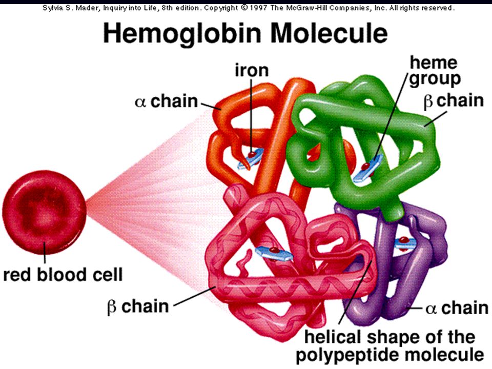
 Hb is found in RBCs its main function is to transport O2 to tissues. Structure: 2 parts : heme + globin Globin: four globin chains (2 α.>")


 Course code: MLHE-201 Supervisor: Prof>")
 –Formed elements 45%– rbc’s, wbc’s, platelets –Buffy coat – wbc and platelets.>")


