Download presentation
Presentation is loading. Please wait.

1
Iron Metabolism HMIM224

2
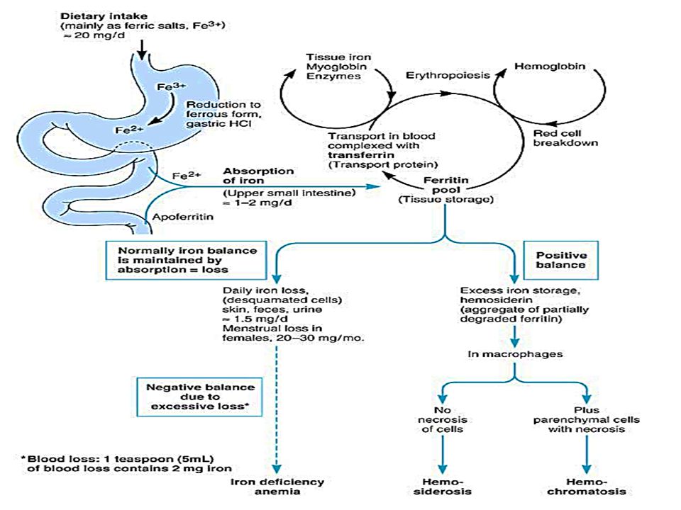
Introduction Iron is an essential element present mainly in heme of hemoglobin, myoglobin, cytochromes & in iron storage proteins ferritin & hemosidrin. An adult human has ~ 4 grams of iron in his body. Clinical importance: - Iron deficiency in the body may lead to iron deficiency anemia (microcytic hypochromic anemia) - Overdose of iron may cause haemosiderosis.
 - Overdose of iron may cause haemosiderosis.")
3
Dietary iron Recommended Dietary Allowance (RDA) : - Adults: 10 mg/day - Females below 50 years & during lactation: increased up to 15 mg/day - Females during pregnancy: increased up to 30 mg/day Sources of dietary iron: - Animal sources: liver, spleen, meat (rich sources) - Plant sources: molasses, dates, vegetables & whole cereals
 : - Adults: 10 mg/day - Females below 50 years & during lactation: increased up to 15 mg/day - Females during pregnancy: increased up to 30 mg/day Sources of dietary iron: - Animal sources: liver, spleen, meat (rich sources) - Plant sources: molasses, dates, vegetables & whole cereals")
4
Absorption of iron Site of absorption: mainly the duodenum On an average diet containing 10 – 15 mg of iron, only about 5 – 15% are absorbed. Factors affecting absorption of iron: 1- Amount of iron ingested: increase in amount of iron in diet, increase amount absorbed 2- State of iron: Iron is liberated from organic complexes of diet (ferritin) by gastric HCl into organic salts. Then, Fe3+ liberated is reduced to Fe2+ by reducing substances in food as ascorbic acid (vitamin C). Iron of heme in meat is better absorbed (while still in heme molecule) 3- Solubility of iron: The absorption of iron is reduced by factors that decrease its solubility. These factors are high pH, excess phosphates , oxalates, phytates & unabsorbed fatty acids in GIT.
 by gastric HCl into organic salts. Then, Fe3+ liberated is reduced to Fe2+ by reducing substances in food as ascorbic acid (vitamin C). Iron of heme in meat is better absorbed (while still in heme molecule) 3- Solubility of iron: The absorption of iron is reduced by factors that decrease its solubility. These factors are high pH, excess phosphates , oxalates, phytates & unabsorbed fatty acids in GIT.")
5
Distribution of iron in the body
Total iron in the body is ~ 4 grams This amount is available in two forms: 1- Functional forms (75%): 1- Hemoglobin (67%) 2- Myoglobin (7.5%) 3- Respiratory enzymes (0.5%): as cytochromes, etc 2- Non-functional forms (25%): Free iron is very toxic. So, iron is bound to proteins (non-heme metaloproteins) that allows it to be transported & stored in non-toxic forms. 1- Transferrin (0.1%): for transport of iron in blood 2- Ferritin & hemosidrin (24.9%): for storage of iron in tissues
: 1- Hemoglobin (67%) 2- Myoglobin (7.5%) 3- Respiratory enzymes (0.5%): as cytochromes, etc. 2- Non-functional forms (25%): Free iron is very toxic. So, iron is bound to proteins (non-heme. metaloproteins) that allows it to be transported & stored in non-toxic forms. 1- Transferrin (0.1%): for transport of iron in blood. 2- Ferritin & hemosidrin (24.9%): for storage of iron in tissues.")
6
Iron storage in the body
Ferritin: - is the chief storage form of iron in tissues. is available in liver, spleen, bone marrow & intestinal mucosal epithelium. is composed of a protein shell with a core containing iron as ferric form. Its binding sites are saturated by 23% with iron. Hemosiderin: - Similar to ferritin but with binding sites saturated by 35% iron. - Increased in cases of excess iron in the body.

7
Blood iron 1- in Hemoglobin of RBCs: Hemoglobin contains 3.4 mg iron /gm of hemoglobin Hemoglobin is ~ 15 gm/100 ml blood Amount of iron in hemoglobin is ~ 50 mg/100 ml blood 2- in plasma: a) Transferrin: Iron is carried in blood by transferrin, which carries two atoms of Fe3+ per molecule. Only about 30% of transferrin is saturated with iron (called Total Iron Binding capacity, TIBC) Transferrin is synthesized in the liver & runs with the b-globulin in electrophoresis. In iron deficiency anemia, plasma iron is reduced while TIBC tends to increase. In liver disease, both plasma iron & TIBC are decreased b) Plasma ferritin: Plasma contains very low concentrations of ferritin (20 – 250 mg/L) Plasma ferritin is a good index of iron storage It is decreased in iron deficiency anemia & is increased in hemosiderosis.
 Transferrin: Iron is carried in blood by transferrin, which carries two atoms of Fe3+ per molecule. Only about 30% of transferrin is saturated with iron (called Total Iron Binding capacity, TIBC) Transferrin is synthesized in the liver & runs with the b-globulin in electrophoresis. In iron deficiency anemia, plasma iron is reduced while TIBC tends to increase. In liver disease, both plasma iron & TIBC are decreased b) Plasma ferritin: Plasma contains very low concentrations of ferritin (20 – 250 mg/L) Plasma ferritin is a good index of iron storage It is decreased in iron deficiency anemia & is increased in hemosiderosis.")
8
Excretion of iron 1- in feces (90 – 95%):
Fecal iron is mainly unabsorbed iron. 2- in urine & sweat (5 - 10%) Daily loss of iron in urine & sweat is about 0.5 – 1 mg of iron. 3- in menstruation & milk (5 - 10%) About 15 – 30 mg of iron (in the form of hemoglobin) is lost in menstruation per month (0.5 – 1 mg/day). Lactation leads to loss of 0.5 – 1 mg of iron per day
 Daily loss of iron in urine & sweat is about 0.5 – 1 mg of iron. 3- in menstruation & milk (5 - 10%) About 15 – 30 mg of iron (in the form of hemoglobin) is lost in menstruation per month (0.5 – 1 mg/day). Lactation leads to loss of 0.5 – 1 mg of iron per day.")
10
Laboratory assessment of iron status
1- Plasma iron 2- Plasma ferritin 3- Plasma transferrin, Total Iron Binding Capacity (TIBC) & TIBC Saturation
 & TIBC Saturation")
11
Laboratory assessment of iron status (cont.)
1- Plasma iron: -Measurement of plasma iron is of limited diagnostic value as: 1- Level fluctuate widely in healthy due to many factors (as diurnal rhythm, menstruation, oral contraceptive pills intake, pregnancy, etc) 2- Measurement of plasma iron do not provide an adequate index of iron status as: In cases of iron deficiency anemia, plasma iron is reduced late when iron is entirely depleted from iron stores. In iron overdose, plasma iron is elevated late when iron stores are seriously overloaded with iron. 3- Plasma iron alters in cases not associated with changes in iron stores as in acute infections or trauma, in which plasma iron is rapidly reduced to low levels while iron stores are not affected
 2- Measurement of plasma iron do not provide an adequate index of iron status as: In cases of iron deficiency anemia, plasma iron is reduced late when iron is entirely. depleted from iron stores. In iron overdose, plasma iron is elevated late when iron stores are seriously overloaded. with iron. 3- Plasma iron alters in cases not associated with changes in iron stores. as in acute infections or trauma, in which plasma iron is rapidly reduced to low levels. while iron stores are not affected.")
12
Laboratory assessment of iron status (cont.)
2- Plasma ferritin: Plasma ferritin is closely related to body iron stores whether low, normal or high. (while plasma iron becomes abnormal only in presence of gross abnormalities) Low (or upper normal) plasma ferritin indicates depleted iron stores. High plasma ferritin indicates iron overdose (hemosiderosis). N.B. Plasma ferritin is one of the acute phase reactants. So, it is elevated in acute disorders as infections….etc
 Low (or upper normal) plasma ferritin indicates depleted iron stores. High plasma ferritin indicates iron overdose (hemosiderosis). N.B. Plasma ferritin is one of the acute phase reactants. So, it is elevated in acute. disorders as infections….etc.")
13
Laboratory assessment of iron status (cont.)
3- Plasma transferrin & Total Iron Binding Capacity (TIBC): - Transferrin level is reduced in protein malnutrition, chronic liver diseases, acute infections & neoplasm. (low protein synthesis by the liver) - Transferrin synthesis is increased in iron deficiency (as a compensatory mechanism) Transferrin can be measured: - Directly - Indirectly by Total Iron Binding capacity (TIBC): TIBC is the ability of transferrin to bind iron. 30-40% of binding sites of transferrin are occupied by iron.
: - Transferrin level is reduced in protein malnutrition, chronic liver diseases, acute infections & neoplasm. (low protein synthesis by the liver) - Transferrin synthesis is increased in iron deficiency (as a compensatory mechanism) Transferrin can be measured: - Directly. - Indirectly by Total Iron Binding capacity (TIBC): TIBC is the ability of transferrin to bind iron % of binding sites of transferrin are occupied by iron.")
14
Iron deficiency Causes of iron deficiency:
Commonest single nutrient deficiency worldwide. Causes of iron deficiency: 1- Deficient intake of iron: ingestion diet poor in iron for a long time 2- Increased requirements of iron : as in pregnancy, lactation & menstruation 3- Impaired absorption of iron: due to intestinal causes (malabsorption syndrome) 4- Excessive loss of iron: in cases of chronic bleeding (from GIT bleeding etc..) Laboratory & clinical manifestations: 1- low plasma ferritin 2- Then, increase transferrin (& TIBC) 3- Then, low plasma iron 4- Finally, anemia is evident by lab investigation (microcytic hypochromic anemia) & by clinical manifestations
 4- Excessive loss of iron: in cases of chronic bleeding (from GIT bleeding etc..) Laboratory & clinical manifestations: 1- low plasma ferritin. 2- Then, increase transferrin (& TIBC) 3- Then, low plasma iron. 4- Finally, anemia is evident by lab investigation (microcytic hypochromic anemia) & by. clinical manifestations.")
15
Iron overload (iron toxicity)
The body is unable to excrete large load of iron (bound to proteins) Amount of iron in the body is controlled by regulating its absorption from GIT rather than its excretion. Some people can absorb large amounts of iron, from intestine (20- 45% of intake), leading to accumulation of excessive amounts of iron in tissues, a condition known as hemosiderosis (hemosiderin accumulates in tissues). Haemochromatosis means hemosiderosis with injury to involved tissues.
 Amount of iron in the body is controlled by regulating its absorption from GIT rather than its excretion. Some people can absorb large amounts of iron, from intestine (20- 45% of intake), leading to accumulation of excessive amounts of iron in tissues, a condition known as hemosiderosis (hemosiderin accumulates in tissues). Haemochromatosis means hemosiderosis with injury to involved tissues.")
16
Iron overload (iron toxicity) cont.
Causes of iron overload: 1- Increased intake & absorption Acute overdose: causes fatal symptoms due to the toxic effects of free iron in plasma. Chronic overload: with increased absorption of iron due to any cause for long duration. 2- Parentral administration of iron including repeated blood transfusion. 3- Genetic haemochromatosis. Laboratory Investigations: 1- Plasma iron is elevated (especially in late cases) 2- Transferrin becomes 70-90% saturated with iron (TIBC saturation) 3- Plasma ferritin is elevated.
 2- Transferrin becomes 70-90% saturated with iron (TIBC saturation) 3- Plasma ferritin is elevated.")
Similar presentations


i.>")


 Mean corpuscular volume (MCV)N: 80-100 fl Mean corpuscular hemoglobin concentration.>")