Download presentation
Presentation is loading. Please wait.

1
HAEMOTOLOGICAL DISORDERS IN PREGNANCY
Dr. RAMYA MODERATOR : Dr.PALLAVEE

2
HAEMOTOLOGICAL DISORDERS IN PREGNANCY
ANAEMIA PLATELET DISORDERS HAEMOGLOBINOPATHIES INHERITED COAGULATION DEFECTS

3
ANAEMIA Commonest haematological disorder occur in preg.
Prevalance in pregnant women – 14 % Developed 51% Developing countries 65-75% - India 80 % leading to maternal deaths

4
DEFINITION Reduction in circulating Hb mass
< 12g/dl in non-pregnant women <10 g/dl in pregnant women CDC Anaemia in iron supplemented preg. Woman Hct 33% & Hb 11g/dl – 1st & 3rd trimester Hct 32% & Hb 10.5 g / dl - 2nd trimester

5
WHO grading of anemia Mild g/dl Moderate g/dl Severe < 7 g/dl

6
ICMR GRADING MILD 10 – 10.9 MODERATE 7 – 9.9 SEVERE < 7 VERY SEVERE
Range in g/dl MILD 10 – 10.9 MODERATE 7 – 9.9 SEVERE < 7 VERY SEVERE < 4

7
Hemotological Changes in preg.

8
Physiological Anemia of pregnancy
Plasma volume s 40-50% RBC mass s 30 % As a result Hb concentration decreases by 2g/dl Decreased Hb concn. Is due to haemodilution Criteria of Physiological Anemia 1) Hb 10 gm % 2)RBC 3.2 million cells / cu mm 3)PCV 32% 4)Peripheral Smear – Normal morphology
 Hb 10 gm % 2)RBC 3.2 million cells / cu mm. 3)PCV 32% 4)Peripheral Smear – Normal morphology.")
9
Classification of Anaemia

10
Classification …….

11
Classification …….

12
Classification …….

13
Classification …….

14
ERYTHROPOISES

15
IRON METABOLISM

16
IRON Requirements during Pregnancy
Maternal req. Of total Iron -1000mg 500 mg Mat. Hb. Mass expansion 300 mg Fetus & Placenta 200mg Shed through gut urine, skin

17
DEVELOPMENT OF Iron def. anemia
Iron Deficiency Anemia – 3 stages a)Depletion of Iron stores b)Iron deficient erythropoiesis c)Frank Iron deficiency Anemia
Depletion of Iron stores. b)Iron deficient erythropoiesis. c)Frank Iron deficiency Anemia.")
18
Symptoms of IRON DEFICIENCY ANEMIA
Fatigue Weakness Headache Loss of appetite Dysphagia Palpitations Dyspnea on exertion Ankle swelling Paresthesias Leukoplakia

19
Physical examination Pallor of varying degrees (Mucous membranes , nail beds – Koilonychia or Platynychia Glossitis Stomatitis Heart murmurs Increased JVP Tachycardia Tachypnea Postural hypotension Crepitations- due to lung congestion

20
Depletion of Iron stores
Ferritin <20 ng/ml Hb / Hct. Normal RBC INDICES normal Iron deficient erythropoiesis Transferrin saturation<25% Hb –Normal Serum Iron < 60mg/dl

21
c)Frank Iron deficiency Anemia
ferritin <20 ng/ml Transferrin saturation<25 % Serum iron <60 mg/dl Hb <10g/dl, Hct.<28%

22
Microcytic Hypochromic

23
PROPHYLAXIX WHO mg Elemental iron micro gram Folic acid / day * 6 months & 3 months postpartum National Nutritional Anemia Control Programme of India - 60 mg elemental Iron mcg Folic acid & Prophylactic supplementation * 100 days in 2nd trimester

24
Iron Supplements

25
Ferrous sulphate 300mg Tid orally daily after meals
To be contd for 12 months after anemia is corrected Indicators of iron therapy response Increase in Reticulocyte count (Increases 3-5 days after initiation of therapy ) Increase in Hb levels. Hb increases 0.3 to 1 g/ week 3 .Epithelial changes (esp tongue & nail ) revert to normal Hb concn. Is normal after 6 wks of therapy
 Increase in Hb levels. Hb increases 0.3 to 1 g/ week. 3 .Epithelial changes (esp tongue & nail ) revert to normal. Hb concn. Is normal after 6 wks of therapy.")
26
PARENTERAL ADMINISTRATION
INDICATIONS Intolerance to oral iron Non compliance pt. Inflammatory bowel disease Pt. unable to absorb iron orally Patients near term

27
Formulae TDI – Total Dose Infusion
Amount of iron needed to restore Hb conc to normal & additional allowance to provide adequate replenishment of iron stores Formulae 1 Total Dose ( mg ) = ( normal Hb – Pts Hb ) * (body wt. in kg ) * 2.21 2 Total Iron Dose (mg ) = 2.3 * wt. kg before preg * D (Target Hb) mg for body store
 = ( normal Hb – Pts Hb ) * (body wt. in. kg ) * Total Iron Dose (mg ) = 2.3 * wt. kg before preg * D (Target Hb) mg for body store.")
28
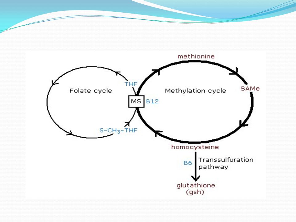
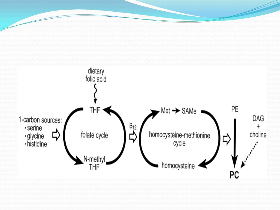
MEGALOBLASTIC ANAEMIA
Incidence – 0.2 – 5 % Caused by folic acid deficiency & Vit B12 deficiency

32
Folic Acid Defciency Pathophysiology Preg. Causes fold increase in Folate requirement ( microgram / day ) to meet needs of fetus & placenta. Placenta transports folate actively to fetus even if the mother is deficient. This cause decreased plasma folate levels.
 to meet needs of fetus & placenta. Placenta transports folate actively to fetus even if the mother is deficient. This cause decreased plasma folate levels.")
33
Causes of Folic acid deficiency
1.Diet- Poor intake, prolonged cooking. 2. Malabsorption – Coeliac disease. 3.Increased demand – Pregnancy, cell proliferation (hemolysis ) 4.Drugs – anticonvulsants, contraceptive pill, cytotoxic drugs (Methotrexate ) 5.Diminished storage – Hepatic disorders & Vit C deficiency
 4.Drugs – anticonvulsants, contraceptive pill, cytotoxic drugs (Methotrexate ) 5.Diminished storage – Hepatic disorders & Vit C deficiency.")
34
Diagnostic features of Folic acid deficiency
1.Serum Folate levels – Low <3 ng/ml 2.Erythrocyte Folate levels - <20 ng/ml 3.Peripheral smear – Hypersegmented neutrophils,Oval macrocytes,Pancytopenia

35
Treatment In other conditions Prophylaxis
Pregnancy induced megaloblastic anaemia- Folic acid, nutritious diet & Iron . Supplementation of 1mg of folic acid daily can improve MA by 7 to 10 days Folic acid should be given with iron Ascorbic acid 100mg Tid enhances action In other conditions Recommended folic acid dose – 5mg /day orally daily Prophylaxis WHO – 400 micrograms folic acid daily to prevent neural tube defects

36
Vit – B12 Deficiency

37
Pathophysiology CAUSES of Vit B12 def.
Vit B12 absorption is unaltered during pregnancy Tissue uptake is increased Decreased serum B12 Recommended B12 intake – 3 microgram /day. CAUSES of Vit B12 def. Strict Veg. diet Use of proton pump inhibitors Metformin. Gastritis Gastrectomy Ileal bypass Crohn’s H. Pylori infection

38
Pathogenisis of PERNICIOUS ANEMIA
GASTRIC ATROPHY ? autoimmune Reduced IF secretion Failure of absorption of dietary Vit B12 Deficiency of Vit B12 Pathogenisis of PERNICIOUS ANEMIA Gastric juice IF Antibody

39
Clinical manifestations
Macrocytic Megaloblastic Anemia Glossitis Peripheral neuropathy Subacute combined degeneration of the Spinal cord

40
Treatment DIAGNOSIS Ser.Vit B12 levels ,100 pg /ml
Radio active Vit B12 absorption test . ( Schilling Test ) Treatment 1000 microgram parenteral cyanocobalamin every wk * 6 weeks Pernicious Anaemia – Oral Vit B12 Total Gastrectomy – 1000 microgram Vit B12 im every month. Partial gastrectomy – Ser. Vit B12 levels measured.
 Treatment microgram parenteral cyanocobalamin every wk * 6 weeks. Pernicious Anaemia – Oral Vit B12. Total Gastrectomy – 1000 microgram Vit B12 im every month. Partial gastrectomy – Ser. Vit B12 levels measured.")
41
ANAEMIA ASSOC. WITH CHRONIC INFECTIONS / DISEASE
Common in developing countries Poor response to Haematinics unless primary cause is treated Worm infestations is common ( Diagnosed by stool examination ) Urinary tract inf, & asymptomatic bacteriuria in preg. Is assoc. with refractory anaemia Chronic renal disorders = due to erythropoietin def. Treated with recombinant Erythropoitin
 Urinary tract inf, & asymptomatic bacteriuria in preg. Is assoc. with refractory anaemia. Chronic renal disorders = due to erythropoietin def. Treated with recombinant Erythropoitin.")
42
Anaemia from acute blood loss
In preg. Abortion , ectopic preg, hydatidform mole, PPH Treatment. Blood transfusion Indicated patient – symptomatic Not indicated – If hemodynamically stable, Hb < 7 g/dl, able to ambulate without adverse symptoms & not septic.

43
Acquired hemolytic anemia
AUTOIMMUNE HEMOLYTIC ANEMIA AUTOANTI-BODIES OF iGg OR WARM ANTIBODIES AGAINST Red cell antigens, causes premature destruction of RBC”s ETIOLOGY Lymphomas,Leukemias , Connective tissue diseases, Infections , Chronic. Inflammatory diseases & drug induced antibodies

44
Diagnosis Direct Coomb’s Test Blood smear – Spherocytosis & Reticulocytosis TREATMENT Prednisone 1mg / kg / day orally Azathioprine Splenectomy

45
2)Preg. Induced hemolytic anemia
Unexplained hemolytic anemia uring pregnancy is rare Severe hemolysis occurs early in pregnancy & resolves within months after delivery No evidence of immune mechanism or defects in RBCs Prednisone given untill delivery 3) Paroxysmal Nocturnal Hemoglobinuria Acquired hemolytic anemia Arises from one abnormal clone of cells like neoplasm Anemia is insiduous in onset & hemoglobinuria develoes at irregular intervals
 Paroxysmal Nocturnal Hemoglobinuria. Acquired hemolytic anemia. Arises from one abnormal clone of cells like neoplasm. Anemia is insiduous in onset & hemoglobinuria develoes at irregular intervals.")
46
Hemolysis may be initiated by transfusion , infections or surgery
40% suffer venous thrombosis, renal failure , HTN & Budd Chiari syndrome. Prophylactic anticoagulation is required Bone marrow transplantation – Definitive treatment Effect on pregnancy Serious & unpredictable Maternal mortality 10 – 20% Venous thrombosis occurs during post partum

47
APLASTIC ANAEMIA Rarely seen in preg.
Marked decrease in marrow stem cels ETIOLOGY Infections Irradiation Leukemia Immunological disorders

48
May be Immunological mediated or autosomal recessive inheritance
30% cases Anaemia improves once pregnancy is terminated. Complications Infection Haemorrhage

49
Diagnosis Blood Values – Anemia Leucopenia Thrombocytopenia
Bone Marrow - Hypocellular

50
Management Supportive care – Cont. Infection surveillance & anti microbial therapy Red cell transfusions to maintain Hct. > 20 Granulocyte transfusion given only during Infections Platelet transfusion to control haemorrhage. Glucocorticoid therapy may be helpful IN SEVERE cases Bone marrow or Stem Cell Transplantation Vaginal delivery is preferred

51
Effect of anaemia in preg.
In MOTHER During preg. Pre eclampsia Infectuion Heart failure Pretem labour Labour Uterine inertia Postpartum Haemorrhage Cardiac failure Shock Puerperium Puerperal sepsis Subinovulation Failure of lactation Puerperal venous thrombosis Pulmonary embolism

52
Fetus Amount of iron transferred to fetus is unaffected even if mother is in iron deficient state Prematurity Low birth weight babies Intra uterine deaths due to severe maternal anoxemia

53
Effect of pregnancy in anaemia
Pt. Mildly anemic progresses to marked Anaemia Pt. Who is severely anemic becomes symptomatic by the end of 2nd trimester

54
DIAGNOSIS OF ANEMIA DURING PREGNANCY
Pt. with Anemia Hb<11, Hct <0.33 Investigations- MCV, PS ,Retic count MCV <80 Low PS –Microcytic Serum Iron studies Serum Iron reduced TIBC-Increased Ser. Ferritin – reduced IRON DEF ANEMIA TREATMRNT WITH ORAL / PARENTERAL IRON MCV 80 – 90 PS – Normal RETIC COUNT INCREASED -Hemolytic Hemoglobin opathies Autoimmune Cases Drug induced RETIC – NORMAL / DECREASED Drugs Bone marrow path Chronic diseases MCV >99 PS-Macrocytic Ser.B12 & Folic acid levels Folate < 3ng/ml Vit B12 <80pg/ml Therapy with 1 mg/day – Folate 1000ug im B12 every wk * 6 wks foll by every month DIAGNOSIS OF ANEMIA DURING PREGNANCY

55
PLATELET DISORDERS Thrombocytopenia - Gestational - Immune mediated
Mild – 1,50,000 – 1,00,000 Moderate – 1,00,000 – 50,000 Severe - < 50,000 Abnormalities of Platelet function

56
Causes of Thrombocytopenia during Pregnancy
COMMON CAUSES Gestational Thrombocytopenia Severe Pre-eclampsia HELLP syndrome Immune thrombocytopenic Purpura Disseminated Intravascular coagulation RARE CAUSES Lupus anticoagulant/APA syndrome SLE Hemolytic Uremic Syndrome Type 2b Von- Willebrand’s syndrome Folic acid def. HIV infections Hemotoligical malignancies May – Hegglin syndrome – Congenital Thrombocytopenia

57
Gestational Thrombocytopenia
Benign Common disorder Appears in 8% of all preg. Unknown cause Rarely drops < 70,000 /mm3 FEATURES Diagnosis of Exclusion – No specific test available Mild Thrombocytopenia , Count > 70,000 – 1 lakh No maternal bleeding No past history of thrombocytopenia Occurs in 3rd trimester No assoc. fetal thrombocytopenia Spont. Resolution after delivery May reccur in subsequent pregnancies

58
Management Majority cases treated as normal
In mod. To severe cases – Reluctance of Anaesthesiologists to give spinal or epidural if Platelets = < 80,000 /cu mm Treatment with steroids & IgG or platelet transfusion before delivery Cord sample should be taken Samples taken on day 1 & 4 CS reseved for obstetric indications

59
Immune Thrombocytopenia
Chronic condition , Incidence 1 in 1000 to 1 in pregnancies Charecterised by autoantibody mediated destruction of maternal platelets MECHANISM Autoanti bodies react with platelet Glycoprotein complex & antibody coated platelets are phagocytosed by Macrophages

60
SYMPTOMS Usually asymptomatic , sometimes Bleeding , Petechiae DIAGNOSIS Plat count < 50,000 / cu mm with past h/o bleeding disorders No specific diagnostic test

61
Prepregnancy counselling for ITP ( RCOG 2009 )
May relapse or worsen If treatment reqd, it will carry for both maternal & fetal risks Increased risk of Hemorrhage at delivery Epidural Anaesthesia is not possible Risk prediction in neonate is not possible. High risk if sibling has thrombocytopenia or mother has undergone splenectomy Maternal deaths / serious outcome – RARE Risk of Intracranial Haemorrhage in fetus is very low.

62
Management Adequate Plat. Count should be maintained
Counts monitored throughout pregnancy If > 30,000 – No treatment If< 30, ) Prednisolone 1 -2 mg / kg oral daily, 2) IV IG – 2g/kg over 2 to 5 days , If no response then 3) Splenectomy 4) Immunosuppressive drugs like a) azathioprine b) Cyclophosphamide , Cyclosporine
 Prednisolone 1 -2 mg / kg oral daily, 2) IV IG – 2g/kg over 2 to 5 days , If no response then. 3) Splenectomy. 4) Immunosuppressive drugs like. a) azathioprine. b) Cyclophosphamide , Cyclosporine.")
63
Management Of delivery
Platelet > 50,000 / cu mm – Vaginal or operative delivery Platelet 50,000 Platelets standby CS not routinely recommende Measures should be taken to avoid trauma to baby head Cord sample taken, If low confirmed by capillary sample If count low, further day 1 & 4 is collected Inj im Vit K avoided till count is known

64
Fetal & Neonatal Effects
PA IgG antibodies crosss placenta causes fetal & neonatal Thrombocytopenia Maternal treatment do not have effect o fetal count Role of Intracaranial hemorrhage < 1 %

65
MICROANGIOPATHIES Thrombotic thrombocytopenic purpura
Rare – life threatening Signs & symptoms ( PENTAD ) Microangiopathic hemolytic anemia Thrombocytopenia Neurological symptoms Renal dysfunction Fever
 Microangiopathic hemolytic anemia. Thrombocytopenia. Neurological symptoms. Renal dysfunction. Fever.")
66
ETIOLOGY Severe def. of VON WILLIBRAND FACTOR ( cleaving protein ( ADAM TS13) Acqd autoantibody Congenital Genetic defect Incidence 1 in pregnancies Time of onset of TTP is variable 1st trimester to several wks post partum Maternal mortality is high

67
MANAGEMENT ACQUIRED Plasma Exchange
Fresh frozen Plasma infused daily until Platelets turn to Normal Rituximab, Monoclonal antibodies against CD20 CONGENITAL FFP Platelet transfusion contraindicated

68
HEMOLYTIC UREMIC SYNDROME
Microangiopathic hemolytic anaemia & thrombocytopenia with predominant Renal involvement Due to endothelial damage by bacterial or viral infections In Preg. Response is poor for plasma exchange

69
THROMBOCYTOSIS Defined as persistant Platelet count >4.5 lakhs / cumm CAUSES 1 )Secondary or Reactive ( > 80000) a)Iron def. b) Infections c) Splenectomy d) Surgery & Trauma ( bone fractures ) e) Malignancy 2) Essential Thrombocytosis > I million a) Idiopathic b) Myelodysplastic syndromes
Iron def. b) Infections. c) Splenectomy. d) Surgery & Trauma ( bone fractures ) e) Malignancy. 2) Essential Thrombocytosis > I million. a) Idiopathic. b) Myelodysplastic syndromes.")
70
SIGNS & SYMPTOMS Usually asymptomatic Arterial & venous Thrombosis Hepatomegaly Bone marrow – Hyperplastic with gross increase in megakaryocytes Blood picture ->1 million Leucocytosis Anemia or mild polycythemia Anisocytosis & Poikilocytosis

71
In Pregnancy – Spont. Abortion , fetal demise & preeclampsia.
TREATMENT Aspirin , Dipyramidole, Heparin, Plateletpheresis PROGNOSIS depends on underlying disease Death due to either thrombosis / hemorrhage / comp of Myeloproliferative disorders/ marrow failure.

Similar presentations


 Nicola Davis.>")

