Download presentation
Presentation is loading. Please wait.

1
Abdomen Borders: Superiorly: xiphoid process, the lower six costal cartilages, and the anterior ends of the lower six ribs Inferiorly: pubic symphysis and the pubic crest, the anterior superior iliac spine, and the iliac crest. Regions: Epigastric Hypochondriac (2) Umpilical Lumbar (2) Hypogastric Iliac (2) These regions are devided by the following lines Mid clavicular lines Transpyloric line- 1/2 of the distance between the jugular notch and the pubic crest Intertubercular line
 Umpilical. Lumbar (2) Hypogastric. Iliac (2) These regions are devided by the following lines. Mid clavicular lines. Transpyloric line- 1/2 of the distance between the jugular notch and the pubic crest. Intertubercular line.")
2
Muscles External Oblique M. Internal Oblique M. Transversus Abdominis M. Rectus Abdominis M. O: I: Inn: F:

3
Pyramidalis M. Cremaster M. Quadratus Lamborum M.

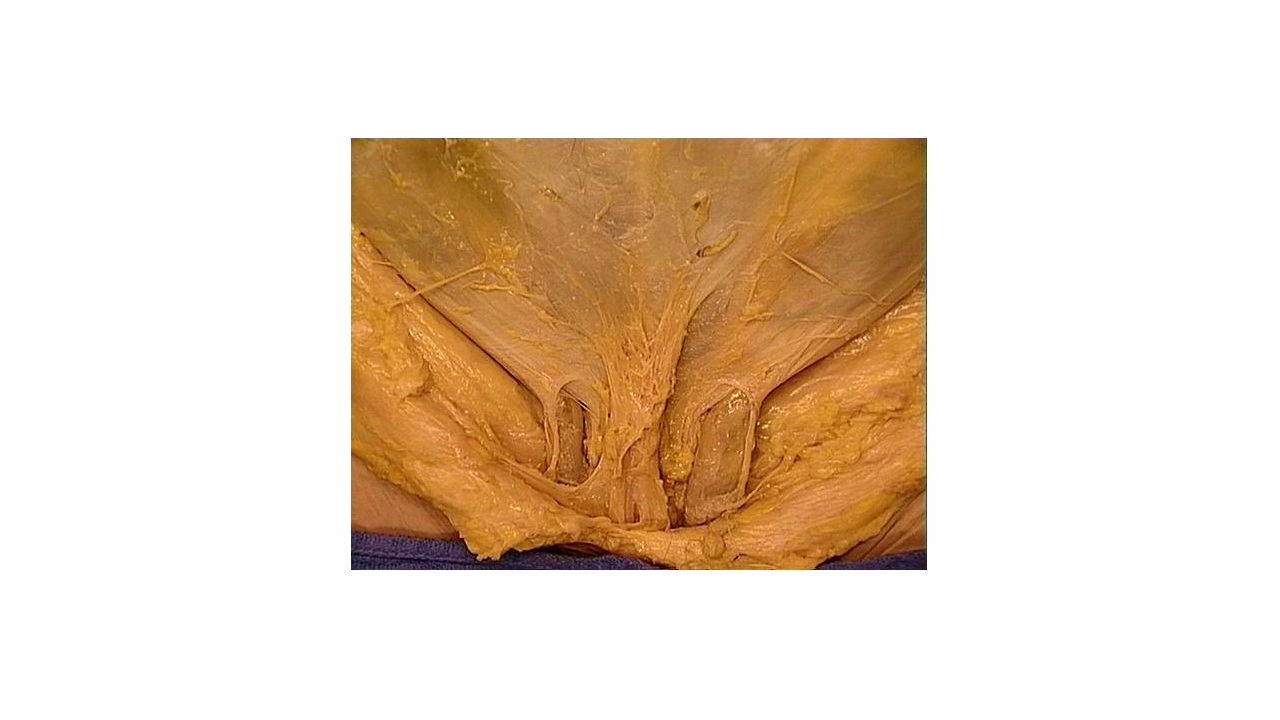
5
Sheath of Rectus Abdominis M.
Both Recti abdominis muscles are integrated into a fibrous sheath , vagina recti abdominis. It is formed at the Superior 2/3 by Anteriorly: External Oblique M. Aponeurosis and the one leaf of Internal Oblique M. Aponeurosis Posteriorly: One leaf of Internal Oblique M. Aponeurosis, Transverse abdominal M. Aponeurosis and Transversalis Fascia It is formed in at the Inferior 1/3 by Anteriorly: External Oblique Aponeurosis , the whole Internal oblique Aponeurosis and Transverse abdominal M. aponeurosis Posteriorly: Transversalis fascia The superior and inferior part are devided by the arcuate line

7
In the medial line both layers of vagina recti abdominis of both sides are connecting together and create a fibrous band – linea alba . This line runs from from xiphoid process to pubic symphysis. Approximately in the half of Linea Alba length there is an umbilical scar -umbilicus

9
Fasciae Superficial abdominal Fascia: outer side of the abdominal wall= Camper's fascia - fatty superficial layer Subcutaneous Abdominal Fascia: Scarpa's fascia - deep fibrous layer. Fascia transversalis: inner side of abdominal wall

10
The inner surface of abdominal wall contains several ligaments separated by fossae:
Ligament/fold Remnant of Lateral fossa Hernia median umbilical ligament urachus supravesical fossa - medial umbilical ligament umbilical artery medial inguinal fossa direct inguinal hernia lateral umbilical fold inferior epigastric vessels lateral inguinal fossa indirect inguinal hernia

11
Inguinal Canal It opens medially under the abdominal skin as Superficial inguinal ring and laterally as deep inguinal ring which leads to peritoneal cavity. Superficial inguinal ring is in the aponeurosis of external oblique muscle and it is demarcated by crus mediale and crus laterale which are crossed by intercrural fibers. Deep inguinal ring is an opening located in aponeurosis of transversus abdominis muscle and internal oblique muscle. Its medial border is formed by interfoveolar ligament. It serves for passing of spermatic cord.

12
Inguinal Canal walls Anterior: Aponeurosis of external oblique M. Superior: Internal Oblique M. and Transversus abdominis M. Inferior: Inguinal ligament

17
Dissection Step 1 Skin insicion

18
Step 2 Skin Subcutaneous Layer: Camper’s and Scarpa’s fascia Find and clean anterior and lateral cutaneous nerves (<intercostal nerves) Find and clean Superficial epigastric vessels, Superficial iliac Circumflex vessels and External pudendal vessels

19
Camper’s Fascia Scarpa’s Fascia

20
Superficial Epigastric artery and vein (<Femoral artery/vein)
Superficial iliac circumflex artery and vein (< Femoral artery/vein) Anterior and Lateral Cutaneous nerves (<Intercostal Nerves) Superficial External Pudendal artery and vein (< Femoral artery/vein)
 Anterior and Lateral Cutaneous nerves (<Intercostal Nerves) Superficial External Pudendal artery and vein (< Femoral artery/vein)")
21
Cutaneous branches

22
Step 3 Clean the surface of external Oblique M. Find and clean Superficial ring of inguinal canal Identify the spermatic cord/ round ligament Find and clean Ilioinguinal nerve (laterally of spermatic cord) and the genital branch of genitofemoral nerve( medially to spermatic cord) Ilioinguinal Nerve Iliohypogastric
 and. the genital branch of genitofemoral nerve( medially to spermatic cord) Ilioinguinal Nerve. Iliohypogastric.")
23
Step 4 Cut the external Oblique M. in the mid axillary line to the anterior iliac spine Separate the external oblique from the internal oblique with your fingers but preserve the nerves and the vessels Find and clean the subcostal , ilioinguinal and iliohypogastric nerves and the deep iliac circumflex vessels Iliohypogastric

25
Step 5 Clean and cut the internal oblique in the same way as external oblique Separate it with transversus abdominis by your fingers Clean the rectus sheath Identify the Fundiform ligament of the penis Fundiform ligament: Scarpa's fascia contributes to formation of the fundiform ligament of the penis.

26
Step 6 Open the rectus sheath by a median incision from the sternum to pubis symphysis Reflect the sheath Loose the rectus abdominis and identify the superior and inferior Epigastric vessels behind the rectus abdominis

27
Superior Epigastric Artery and Vein (<Internal thoracic artery/vein)
Inferior Epigastric Artery and Vein (<External Iliac artery/vein) Deep Circumflex Iliac Artery and vein (< External Iliac artery/vein)
 Deep Circumflex Iliac Artery and vein (< External Iliac artery/vein)")
28
The space that has been opened is the major portion of the peritoneal cavity, called the greater peritoneal sac. This space is located between the inner surface of the abdominal wall and the outer surface of the abdominal viscera. The greater peritoneal sac is bounded between the parietal and visceral peritoneum. It communicates through the epiploic foramen (of Winslow) with the smaller, lesser peritoneal sac (also called omental bursa). The lesser peritoneal sac is the largest recess of the peritoneal cavity and is located posterior to the stomach. Its boundaries will be described later. The Falciform ligament of the liver is formed as the visceral peritoneum that covers the liver and is reflected onto the internal surface of the anterior abdominal wallas ligamentum teres

29
The peritoneum is divided into two parts: the visceral peritoneum, which covers the surface of the visceral organs, and the parietal peritoneum, which lines the internal surface of the walls of the abdominal and pelvic cavity. Inside the abdominal cavity the peritoneum forms folds or reflections, referred to as mesenteries and peritoneal ligaments. Mesenteries attach organs to the posterior abdominal wall. Peritoneal ligaments generally attach one organ to another. Peritoneal ligaments and mesenteries hold abdominal organs in place, and serve as conduits through which nerves, blood and lymphatic vessels pass to and from the abdominal organs. An organ, which is surrounded by the peritoneum and has a supporting mesentery, is defined as an intraperitoneal organ (e.g. spleen, liver, and transverse colon). An organ, which is only partially covered by the peritoneum and has no supporting mesentery, is defined as a retroperitoneal or extraperitoneal organ (e.g. pancreas, kidney, ascending colon, and descending colon).
. An organ, which is only partially covered by the peritoneum and has no supporting mesentery, is defined as a retroperitoneal or extraperitoneal organ (e.g. pancreas, kidney, ascending colon, and descending colon).")
31
The lesser omentum is actually a combination of three peritoneal ligaments:
hepatoesophageal ligament hepatogastric ligament hepatoduodenal ligament The hepatoduodenal ligament contains the proper hepatic artery, common bile duct, and portal vein. These three structures collectively are also known as the portal triad.

32
the omental foramen (epiploic foramen, foramen of Winslow is the passage of communication, or foramen, between the greater sac (general cavity (of the abdomen)), and the lesser sac.
), and the lesser sac.")
33
The greater omentum is the largest peritoneal fold
The greater omentum is the largest peritoneal fold. It consists of a double sheet of peritoneum.The two layers which descend from the greater curvature of the stomach and commencement of the duodenum pass in front of the small intestines, sometimes as low down as the pelvis; they then turn upon themselves, and ascend again as far as the transverse colon, where they separate and enclose that part of the intestine. The left border of the greater omentum is continuous with the gastrolienal ligament; its right border extends as far as the commencement of the duodenum. The greater omentum is often defined to encompass a variety of structures. Most sources include the following two Gastrocolic ligament - to transverse colon (occasionally on its own considered synonymous with "greater omentum Gastrosplenic ligament - to spleen The splenorenal ligament (from the left kidney to the spleen) is occasionally considered part of the greater omentum
 is occasionally considered part of the greater omentum.")
34
Blood Supply

35
Coeliac Artery

36
Superior Mesenteric Artery

37
Inferior Mesenteric

38
Abdominal aorta

39
Gastric

42
Stomach Shape Most of the stomach projects to the Left hypochondriac region. The size of the stomach is variable, about 25 cm long The volume is about 1 L, maximum capacity is about 2-3 L. Cardia – surrounds the cardiac orifice, through which the stomach receives content through the oesophageus, left to the cardia there is a wide part that projects superiorly the fundus Fundus ventriculi (fundus of stomach) – is separated from the oesophageus and cardia via the incisura cardiaca Fundus continues downwards and to the right into the corpus ventriculi (body of stomach) which it turns to the right and horizontally verges into the Pyloric Part. Pylorus has a sphincter which opens only when if the content of the stomach is evacuated into the small intestine ostium pyloricum – the opening through which the stomach communicates with the duodenum, and surrounded by the pylorus Angular Notch(insicura angularis) - deeper notch in the lesser curvature that indectates the border between body of the stomach and it's pyloric part Cardiac Notch (incisura cardiaca)- seperates the cardia from the fundus of the stomach
 – is separated from the oesophageus and cardia via the incisura cardiaca Fundus continues downwards and to the right into the corpus ventriculi (body of stomach) which it turns to the right and horizontally verges into the Pyloric Part. Pylorus has a sphincter which opens only when if the content of the stomach is evacuated into the small intestine. ostium pyloricum – the opening through which the stomach communicates with the duodenum, and surrounded by the pylorus. Angular Notch(insicura angularis) - deeper notch in the lesser curvature that indectates the border between body of the stomach and it s pyloric part. Cardiac Notch (incisura cardiaca)- seperates the cardia from the fundus of the stomach.")
43
Anterior Part- Anterior wall of the stomach directs towards the liver, anterior abdominal wall in the range of epigastric region Posterior Part- Posterior wall is directed towards diaphragm, left kidney, adrenal gland, spleen, pancreas and mesocolon transversum . Lesser curvature - The smaller margin that directs superiorly and to the right Greater curvature - the larger margin that directs posteriorly and to the left

44
The mucosa is folded into Plicae gastricae (gastric folds), which are longitudinal or irregular. They disappear when the stomach is full but there are several constant folds along the lesser curvature that form sulcus salivarius. There are 3 types of glands in the stomach. cardiac gastric glands: glands in the beginning of the stomach, produce mucus. intermediate (Tubulous glands) gastric glands produce gastric juice, succus gastricus, containing HCl pyloric gastric glands: in the pyloric part, produce mucus . All the gasrtic glands open into the orifices, foveolae gastricae, which are usually in the centers of small areas, about 2-6 mm in diameter, called Areae gastricae. This areas are surrounded by small grooves. Endocrine cells in the pyloric mucosa produce the Gastrin hormone, that increases secretion of gastric juice. Musculature – the musculature is formed by 3 layer. Oblique fibers: direct from cardia towards greater curvature of the stomach. Circular layer– forms cardiac sphincter and m. sphincter pylori Longitudinal layer– the outer most layer of the musculature. Continue to the longitudinal musculature of the small intestine.
 gastric glands produce gastric juice, succus gastricus, containing HCl. pyloric gastric glands: in the pyloric part, produce mucus . All the gasrtic glands open into the orifices, foveolae gastricae, which are usually in the centers of. small areas, about 2-6 mm in diameter, called Areae gastricae. This areas are surrounded by small. grooves. Endocrine cells in the pyloric mucosa produce the Gastrin hormone, that increases secretion of. gastric juice. Musculature – the musculature is formed by 3 layer. Oblique fibers: direct from cardia towards greater curvature of the stomach. Circular layer– forms cardiac sphincter and m. sphincter pylori. Longitudinal layer– the outer most layer of the musculature. Continue to the longitudinal. musculature of the small intestine.")
45
The duodenum The duodenum is connected with the stomach through the pyloric opening and with the jejunum (small intestine) at the duodenojejunal flexure. The duodenum is cm long and 3-4cm wide. It consists of four parts: Superior Part (4-5cm): It is connected to the stomach at the level of L1 vertebra. It is enlarged at this level forming the ampulla of duodenum. It is located intraperiotonially. Then it turns caudally (superior duodenal flexure) and verges into descending part. Descending Part (6-10cm): it descends along the right margin of the lumbar vertebral column, L2-L3. Caudally at the right side of L3 and passes into the inferior/horizontal part forming the inferior duodenal flexure. It is located retroperitonially. Horizontal (inferior) part (7-8cm): It lies in front of L3 body. It is located retroperitonially Ascending part: Slowly turns upwards and then turns downwards and onto the right side of L2 verges into jejunum forming the duodenojejunal flexure. The position this flexure is provided by a fibrous strip (Treitz’s ligament) which contains smooth muscles (M suspensory of duodenum) which starts from crus mediale sinistrum of diaphragm (Left Medial foot). It is located retroperitonially. Ampulla Fibrous strip contains Suspensory Muscle
 at the duodenojejunal flexure. The duodenum is cm long and 3-4cm wide. It consists of four parts: Superior Part (4-5cm): It is connected to the stomach at the level of L1 vertebra. It is enlarged at this level forming the ampulla of duodenum. It is located intraperiotonially. Then it turns caudally (superior duodenal flexure) and verges into descending part. Descending Part (6-10cm): it descends along the right margin of the lumbar vertebral column, L2-L3. Caudally at the right side of L3 and passes into the inferior/horizontal part forming the inferior duodenal flexure. It is located retroperitonially. Horizontal (inferior) part (7-8cm): It lies in front of L3 body. It is located retroperitonially. Ascending part: Slowly turns upwards and then turns downwards and onto the right side of L2 verges into jejunum forming the duodenojejunal flexure. The position this flexure is provided by a fibrous strip (Treitz’s ligament) which contains smooth muscles (M suspensory of duodenum) which starts from crus mediale sinistrum of diaphragm (Left Medial foot). It is located retroperitonially. Ampulla. Fibrous strip contains Suspensory Muscle.")
46
Structure of the duodenal wall
The duodenal mucosa is fitted with numerous transverse circular folds (plicae) which surfaces are covered intestinal villi. Inside the submucosa there are the duodenal glands (Brunner’s) whose secretion is alkaline to neutralize with the acidic secretion of the stomach. On the posterior wall of descending part there is the longitudinal duodenal fold which is conditional on the course of ductus choledochus. This fold terminates distally on the major duodenal papilla (pailla of Vater) (10 cm from pylorus) onto which the ductus choledochus and major pancreatic duct opens out. The common section of these ducts is enlarged in hepatopancreatic ampulla and it against the duodenal lumen it is enclosed by the M. Sphincter of Oddi. Approximately 2 cm above major duodenal papilla is located the minor duodenal papilla onto which the minor pancreatic duct opens out
 which surfaces are covered intestinal villi. Inside the submucosa there are the duodenal glands (Brunner’s) whose secretion is alkaline to neutralize with the acidic secretion of the stomach. On the posterior wall of descending part there is the longitudinal duodenal fold which is conditional on the course of ductus choledochus. This fold terminates distally on the major duodenal papilla (pailla of Vater) (10 cm from pylorus) onto which the ductus choledochus and major pancreatic duct opens out. The common section of these ducts is enlarged in hepatopancreatic ampulla and it against the duodenal lumen it is enclosed by the M. Sphincter of Oddi. Approximately 2 cm above major duodenal papilla is located the minor duodenal papilla onto which the minor pancreatic duct opens out.")
47
Biliary tract

48
Pancreas

49
The small intestine; jejunum, ileum
The small intestine is hung by the mesenterium on the posterior wall of the peritoneal cavity. It is 3-5 meters long. It consists of the jejunum and the ileum. The jejunum joins the duodenum in the duodenojejunal flexure and the ileum opens into the lateral wall of the caecum (iliocaecal opening) This opening is equipped with the ileocaecal valve which obstructs the retrograde passage of the large intestine’s content into the small intestine. The junction between the jejunum and ileum is not quite distinct . Nevertheless there are numerous differences between both parts: JEJUNUM ILEUM upper left part of the (inframesocolic space) Peritoneal cavity lower right part of the (inframesocolic space) Peritoneal cavity wider (3-4 cm) narrower (2-3 cm) more plicae circulares (circular folds) fewer plicae circulares (circular folds) 1-2 arcades Arteries of small intestine 2-3 arcades Arteries of small intestine folliculi lymph. Solitarii folliculi lymph. Aggregati In 2% of all cases there is a blind diverticulum (Meckel’s) inside the intestinal wall . This is a remnant of ductus omphaloentericus .Rarely is open and can continue up to the umbilicus and then in newborn , the ileal content outflows on the body surface (umbilical fistula)
 This opening is equipped with the ileocaecal valve which obstructs the retrograde passage of the large intestine’s content into the small intestine. The junction between the jejunum and ileum is not quite distinct . Nevertheless there are numerous differences between both parts: JEJUNUM. ILEUM. upper left part of the (inframesocolic space) Peritoneal cavity. lower right part of the (inframesocolic space) Peritoneal cavity. wider (3-4 cm) narrower (2-3 cm) more plicae circulares (circular folds) fewer plicae circulares (circular folds) 1-2 arcades Arteries of small intestine. 2-3 arcades Arteries of small intestine. folliculi lymph. Solitarii. folliculi lymph. Aggregati. In 2% of all cases there is a blind diverticulum (Meckel’s) inside the intestinal wall . This is a remnant of ductus omphaloentericus .Rarely is open and can continue up to the umbilicus and then in newborn , the ileal content outflows on the body surface (umbilical fistula)")
50
The large intestine (colon)
On the wall of large intestine there are small pouches which are called haustra and they are made by the sacculation of large intestine.There are also numerous small appendices which contain fatty tissue, the epiploic appendices(appences epiploicae). In the cavital side the large intestine has semilunar folds (plicae semilunaris) but WITHOUT VILLI. The submucosa separates the mucosa from the musculature and contains vascular and nerve plexuses Musculature has two layers: circular musculature – inner layer longitudinal musculature-outer layer, it is more developed in the three stripes called taeniae: Omental taenia: attaches on the anterior side of the colon and it is also connected with greater omentum Mesocolic taenia: attaches on the posterior side of the colon and it is also connected with the posterion abdominal wall Free taenia (taenia libera): attaches on the inferior side of the colon and it is free Taeniae are shorter than the large intestine and so they pull the large intestine and this is why the haustrae are formed
. In the cavital side the large intestine has semilunar folds (plicae semilunaris) but WITHOUT VILLI. The submucosa separates the mucosa from the musculature and contains vascular and nerve. plexuses. Musculature has two layers: circular musculature – inner layer. longitudinal musculature-outer layer, it is more developed in the three stripes called taeniae: Omental taenia: attaches on the anterior side of the colon and it is also connected with greater omentum. Mesocolic taenia: attaches on the posterior side of the colon and it is also connected with the posterion abdominal wall. Free taenia (taenia libera): attaches on the inferior side of the colon and it is free. Taeniae are shorter than the large intestine and so they pull the large intestine and this is why the haustrae are formed.")
51
The large intestine (colon)
The large intestine consists of several parts: Caecum with the appendix vermiformis Ascending Colon retroperitoneally Transverse Colon intraperitoneally Descending Colon retroperitoneally Sigmoid Colon intraperitoneally Rectum The ascending colon turns to the left, forming the right colic flexure (hepatic flexure) The transverse colon turns caudally, forming the left colic flexure (splenic flexure)
 The transverse colon turns caudally, forming the left colic flexure (splenic flexure)")
52
The caecum and the appendix
The caecum is an enlarged part of large intestine. Sometimes is free which means that is movable because it doesn’t attach directly to the posterior abdominal wall but it hangs at its position by a peritoneal fold, which is called mesocaecum. The ileum opens on the medial side of caecum on the ileocaecal opening (ostium). In the cavital side this structure appears as a papilla which works as a valve and is called ileocaecal valve. The ileocaecal valve has a superior and inferior lip. On the postero-inferior side of the caecum arise the appendix vermiformis. This point can be found during the surgery because is the intersection of the three taeniae The appendix is hangs at its position by the mesoappendix. This peritoneal fold contains vessels which supply the appendix. The appendix position is very variable: 40%pelvic position: it passes the linea terminalis and it enters the small pelvis, often it attaches the right ovary and it can be connected with the ovary (appendiculoovarian ligament). This is important because appendicular inflammation can cause inflamation in the ovary and consecutive sterility 10-15%retrocaecal: behind caecum ~10%ileocaecal, retroileal, subcaecal 2-5% praecaecal: in front of caecum a b
. In the cavital side this structure appears as a papilla which works as a valve and is called ileocaecal valve. The ileocaecal valve has a superior and inferior lip. On the postero-inferior side of the caecum arise the appendix vermiformis. This point can be found during the surgery because is the intersection of the three taeniae The appendix is hangs at its position by the mesoappendix. This peritoneal fold contains vessels which supply the appendix. The appendix position is very variable: 40%pelvic position: it passes the linea terminalis and it enters the small pelvis, often it attaches the right ovary and it can be connected with the ovary (appendiculoovarian ligament). This is important because appendicular inflammation can cause inflamation in the ovary and consecutive sterility %retrocaecal: behind caecum. ~10%ileocaecal, retroileal, subcaecal. 2-5% praecaecal: in front of caecum a 3b.")
53
ASCENDING COLON TRANSVERSE COLON
The ascending colon is located It is about cm long and it is the continuation of the ceacum in the right plane of peritoneal cavity. Ascends towards visceral surface of the liver. TRANSVERSE COLON The transverse colon is an intraperitoneal portion of the colon and it is mobile. It hangs at this position by the mesocolon transversum. This peritoneal fold attaches the transverse colon to posterior abdominal wall. It arises from the line on the posterior abdominal wall which passes from right renal hilus crosses the descending part of duodenum continues along with the inferior side of pancreas and ends on the anterior side of left kidney.

54
The transverse colon is free. It is mobile
The transverse colon is free!!!. It is mobile!!! It doesn’t attach to posterior abdominal wall It hangs by the mesocolon transversum

55
DESCENDING COLON SIGMOID COLON
The descending colon is the continuation of the left colic flexure. It is longer than the ascending colon. It is located secondarily retroperitoneally. It descends to the left iliac fossa. SIGMOID COLON Sigmoid colon is the continuation of the descending colon, located in the left iliac fossa. It is S-shaped portion of large intestine. It is located intraperitonially and it is suspended on the peritoneal fold called the mesosigmoideum that attaches the sigmoid colon to the posterior abdominal wall. Firstly, it directs downwards, then medially and finally over the terminal line between the greater and lesser pelvis. it descends to the lesser pelvis where it continues as the rectum.

57
Rectum The rectum is connected with the sigmoid loop at the level of S2-S3. Its length reaches up to cm, its lumen varies between 4 till 8 cm. It consist of two parts the ampulla of the rectum and the anal canal. The ampulla vaults backwards as the sacral bone and it is also curved in frontal plane first rightwards then leftwards and finally rightwards. In the cavital side at the places of curvatures distinctive transversal folds (plicae transversae recti) are located. The most distinctive called Kohlrausch fold(plica) and it lies on the right side 6 cm away from the anus ( it also called plica transversa recti dextra). The rectum is located intraperitonially until the Kohlrausch Fold and then is located subperitonially.
 are located. The most distinctive called Kohlrausch fold(plica) and it lies on the right side 6 cm away from the anus ( it also called plica transversa recti dextra). The rectum is located intraperitonially until the Kohlrausch Fold and then is located subperitonially.")
58
ANAL CANAL The anal canal is shorter and represents the terminal part of the digestive tube. On the surface of rectum the border between the ampulla and the anal canal is created by the transversal incisure-notch ,linea anorectalis (anorectal line) which lies at the level of coccygeal apex.The anal canal opens on the body surface as anus.Inside the cranial part of anal canal the mucosa is organized up to longitudinal anal collumns which are underlined by venous plexuses and stripes of smooth muscles.In between these folds there are grooves which raise into blind recesses, the sinus anales (anal sinuses).The particular sinuses enclose transversally orientd valvulae anales(anal valves) which are connected by a mucosal zone, the pecten analis, which is covered by the stratified squamous epithelium .The caudal boundary of pecten analis represents linea anocutanea .
 which lies at the level of coccygeal apex.The anal canal opens on the body surface as anus.Inside the cranial part of anal canal the mucosa is organized up to longitudinal anal collumns which are underlined by venous plexuses and stripes of smooth muscles.In between these folds there are grooves which raise into blind recesses, the sinus anales (anal sinuses).The particular sinuses enclose transversally orientd valvulae anales(anal valves) which are connected by a mucosal zone, the pecten analis, which is covered by the stratified squamous epithelium .The caudal boundary of pecten analis represents linea anocutanea .")
59
Caudate lobe Liver Left sagittal fissure: Ligamentum venosum Ligamentum teres Right sagittal fissure : IVC gallbladder Transverse fissure: 1)Hepatic artery R/L 2)Portal vein C SR R 2 1 Left lobe Right lobe D G Quadrate lobe
Hepatic artery R/L. 2)Portal vein. C. SR. R Left lobe Right lobe. D. G. Quadrate lobe.")
61
Kidneys

Similar presentations