Download presentation
Presentation is loading. Please wait.

1
“beauty is only skin deep…”
INTEGUMENTARY SYSTEM “beauty is only skin deep…”

2
Body Membranes Epithelial Membranes: covering and lining membranes include: Cutaneous membrane (skin) Mucus membrane Serous membrane They all have an underlying layer of connective tissue

3
Cutaneous and Mucus Membranes

4
Cutaneous Membrane Skin
Superficial layer is keratinized stratified squamous Underlying layer is dense fibrous connective tissue. Exposed to air making it a dry membrane

5
Mucous membrane Composed of epithelium resting on a loose connective tissue membrane called a lamina propria. Lines body cavities that open to the exterior: respiratory, digestive, urinary, and reproductive tracts. Wet or moist membranes that are continuously bathed in secretions or urine

6
Serous Membranes

7
Serous Membrane Epithelium resting on a thin layer of areolar connective tissue. Line membranes that are closed to the exterior (except for the dorsal body cavity and joint cavities) Occur in pairs: Parietal layer: outermost layer, fold in on itself to create the: Visceral layer: layer closest to the organ
 Occur in pairs: Parietal layer: outermost layer, fold in on itself to create the: Visceral layer: layer closest to the organ.")
8
Parietal vs Visceral

9
Connective Tissue Membrane
Synovial Membrane: composed of soft areolar connective and has no epithelial. Line the fibrous capsules surrounding joints Provide a smooth surface and secrete a lubricating fluid Cushion organs moving against each other during muscle activity.

10
Synovial Membrane

11
SKIN surface area: 1.2-2.2 square meters
considered the largest organ of the body 7% of total body weight (9-11 pounds) varies in thickness from mm epidermis replaced every days thick skin-covers palms, fingertips, soles of feet (5 epidermal layers) thin skin-covers the rest of the body (4 epidermal layers)
 varies in thickness from mm. epidermis replaced every days. thick skin-covers palms, fingertips, soles of feet (5 epidermal layers) thin skin-covers the rest of the body (4 epidermal layers)")
12
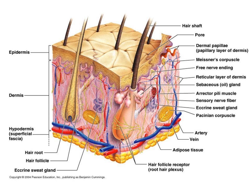
SKIN epidermis dermis hypodermis (subcutaneous tissue)
outermost protective shield epithelial cells nonvascular dermis leathery (hide) fibrous connective tissue vascular hypodermis (subcutaneous tissue) deepest layer adipose tissue (fat cells)
 fibrous connective tissue. vascular. hypodermis (subcutaneous tissue) deepest layer. adipose tissue (fat cells)")
14
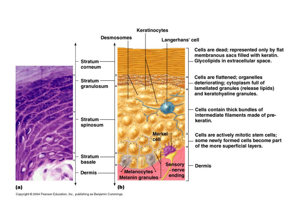
CELLS OF THE EPIDERMIS keratinocytes melanocytes
most numerous epidermal cell produce keratin—fibrous protein waterproof, tough protective covering tightly connected by desmosomes melanocytes found in stratum basale produce pigment melanin—uv protection for keratinocytes

15
cells (cont) Langerhan’s cells Merkel cells macrophages
play role in immunity Merkel cells sensory receptor for touch

16
LAYERS OF THE EPIDERMIS
stratum corneum (horny layer) outermost layer; dead, keratinized cells stratum lucidum (clear layer) found in thick skin only thin translucent band stratum granulosum (granular layer) change in keratinocytes—flattened cells w/ granules stratum spinosum (spiny layer) ‘prickles’ irregular shape of keratinocytes bundles of pre-keratin filaments stratum basale (basal layer) mostly single row of cells; site of mitotic cell division 10-25% are melanocytes
 outermost layer; dead, keratinized cells. stratum lucidum (clear layer) found in thick skin only. thin translucent band. stratum granulosum (granular layer) change in keratinocytes—flattened cells w/ granules. stratum spinosum (spiny layer) ‘prickles’ irregular shape of keratinocytes. bundles of pre-keratin filaments. stratum basale (basal layer) mostly single row of cells; site of mitotic cell division % are melanocytes.")
21
Dermis Second major skin region containing strong, flexible connective tissue Cell types include fibroblasts, macrophages, and occasionally mast cells and white blood cells Composed of two layers – papillary and reticular

22
DERMIS papillary layer reticular layer areolar connective tissue
peglike projection: dermal papillae free nerve endings, Meissner’s corpuscles, Pacinian corpuscles dermal ridgesepidermal ridgesfingerprints reticular layer 80% of the thickness of dermis dense irregular connective tissue important cleavage or tension lines--surgery

23
Functions of the Integumentary System
Protection – chemical, physical, and mechanical barrier Body temperature regulation is accomplished by: Dilation (cooling) and constriction (warming) of dermal vessels Increasing sweat gland secretions to cool the body Cutaneous sensation – exoreceptors sense touch and pain
 and constriction (warming) of dermal vessels. Increasing sweat gland secretions to cool the body. Cutaneous sensation – exoreceptors sense touch and pain.")
24
Functions of the Integumentary System
Metabolic functions – synthesis of vitamin D in dermal blood vessels Blood reservoir – skin blood vessels store up to 5% of the body’s blood volume Excretion – limited amounts of nitrogenous wastes are eliminated from the body in sweat

25
Skin Color Three pigments contribute to skin color
Melanin – yellow to reddish-brown to black pigment, responsible for dark skin colors Freckles and pigmented moles – result from local accumulations of melanin Carotene – yellow to orange pigment, most obvious in the palms and soles of the feet Hemoglobin – reddish pigment responsible for the pinkish hue of the skin

26
Skin Cancer The three major types of skin cancer are:
Basal cell carcinoma Squamous cell carcinoma Melanoma

27
Basal Cell Carcinoma Least malignant and most common skin cancer
Stratum basale cells proliferate and invade the dermis and hypodermis Slow growing and do not often metastasize Can be cured by surgical excision in 99% of the cases

28
Squamous Cell Carcinoma
Arises from keratinocytes of stratum spinosum Arise most often on scalp, ears, and lower lip Grows rapidly and metastasizes if not removed Prognosis is good if treated by radiation therapy or removed surgically

30
Melanoma Cancer of melanocytes is the most dangerous type of skin cancer because it is: Highly metastatic Resistant to chemotherapy

31
Melanoma Melanomas have the following characteristics (ABCD rule)
A: Asymmetry; the two sides of the pigmented area do not match B: Border is irregular and exhibits indentations C: Color (pigmented area) is black, brown, tan, and sometimes red or blue D: Diameter is larger than 6 mm (size of a pencil eraser)
 is black, brown, tan, and sometimes red or blue. D: Diameter is larger than 6 mm (size of a pencil eraser)")
34
Skin and Aging Epidermal replacement of cells slows and skin becomes thinner Skin becomes dry and itchy Subcutaneous fat layer diminishes, leading to intolerance of cold Decreased elasticity and loss of subcutaneous tissue leads to wrinkles Decreased numbers of melanocytes and Langerhans’ cells increase the risk of skin cancer

35
A B C D E

36
Burns First-degree – only the epidermis is damaged
Symptoms include localized redness, swelling, and pain Second-degree – epidermis and upper regions of dermis are damaged Symptoms mimic first degree burns, but blisters also appear Third-degree – entire thickness of the skin is damaged Burned area appears gray-white, cherry red, or black; there is no initial edema or pain (since nerve endings are destroyed)
")
38
Start: midterm exam on Thurs—copy topics body systems
Anatomy-Physiology Start: midterm exam on Thurs—copy topics body systems homeostasis—negative & positive feedback mechanisms directional terms & body planes cells—plasma membrane, cell transport, membrane junctions osmosis—hypo, hyper, isotonic solutions tissue types integumentary system functions epidermal layers dermis skin appendages—hair, nails, sweat glands skin cancer Objectives: notes: integumentary system work on Chap. 5 study guide—due Thurs

Similar presentations