Download presentation
Presentation is loading. Please wait.

1
Giardia duodenalis (G. lamblia; G. intestinalis) –Giardiasis. –Most distinctive of the flagellates. –Has both a trophozoite and cyst stage.

2
Giardia duodenalis Trophozoite Trophozoites are binucleated (looks like a face). 12-15 μm.
 μm.")
3
Giardia duodenalis Trophozoite Trophozoites are binucleated (looks like a face). 12-15 μm. Ventral surface bears adhesive disk to adhere to surface of intestinal cell.

4
Giardia duodenalis Trophozoite Trophozoites are binucleated (looks like a face). 12-15 μm. Ventral surface bears adhesive disk to adhere to surface of intestinal cell. 8 flagella (2 anterior, 2 posterior, 2 ventral, and 2 caudal) - all arise from kinetosome.
 - all arise from kinetosome..")
5
Giardia duodenalis Trophozoite Trophozoites are binucleated (looks like a face). 12-15 μm. Ventral surface bears adhesive disk to adhere to surface of intestinal cell. 8 flagella (2 anterior, 2 posterior, 2 ventral, and 2 caudal) - all arise from kinetosome. Median bodies occur behind adhesive disk - function is unknown.
 - all arise from kinetosome. Median bodies occur behind adhesive disk - function is unknown..")
6
Giardia duodenalis Trophozoite Light microscope photos of trophozoites

7
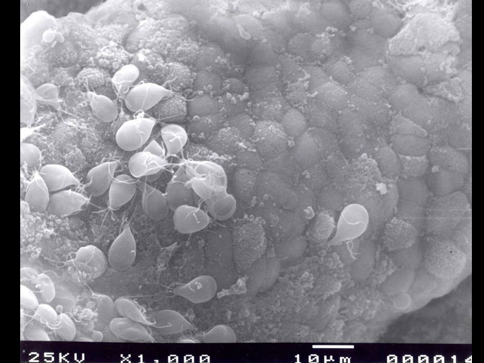
Giardia duodenalis Lives in the upper part of the small intestine (duodenum, jejunum, and upper ileum). Here the trophozoites attach to the epithelial cells.

8
Giardia duodenalis Trophozoite Scanning EM view of trophozoite surface showing the adhesive disk. ventral dorsal

9
Feeds on mucous that forms in response to irritation.

10
Also absorbs vitamins and amino acids.

11
Feeds on mucous that forms in response to irritation. Also absorbs vitamins and amino acids. Interferes with absorption in host especially lipids.

12
Feeds on mucous that forms in response to irritation. Also absorbs vitamins and amino acids. Interferes with absorption in host especially lipids. Giardia can also interfere with vitamin/nutrient absorption. –Vitamin A vision –Vitamin D rickets: Both of these are due to long standing infections.

13
Cyst of Giardia duodenalis The cyst forms as trophozoites become dehydrated when they pass through the large intestine.

14
Cyst of Giardia duodenalis The cyst forms as trophozoites become dehydrated when they pass through the large intestine. Morphology: ovoid in shape; 8-12 µm long x 7-10 µm wide thin cyst wall. Four nuclei present, often concentrated at on end. Flagella shorten and are retracted within cyst. Axonemes provide internal support.

15
Cyst of Giardia duodenalis Cyst may remain viable in the external environment (usually water) for many months.
 for many months.")
16
Cyst of Giardia duodenalis Cyst may remain viable in the external environment (usually water) for many months. -14 billion cysts can be passed in 1 stool sample -Moderate infections: 300 million cysts.

17
Cyst of Giardia duodenalis

18
Symptoms Range from none abdominal discomfort causing acute or chronic diarrhea and other GI signs. Gray, greasy, voluminous malodorous diarrhea! Flatulence.

19
Giardia duodenalis Giardia trophs are attracted to bile salts: so sometimes you can get infections in bile ducts and gall bladder, causing jaundice and colic. This is irritating but not life threatening infection like E. histolytica.

20
Pathogenesis and Pathology Nutrient malabsorption and physical blockage and damage to microvilli. Trophs attach to small intestine cause damage (mechanical and toxins).
..")
24
Giardia trophozoite Trophozoite attaches to surface of epithelial cells with its adhesive disk.

25
Pathogenesis and Pathology 1) Fat/CHO digestion decreases and causes maldigestion.
 Fat/CHO digestion decreases and causes maldigestion.")
26
Pathogenesis and Pathology 1) Fat/CHO digestion decreases and causes maldigestion. 2) Absorption decreases due to villus blunting causing malabsorption.
 Absorption decreases due to villus blunting causing malabsorption..")
27
Pathogenesis and Pathology 1) Fat/CHO digestion decreases and causes maldigestion. 2) Absorption decreases due to villus blunting causing malabsorption. 3) Malabsorption and maldigestion causes diarrhea.
 Absorption decreases due to villus blunting causing malabsorption. 3) Malabsorption and maldigestion causes diarrhea..")
28
Pathogenesis and Pathology 4) Physical damage: clubbing of villi; decreases villus-to-crypt ratio; brush borders of cells are irregular.
 Physical damage: clubbing of villi; decreases villus-to-crypt ratio; brush borders of cells are irregular.")
29
Epidemiology Get infected by ingesting cysts through contaminated water.

30
Epidemiology Get infected by ingesting cysts through contaminated water. Most common intestinal flagellate of people.

31
Epidemiology Get infected by ingesting cysts through contaminated water. Most common intestinal flagellate of people. World wide distribution; prevalence ranges from 2.4-67.5%.

32
Epidemiology Get infected by ingesting cysts through contaminated water. Most common intestinal flagellate of people. World wide distribution; prevalence ranges from 2.4- 67.5%. Reservoir hosts can play a significant role.

33
Reservoir Hosts Transmission from animals to humans is controversial; dependent on strain or type involved.

34
Human Infections There are hot spots: Vacations and Travels Camping.

35
Human Infections There are hot spots: Vacations and Travels Camping. Colorado ski resorts are notorious for outbreaks drinking from Mountain Springs, washing utensils/drinking water that is not treated.

36
Human Infections There are hot spots: Vacations and Travels Camping. Colorado ski resorts are notorious for outbreaks drinking from Mountain Springs, washing utensils/drinking water that is not treated. Day care centers.

37
Diagnosis Trophs in diarrheic feces; cysts in formed feces. At least 3 exams (one every other day) before judge negative. ELISA tests: detect soluble antigen.
 before judge negative. ELISA tests: detect soluble antigen..")
38
Treatment and Prognosis Drug of choice is Flagyl. Giardia thrives in people not necessarily hard to treat, but keeping those who were infected from becoming reinfected.

39
Blood and Tissue Flagellates Phylum Euglenoidea Known as Hemoflagellates or Kinetoplastids. Some have forms that live in the alimentary canal of insects such as flies, bugs, etc.

40
Adaptation to Parasitism Most parasites came from free-living forms.

41
Adaptation to Parasitism Most parasites came from free-living forms. They became parasites when hosts ingested them and they survived the process.

42
Adaptation to Parasitism Most parasites came from free-living forms. They became parasites when hosts ingested them and they survived the process. They were then selected for and adapted to colonize hosts.

43
Adaptation to Parasitism This is not the case for blood and tissue flagellates.

44
Adaptation to Parasitism This is not the case for blood and tissue flagellates. Because most insect species have flagellates that live within them and these share characters with human blood and tissue flagellates.

45
Adaptation to Parasitism This is not the case for blood and tissue flagellates. Because most insect species have flagellates that live within them and these share characters with human blood and tissue flagellates. Therefore biting insects probably gave these parasites to us!

46
Blood and Tissue Flagellate Anatomy and Life Stages There are seven ontogenetic stages, but not all species have all seven. These stages are continuous.

47
Life-cycle stages of trypanosomatidae. A. promastigote; b. ophistomastigote; c. epimastigote; d. trypomastigote; e. choanomastigote; f. amastigote; g. paramastigote; K. kinetoplast; N. nucleus; F. flagellum.

48
You will be responsible for 4 of them. Remember not all 4 stages will be found in each species life cycle.

49
Promastigote Flagellum Kinetosome Kinetoplast Nucleus anterior posterior

50
Epimastigote Undulating membrane anterior posterior

51
Trypomastigote Undulating membrane anterior posterior

52
Amastigote Flagellum Kinetosome Kinetoplast Nucleus anterior posterior These are intracellular, stages that occur within cells.

53
Leishmaniasis Infection with Leishmania spp. –Disease of the Reticulo-Endothelial Cells.

54
Leishmaniasis Infection with Leishmania spp. –Disease of the Reticulo-Endothelial Cells. Reticulo-Endothelial System- is diffuse in the body and made up of all phagocytes except for leucocytes.

55
Leishmaniasis Infection with Leishmania spp. –Disease of the Reticulo-Endothelial Cells. Reticulo-Endothelial System- is diffuse in the body and made up of all phagocytes except for leucocytes. Macrophage: is a standard reticulo-endothelial cell.

Similar presentations


>")


 Giardia lamblia ( 蓝氏贾第鞭毛虫 ) Intestinal flagellate Giardia lambilia lives in small intestine Giardiasis Diarrhea “traveler’s diarrhea”>")


