Download presentation
Presentation is loading. Please wait.

1
Microscopy and Cytology

2
Introduction to Microscopes

3
Microscopy Permits Visualization of Objects Too Small to Be Normally Seen

4
Types of Microscopes Light microscopes Electron microscopes
Simple light microscope Compound light microscope Dissecting light microscope Electron microscopes Transmission electron microscope Scanning electron microscope Ultra high power microscope Scanning-tunneling microscope Atomic force microscope

5
Simple vs. Compound Microscope
Simple – One Lens Compound – Multiple Lenses

6
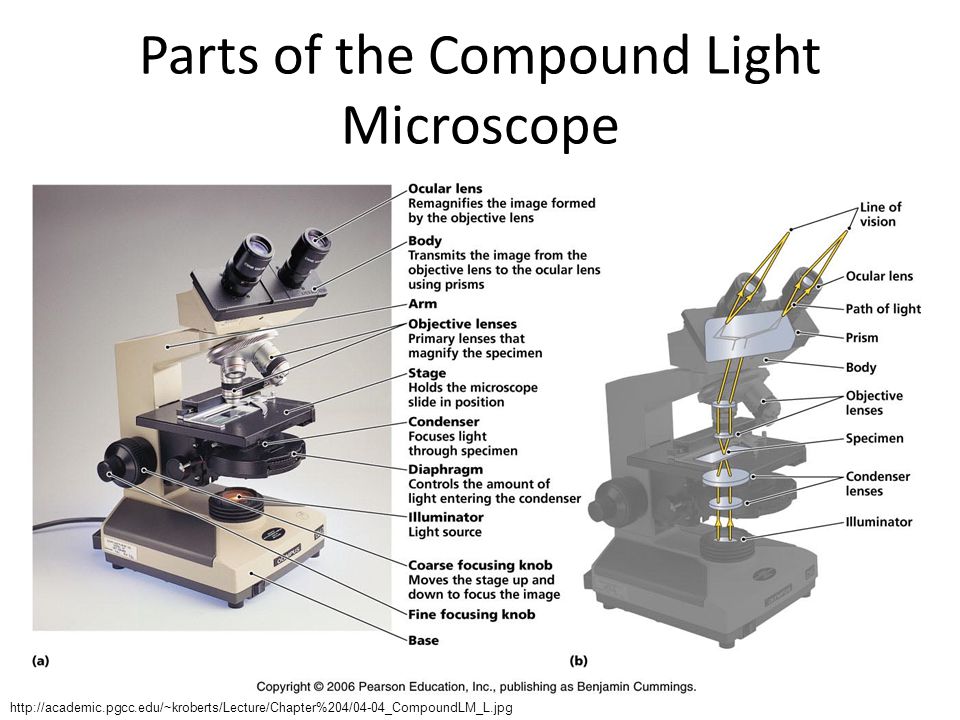
Parts of the Compound Light Microscope
7
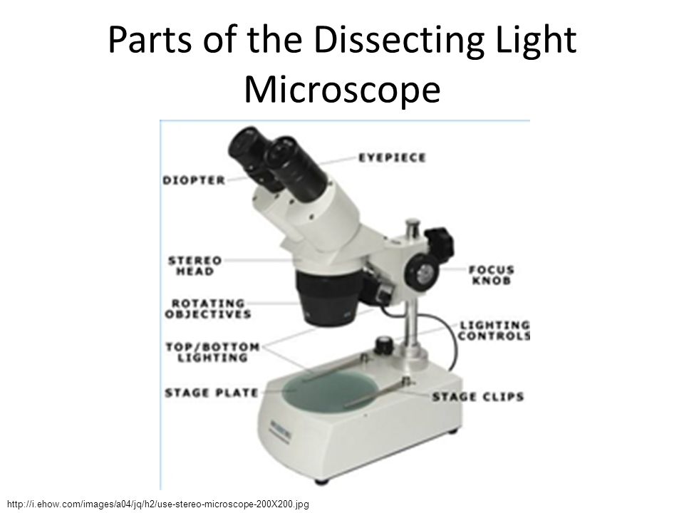
Parts of the Dissecting Light Microscope
8
Electron Microscopes Magnify Extremely Small Objects

9
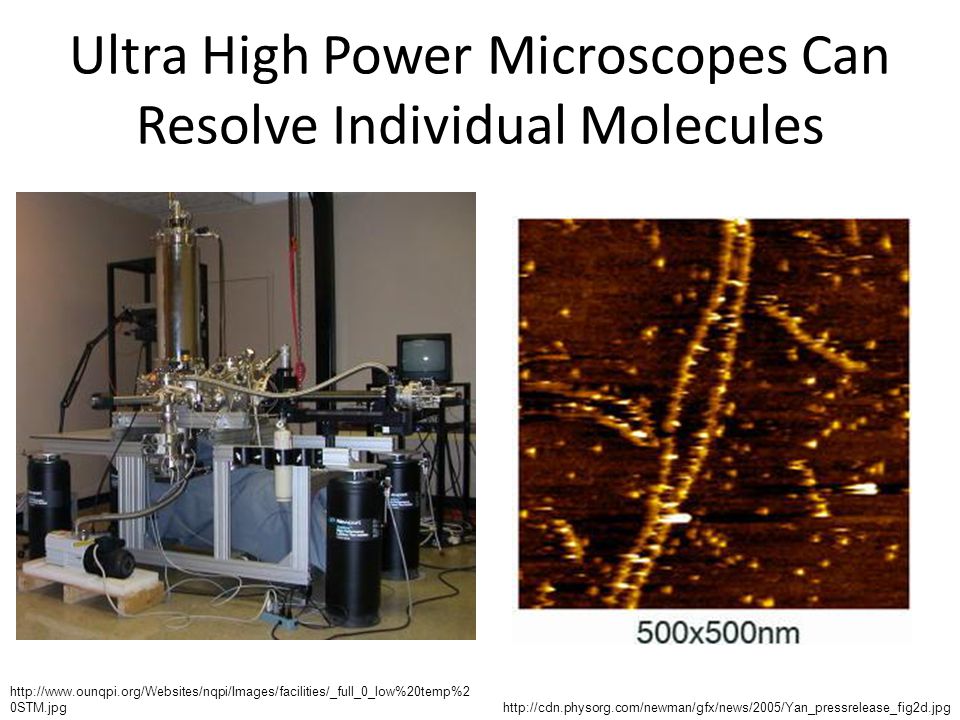
Ultra High Power Microscopes Can Resolve Individual Molecules
10
Principles of Microscopy

11
Important Concepts in Microscopy
Magnification Resolving Power Contrast Viewing Field Image orientation Depth of focus Size of the field of view Working distance

12
Magnification How much bigger the object under the microscope looks
Depends on the lens or lenses Total magnification = product of lens magnifications Oculars: 10X Objective lenses: 4X, 10X, 40X, 100X Totals: 40X, 100X, 400X, 1000X

13
Resolving Power Ability to tell the difference between two objects that are close together Higher resolution lets us see smaller things clearly Depends on: Light wavelength – shorter is better (blue filter) Refractive index – keeping constant is better (immersion oil)
 Refractive index – keeping constant is better (immersion oil)")
14
Oil Immersion Improves Resolution

15
Contrast Ability to tell the difference between objects and background
Can be improved using stains Bauman, R.W. (2010). Microbiology with Diseases by Taxonomy (3rd ed.) New York, NY: Benjamin Cummings.
. Microbiology with Diseases by Taxonomy (3rd ed.) New York, NY: Benjamin Cummings.")
16
Considerations for the Viewing Field
Orientation – Image is inverted and reversed Depth of focus – How much thickness of the sample is in focus Smaller as magnification increases Parfocal – stays in focus as magnification increases Field of view – How much area of the slide is seen Parcentral – stays centered as magnification increases Working distance – How far the objective lens is from the slide

17
Microscope Care

18
Use the Coarse Focus Knob for the Lowest Power Only

19
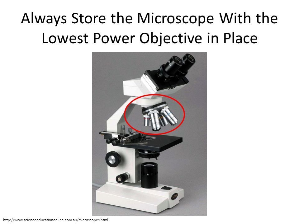
Always Store the Microscope With the Lowest Power Objective in Place
20
At the Beginning of the Day…
Remove the dust cover from the microscope. Inspect for damage. Report anything you find! Plug in the microscope. Clean all lenses with lens paper ONLY. DO NOT clean lenses with anything other than lens paper! Inform instructor if you find oil on a lens. Rotate the 4X objective into position above the stage. Center the stage, and roll it down to the lowest position. Turn on the microscope light source.

21
Use of the Oil Immersion Lens
Find specimen and focus on 4X using coarse and then fine focus knobs. Move up to 10X and focus using FINE FOCUS KNOB only. Move up to 40X and focus using FINE FOCUS KNOB only.. Slide 40X objective partly out of the way. Place ONE drop of immersion oil on slide. Gently slide 100X (oil immersion) objective into place. Focus using FINE FOCUS KNOB only!
 objective into place. Focus using FINE FOCUS KNOB only!")
22
Use of the Oil Immersion Lens
When finished observing under oil immersion: Rotate from 100X objective to 4X objective and remove slide. Clean oil from slide using lens cleaner and lens paper. Carefully clean oil from the oil immersion lens using lens cleaner and lens paper at the end of each class.

23
At the End of the Day… Remove slides from the microscope stage.
Turn off the microscope light source. Clean oculars, ALL lenses, stage, and base with lens cleaner and wipe with lens paper. Rotate the nosepiece until the 4X objective is in place. Center the stage, and roll it to the lowest position. Unplug the microscope. Cover the microscope with the dust cover.

24
NEVER CLEAN THE MICROSCOPE WITH ANYTHING OTHER THAN LENS PAPER!

25
Introduction to Cytology

26
Cytology is the Study of Cells
Cell = smallest unit of life Composed of water and macromolecules H, C, O, N are most predominant elements Two types of cells Prokaryotic cells Eukaryotic cells Organisms can be one or many cells Unicellular – Single-celled organism Multicellular – Organism composed of many cells

27
Robert Hooke and the Cell Theory
The cell is the smallest unit of life. All living organisms are composed of cells. All cells arise from other cells.

28
Important Features of Prokaryotic Cells
External Structures Internal Structures Capsule Cell wall Plasma membrane Flagella Pili Cytoplasm Nucleoid (chromosome) Ribosomes
 Ribosomes.")
29
Overview of a Prokaryotic Cell

30
Bacterial Cell Morphologies
Coccus (Sphere) Bacillus (Rod) Spiral
 Bacillus (Rod) Spiral")
31
Important Features of Eukaryotic Cells
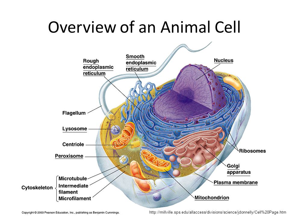
External Structures Internal Structures Cell wall (plants) Plasma membrane Flagella Cilia Cytoplasm Membranous organelles Nucleus Mitochondria Chloroplasts (plants) Endoplasmic reticulum (R/S) Golgi apparatus Lysosomes Peroxisomes Nonmembranous organelles Nucleoli Ribosomes Cytoskeleton Centrioles (animal cells)
 Plasma membrane. Flagella. Cilia. Cytoplasm. Membranous organelles. Nucleus. Mitochondria. Chloroplasts (plants) Endoplasmic reticulum (R/S) Golgi apparatus. Lysosomes. Peroxisomes. Nonmembranous organelles. Nucleoli. Ribosomes. Cytoskeleton. Centrioles (animal cells)")
32
Overview of an Animal Cell
33
Overview of a Plant Cell

35
Introduction to Microscope Use
Light microscope Exercise 7.1 – field of view Exercise 7.2 – depth of focus Exercise 7.3 – image orientation Dissecting microscope Exercise 7.4 – introduction to dissecting microscopes

36
Cytology PREPARE ALL SLIDES FIRST! Exercise 7.5 – Models
Exercise 7.6 – Wet mounts Cyanobacteria – prepared slide Elodea leaf + safranin Onion epidermis + iodine Cheek cells + methylene blue Ear swab + Romanowsky stain Exercise 7.7 – Prepared bacterial slides

37
Elodea Leaf Drop of water on slide Transfer Elodea leaf into drop
Place one edge of coverslip against drop Gently lower coverslip over drop Drop of safranin on slide next to coverslip – diffuse in 4X 10X 40X

38
Onion Epidermis Drop of water on slide
Transfer onion epidermis into drop Place one edge of coverslip against drop Gently lower coverslip over drop Drop of iodine on slide next to coverslip – diffuse in 4X 10X 40X

39
Cheek Cells Drop of methylene blue on slide
Scrape inside of cheek with toothpick Swirl into stain drop Place one edge of coverslip against drop Gently lower coverslip over drop 4X 10X 40X

40
Ear Swab Slide Preparation Staining Procedure
Roll wet, sterile swab over top of ear and roll onto clean slide Repeat 2 more times Air-dry (10+ minutes) Hold slide with clothespin 1 second per dip, 10 dips per jar Order of stains: Alcohol (light blue) Eosin (red) Methylene blue (blue) Distilled water Blot with bibulous paper 4X 10X 40X 100X w/oil
 Hold slide with clothespin. 1 second per dip, 10 dips per jar. Order of stains: Alcohol (light blue) Eosin (red) Methylene blue (blue) Distilled water. Blot with bibulous paper. 4X 10X 40X 100X w/oil.")
41
Order of Experiments Prepare all slides (Exercise 7.6)
Introduction to microscopy (Exercises ) View wet mount slides and prepared bacterial slides under microscope (Exercise ) Exercise 7.5 can be performed whenever you have spare time.
 View wet mount slides and prepared bacterial slides under microscope (Exercise ) Exercise 7.5 can be performed whenever you have spare time.")
Similar presentations


 Electron (up to X 200,000) Scanning Electron Microscope (SEM) Transmission Electron Microscope (TEM)>")


 Spring 2010 Prof. AnnMarie Armenti, MS.>")


 – Late stamp is OK.>")


 Arm Stage Coarse Adjustment Knob Fine Adjustment Knob Always carry a microscope with one.>")
