Download presentation
Presentation is loading. Please wait.

1
Instructor: Li Li Office: 0850 physiological sciences Email: tyvm80@163.com Jining medical college The Kidneys And Regulation Of Water And Inorganic Ions Department of Physiology

2
The Kidneys And Regulation Of Water And Inorganic Ions ⑴ Basic principles of renal physiology ⑵ Regulation of sodium, water, and potassium balance ⑶ Calcium regulation ⑷ Hydrogen regulation

3
Basic Principles Of Renal Physiology ⑴ Renal Functions ⑵ Structure Of The Kidneys And Urinary System ⑶ Renal Blood Flow And Its Regulatin ⑷ The Urine Formation By The Kidneys ⑸ Concentrated And Diluted Urine By Kidneys

4
Basic Principles Of Renal Physiology Renal Functions ① Regulate the water concentration, inorganic ion composition, and volume of the internal environment. ② Excrete metabolic waste products and some foreign chemicals into the urine. ③ Gluconeogenesis. ④ Act as endocrine glands.

5
plasma Interstitial fluid Extracellular fluidInternal environment Intracellular fluid Figure Internal environment.

6
Figure Position of the kidney.

7
Figure Urinary system.

8
Basic Principles Of Renal Physiology Structure Of The Kidneys And Urinary System Renal nephron (fuctional unit of the kidney) Renal corpuscle glomerulus Bowman’s capsule Renal tubule proximal tubule loop of Henle distal tubule What is the main difference between the cortical and juxamedullary nephrons?
 Renal corpuscle glomerulus Bowman’s capsule Renal tubule proximal tubule loop of Henle distal tubule What is the main difference between the cortical and juxamedullary nephrons")
9
Figure Structure of the kidney.

10
Figure : The nephron. Proximal convoluted tubule Loop of Henle Collecting duct Afferent arteriole Efferent arteriole Renal corpuscle Distal tubule

11
Figure The Nephron.

12
Figure The Kidney Blood Flow.

13
Figure The nephron Blood Flow.

14
Basic Principles Of Renal Physiology Structure Of The Kidneys And Urinary System The blood flow of renal nephron ③ kidneys have two sets of capillaries – the glomerular capillaries and the peritubular capillaries. ② the nephron have two sets of arterioles: the afferent and efferent arterioles. ① The kidneys receive about 22% of the cardiac output, and the renal cortex receives most of the kidney’s blood flow. The high blood flow supplies enough plasma for the high rates of glomerular filtration.

15
Figure Urinary system.

16
Figure The urine formation.

17
Figure The filtration membrane. afferent arteriole efferent arteriole podocyte capillary tubule Bowman’s capsule capillary endothelium Basement membrane The visceral lalyer of Bowman’s capsule

18
Basic Principles Of Renal Physiology Structure Of The Kidneys And Urinary System The filtration membrane ① the single-celled capillary endothelium which is fenestrated with pores that are 70-90nm in diameter(keep the blood cells from passing). ② a noncellular proteinaceous layer of basement membrane which is negatively charged(keep the protein from passing). ③ visceral layer of Bowman’s capsule(podocyte).
. ③ visceral layer of Bowman’s capsule(podocyte)..")
19
Permeability of filtration membrane diameter(nm) Cationic molecule Neutral molecule Anionic molecule Figure The permeability of filtration membrane.
 Cationic molecule Neutral molecule Anionic molecule Figure The permeability of filtration membrane.")
20
Basic Principles Of Renal Physiology Structure Of The Kidneys And Urinary System Permeability of filtration membrane ① The filtration membrane is selective in determining which molecules will filter, based on their size and electrical charge. ② Generally, filtration is inversely proportional to diameter. ③ The molecules with the same size, the filtration permeability is in this order: cationic molecule>neutral molecule>anionic molecule.

21
Figure Juxtaglomerular apparatus.

22
Basic Principles Of Renal Physiology Structure Of The Kidneys And Urinary System Juxtaglomerular apparatus ① macula densa: Near the end of the ascending limb of each loop of Henle, the tubular epithelium is modified histologically to form the macula densa, which can sense changes in volume delivery to the distal tubule by way of signals. ② juxtaglomerular cells: The wall of the afferent arteriole contains secretory cells known as juxtaglomerular cells, which are the major storage and release sites for rennin.

23
Figure: Renal Corpuscle

24
Figure: Innervation of the renal vessels.

25
Basic Principles Of Renal Physiology Structure Of The Kidneys And Urinary System Innervation of the renal vessels ① All the blood vessels are richly innervated by sympathetic fibers. ② Besides all the blood vessels, the sympathetic fibers are also distributed to the proximal and distal tubules and the juxtaglomerular cells. ③ So when the sympathetic fibers are stimulated, the renal blood flow and the rlease of rennin and the absorption of substances in the tubule will change.

26
Basic Principles Of Renal Physiology Renal Blood Flow And Its Regulation Autoregulation Of GFR And Renal Blood Flow Definition: although changes in arterial pressure have some influence on renal blood flow, the kidneys have effective mechanisms for maintaining renal blood flow and glomerular filtration rate relatively constant over an arterial pressure range between 80 amd 180mmHg. Function: maintain a relatively constant GFR and allow precise control of renal excretion of the waste substances. Mechanism: ① Role of tubuloglomerular feed-back. ② Myogenic autoregulation of renal blood flow and GFR.

27
Basic Principles Of Renal Physiology Renal Blood Flow And Its Regulation Autoregulation Of GFR And Renal Blood Flow Mechanism: ② Myogenic autoregulation of renal blood flow and GFR. ① Role of tubuloglomerular feed-back: GFR↓ → Reabsorption of NaCl in ascending loop of Henle ↑→ concentration of NaCl at the macula densa ↓→ resistance of the afferent arterioles ↓+ rennin release →GFR↑ (go back to normal) Arterial blood pressure↑→stretch arteriolar wall more→the vascular smooth muscle contract→resistance of arteriole↑→prevent excessive increase in renal blood flow
 Arterial blood pressure↑→stretch arteriolar wall more→the vascular smooth muscle contract→resistance of arteriole↑→prevent excessive increase in renal blood flow.")
28
Basic Principles Of Renal Physiology Renal Blood Flow And Its Regulation Nervous and Hormonal Regulation of GFR and Renal Blood Flow Nervous regulation: sympathetic fibers is stimulated →mardked decrease in renal blood(α1,α2,β1 receptors). Hormomal regulation: PGI2, NO, Adenosine.

29
Figure The urine formation.

30
Basic Principles Of Renal Physiology The Urine Formation By The Kidneys ① glomerular filtration. ② tubular reabsorption of the filtrate. ③ tubular secretion.

31
Basic Principles Of Renal Physiology The Urine Formation By The Kidneys Glomerular filtration ① Urine formation begins with the filtration of plasma from the glomerular capillaries into Bowman’s space, this process is termed glomerular filtration. ② The filtrate is called glomerular filtrate (ultrafiltrate), it is cell-free and except for plasma proteins, contains all the substances in plasma in virtually the same concentration as in plasma.
, it is cell-free and except for plasma proteins, contains all the substances in plasma in virtually the same concentration as in plasma..")
32
Basic Principles Of Renal Physiology The Urine Formation By The Kidneys Glomerular filtration rate(GFR): The volume of ultrafiltrate filtered from the glomerulus into Bowman’s space by the two kidneys per unit time(minute) is called GFR. Glomerular filtration Filtration fraction(FF): The ratio of GFR to renal plasma flow is termed filtration fraction(FF). GFR is determined by effective filtration pressure(EFP) and filtration coeffecient (K).
: The ratio of GFR to renal plasma flow is termed filtration fraction(FF). GFR is determined by effective filtration pressure(EFP) and filtration coeffecient (K)..")
33
Basic Principles Of Renal Physiology The Urine Formation By The Kidneys Glomerular filtration Effective filtration pressure(EFP): The difference of force that favors filtration and opposes filtration. EFP= P GC -P BC -π GC P GC : The glomerular capillary hydrostatic pressure. P BC : The hydrostatic pressure exerted by the fluid in Bowman’s space. π GC : The colloid osmotic force that results from the presence of protein in the glomerular capillary plasma.

34
Figure Effective filtration pressure.

35
Basic Principles Of Renal Physiology The Urine Formation By The Kidneys Glomerular filtration Characteristics of EFP: EFP decreases gradully from the beginning to the end of the glomerular capillaries, because of the colloid osmotic pressure in the glomerular capillaries increases gradully. So from the beginning to the end of the glomerular capillaries, there is a site in which the EEP is zero.

36
Basic Principles Of Renal Physiology The Urine Formation By The Kidneys Glomerular filtration Filtration coeffecient (K): ① Mesangial cell contraction (many agents such as Angiotensin Ⅱ, endothelins, vasopressin can alter Mesangial cell contraction). ② Size of pores. ③ Thickness of membrane. ④ Electrical charge.

37
Basic Principles Of Renal Physiology The Urine Formation By The Kidneys Glomerular filtration Determinants of the Glomerular Filtration Rate: ① Glomerular capillary hydrostatic pressure which is determined by arterial blood pressure, afferent arteriolar resistance and efferent arteriolar resistance. ② Glomerular capillary colloid osmotic pressure which is determined by arterial colloid osmotic pressure and the fraction of the plasma filtered by the glomerular capillaries. ③ Bowman’s capsule hydrostatic pressure. ④ Renal plasma flow. ⑤ Glomerular capillary filtration coefficient.

38
Figure The urine formation.

39
Figure : The nephron. Proximal convoluted tubule Loop of Henle Collecting duct Afferent arteriole Efferent arteriole Renal corpuscle Distal tubule

40
Figure Structure of the kidney.

41
When the opposing force equals the forces favoring filtration, filtration stops. Figure: Filtration balance.

42
Basic Principles Of Renal Physiology The Urine Formation By The Kidneys Tubular Reabsoption Definition: Movement of a substance from tubular lumen to interstitial fluid. Characteristics: ① Reabsorption of waste products is relatively incomplete, and reabsorption of most useful plasma components is relatively complete. ② Reabsorption of the most nutrients such as glucose is not controlled physiologically, the reabsorption rates for water and many ions are under physiological control.

43
SubstanceAmount filtered per day Amount excreted per day Percent reabsorbed Water, L Sodium, g Glucose, g Urea, g 1801.899 630 3.299.5 1800 100 54 30 44 Table: Average values for several components that undergo filtration and reabsorption

44
Basic Principles Of Renal Physiology The Urine Formation By The Kidneys Tubular Reabsoption Transport processes: active transport(primary and secondary active transport) and passive transport (protein-mediated diffusion and osmosis of water and solvent drag). Route: transcellular route and paracellular route.

45
Basic Principles Of Renal Physiology The Urine Formation By The Kidneys N a +, Cl -, and water reabsorption Tubular Reabsoption Proximal tubules ① 65% of the filtered loads of the sodium and water and slightly lower percentage of chloride are reabsorbed. ② In the first half of the proximal tubule, sodium is transported by co-transport along with glucose, amino acids and other solutes by the way of transcellular route, and Cl - can not be reabsorbed. ③ In the second half of the proximal tubule, sodium ions are mainly passively transported along with chloride ions by the way of paracellular route. ④ Water is passively reabsorbed by osmosis through transcellular and paracellular route.

46
Figure: Morphological characteristics of proximal tubules.

47
Basolateral membrane Sodium pump : active transport Facilitated diffusion G 、 AA H2OH2O Epithelial cells Interstitial fluid lumenBlood There is no Cl - reabsorption in the first half of the proximal tubule. Figure Reabsorption of substance in the first half of the proximal tubule.

48
Na + H+H+ Cl - HCO 3 - K+K+ Cl - ATP Na + K+K+ ① ② LumenBlood Figure Reabsorption of substance in the second half of the proximal tubule. Intercellular junction

49
Figure Reabsorption of substance in the proximal tubule.

50
The Urine Formation By The Kidneys N a +, Cl -, and water reabsorption Tubular Reabsoption Loop of Henle Basic Principles Of Renal Physiology ① In the thin segment of descending limb, water diffuses out by osmosis, and the tubular fluid becomes more and more concentrated and the osmolarity reaches the maximum at the bend of the thin segment of ascending limb. ② In the thin segment of ascending limb, the tubular membrane is impermeable to water, but permeable to solutes, so sodium chloride diffuses into the surrounding interstitial fluid, and the tubular fluid osmotic pressure is gradually deceased. ③ In the thick segment of ascending limb, the tubular membrane is impermeable to water, sodium chloride are actively reabsorbed by the Na + :2Cl - :K + co-transporter.

51
By diffusion Major site NaCl Glomerular corpuscle Bowman’s capsule Proxiaml tubule Distal tubule Loop of Henle water By osmosis Figure Reabsorption of substance in the loop of Henle. Nacl

52
Cl - channel ① ② ③ ④ Epithelial cell lumenInterstitial fluid Figure Reabsorption of substance in the thick segment of ascending limb of loop of Henle.

53
The Urine Formation By The Kidneys N a +, Cl -, and water reabsorption Tubular Reabsoption Early segment of the diatal tubule Basic Principles Of Renal Physiology The tubular membrane is impermeable to water, sodium chloride is actively reabsorbed by Na + -Cl - cotransporter. More diatal tubules and collecting ducts ① N a + enter the epithelial cells by N a + channel and the potential differences between the lumen fluid and epithelial cells caused by sodium movement make chloride ions be passively reabsorbed and become the favoring force of potassium secretion. ② In this part, the reabsorption of sodium and water can be regulated by aldosterone and antidiuretic hormone(ADH) respectively.
 respectively..")
54
lumen blood Figure Reabsorption of substance in the early segment of distal tubule.

55
lumenblood Principal cell Intercalated cell Figure Reabsorption of substance in the more distal tubules and collecting ducts. N a + channel Cl -

56
The Urine Formation By The Kidneys Glucose and amino acid reabsorption Tubular Reabsoption Basic Principles Of Renal Physiology ① The proximal tubule is the sole segment able to actively reabsorb glucose. ② Glucose reabsorption is coupled to sodium by secondary ative transport. ③ The ability of proximal tubule to reabsorb glucose is not infinite, when the plasma level of glucose exceed the ablity of proximal tubule to reabsorb glucose, glucose will appear in the urine, so there is the renal threshold for glucose (the plasma level at which the glucose first appears in the urine) and transpot mximum for gucose (when reabsorption of glucose in all nephrons is saturated, the amounts of glucose in the urine rise in proportion to the rise in glucose concentration in plasma). ④ Reabsorption of amino acids occurs only in proximal tubule and mechanism is the same as that of glucose.
 and transpot mximum for gucose (when reabsorption of glucose in all nephrons is saturated, the amounts of glucose in the urine rise in proportion to the rise in glucose concentration in plasma). ④ Reabsorption of amino acids occurs only in proximal tubule and mechanism is the same as that of glucose..")
57
Basolateral membrane Sodium pump : active transport Facilitated diffusion G 、 AA H2OH2O Epithelial cells Interstitial fluid lumenBlood Figure Reabsorption of glucose and amino acid in the proximal tubule.

58
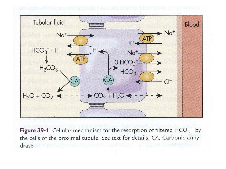
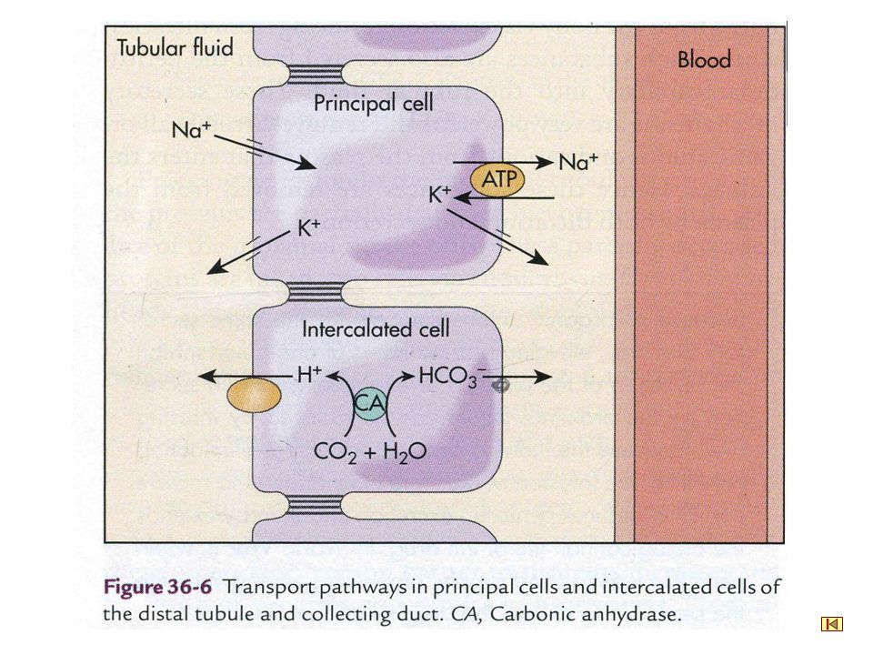
The Urine Formation By The Kidneys Bicarbonate ion reabsorption Tubular Reabsoption Basic Principles Of Renal Physiology ① Proximal tubule plays a major role in reabsorbing bicarbonate ions and reabsorb about 80% bicarbonate ions, and the reabsorption of bicarbonate ions is always coupled to hydrogen secretion. ② The thick segment of ascending limb can also reabsorb bicarbonate ions, and the mechanism is the same as that occurs in the proximal tubule. ③ In the distal tubules and collecting ducts, bicarbonate ions combines H + secreted by proton pump and H + -K + ATPase.

59
Figure: Bicarbonate ion reabsorption in proximal tubule and loop of Henle. lumen epithelial cell interstitial fluid blood

60
Figure: Bicarbonate ion reabsorption in distal tubules and collecting ducts. H 2 O + CO 2 HCO3 - H 2 CO 3 H 2 O + CO 2 HCO3 - H 2 CO 3 CA

64
By diffusion Major site NaCl Glomerular corpuscle Bowman’s capsule Proxiaml tubule Distal tubule Loop of Henle water By osmosis

73
The Urine Formation By The Kidneys Tubular Seretion Basic Principles Of Renal Physiology Definition:Movement of substances from peritubular capillaries into tubular lumen. Characteristics: ① Secretion can occur by diffusion or by transcellular mediated transport. ② The most important substances secreted by the tubules are hydrogen ions, potassium ions and NH 3 or NH 4 +. ③ Tubular secretion is under physiological control.

74
The Urine Formation By The Kidneys Tubular Seretion Basic Principles Of Renal Physiology H + secretion by renal tubules: ① The cells of the proximal and distal tubules secrete hydrogen ions. ② In the proximal tubules, H + is secreted by the Na + -H + counter- transporter, but in the distal tubules, H + is secreted by proton pump and H+-K+ ATP ase, so the secretion of H + in the distal tubules is relatively independent of Na + in the tubular fluid. K + secretion by renal tubules: ① K + secretion is a passive process, the driving force of the process is the high concentration of potassium ions inside the cell created by sodium pump and negative potential in the tubule lumen caused by Na + movement. ② Increased potassium concentrations in the extracellular fluid and increased aldosterone and increased tubular flow rate affect k + secretion.

78
Figure: NH 3 or NH 4 + secretion.

79
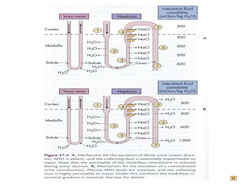
Concentrated And Diluted Urine By Kidneys Basic Principles Of Renal Physiology Diluted urine Definition: The osmotic pressure in the excreted urine that is lower than that in the blood plasma is termed hypo-osmotic urine or dilute urine. Mechanism: The permeability of the later distal tubule and collecting tubule and collecting ducts to water can be regulated by ADH. When there is a large excess of water in the body, the membrane of these sites has decreased permeability to water, but solutes are reabsorbed continuously, so the excreted urine is hypo-osmotic. Water diuresis: Excretion of increased urine volume with low osmotic pressure caused by drinking water is known as water diuresis.

80
By diffusion Major site NaCl Glomerular corpuscle Bowman’s capsule Proxiaml tubule Distal tubule Loop of Henle water By osmosis Figure: changes of osmotic pressure in the loop of Henle.

81
Concentrated And Diluted Urine By Kidneys Basic Principles Of Renal Physiology Concentrated urine Definition: The osmotic pressure in the excreted urine that is higher than that in the blood plasma is termed hyper-osmotic urine or concentrated urine. The ability of the kidneys to concentrate urine is determined by the ADH concentration in the blood plasma and the degree of hyperosmolarity in the renal medullary interstitium.

82
① Active transport of sodium and co-transport of potassium, chloride, and other ions out of the thick segment of the ascending limb of the loop of Henle into the medullary interstitium( plays a primary role). Concentrated And Diluted Urine By Kidneys Basic Principles Of Renal Physiology Concentrated urine Mechanism: ② Diffusion of sodium chloride and other ions from the thin segment of the ascending limb of the loop of Henle( contributes to the hyperosmolarity in the inner medullary interstitium). ③ Diffusion of urea from medullary colleting ducts into the surrounding interstitium and urea cycle ( also contributes to the medullary hyperosmolarity). ④ Diffusion of only small amounts of water from the medullary tubules into the medullary interstitium, far less than reabsorption of solutes into the medullary interstitium.
. ③ Diffusion of urea from medullary colleting ducts into the surrounding interstitium and urea cycle ( also contributes to the medullary hyperosmolarity). ④ Diffusion of only small amounts of water from the medullary tubules into the medullary interstitium, far less than reabsorption of solutes into the medullary interstitium..")
83
Figure : Forming and maintaining of hyperosmolarity in the renal medulla.

84
Concentrated And Diluted Urine By Kidneys Basic Principles Of Renal Physiology Concentrated urine Mechanism for maintaining a hyperosmolarity in the renal medulla: ① The blood vessels in the medulla-termed vasa recta-form hairpin loops that run parallel to the loops at an osmolarity of 300mOsm/L. ② As the blood flows down the loop deeper and deeper into the medulla, sodium and chloride do diffuse into, and water out of, the vessel. ③ However, after the blood flows up the ascending vessel loop, sodium and chloride do diffuse out of, and water into, the vessel. So the hairpin-loop structure of the vasa recta minimizes excessive loss of solute from the interstitium by diffusion.

87
Regulation Of Urine Formation By The Kidneys Basic Principles Of Renal Physiology Effect of solute concentration in the tubular fluid and osmotic diuresis Osmotic diuresis Definition: The presence of large quantities of unreabsorbed solutes in the renal tubules causes an increase in urine volume called osmotic diuresis. Mechanism: ① The amount of water reabsorbed by the renal tubules depends mainly on the osmotic pressure difference between the tubular fluid and surrounding interstitial fluid and the permeability of membrane to water. ② The prsence of large quantities of unreabsored solutes increase the osmotic pressure in the proximal tubules, which inhibit the reabsorption of water and N a +.

88
Regulation Of Urine Formation By The Kidneys Basic Principles Of Renal Physiology Effect of solute concentration in the tubular fluid and osmotic diuresis Osmotic diuresis Mechanism: ③ There is a limit to the concentration gradient against which N a + can be pumped out of the proximal tubules. In the loop, because the limiting concentration gradient for N a + reabsorption is reached, the reabsorption of N a +, K +, Cl - in the ascending limb of the loop is decreased and the medullary hypertonicity is also decreased, reabsorption of water and N a + is decreased.

89
By diffusion Major site NaCl Glomerular corpuscle Bowman’s capsule Proxiaml tubule Distal tubule Loop of Henle water By osmosis

90
Regulation Of Urine Formation By The Kidneys Basic Principles Of Renal Physiology Glomerulotubular balance Definition: The intrinsic ability of the tubules ( mainly in the proximal tubules) to increase the reabsorption in response to increased tubular loads is termed glomerulotubular balance. Significance: The importance of the glomerulotubular balance is to prevent the excretion of water and sodium ions from great variations when GFR changes. Mechanism: GFR↑→Plasma colloid osmotic pressure in the peritubular capillaries↑→The amount of interstitial fluid that flows into the peritubular capillaries↑→Hydrostatic pressure in the interstitium ↓ →Tubular reabsorption↑

91
660 150 510 NaCl H2OH2O Blood pressure↓ Colloid pressure ↑ Figure: Glomerulotubular balance.

92
Regulation Of Urine Formation By The Kidneys Basic Principles Of Renal Physiology Regulation of tubular reabsorption by nervous system ① Sympathetic fibers innervated almost all the blood vessles and renal tubules and jaxtaglomerular apparatus. ② Activation of sympathetic nerves increases the N a + reabsorption by the proximal tubules and thick segment of ascending limb. ③ Stimulation of sympathetic nervous system can indirectly increase Na + reabsorption by releasing rennin.

93
Regulation Of Urine Formation By The Kidneys Basic Principles Of Renal Physiology Regulation of tubular reabsorption by hormones ADH Synthesis: Supraoptic and paraventricular nucei of the hypothalamus. Storage: Secretory granules of axon terminals. Receptor and function: V1 and V2. V1: vascular constriction. V2: increase the permeability of later distal, collecting tubules, and collecting ducts to water. Stimulus: ① Plasma crystal osmotic pressure. ② Cardiovascular reflex caused by decreased arterial pressure and/or decreased blood volume. ③ Other factors such as nausea, nicotine, morphine and alcohol.

94
Figure: Synthesis of ADH.

95
Figure: Mechanism of the function of ADH.

96
Regulation Of Urine Formation By The Kidneys Basic Principles Of Renal Physiology Regulation of tubular reabsorption by hormones ADH Stimulus: ① Plasma crystal osmotic pressure. ② Cardiovascular reflex caused by decreased arterial pressure and/or decreased blood volume. ③ Other factors such as nausea and nicotine and morphine. Plasma crystal osmotic pressure ↑↑→ADH↑↑ Blood pressure↓↓→cardiovascular reflex→ADH↑↑ nausea and nicotine↑↑→ADH↑↑ alcohol↑↑→ADH↓↓ Water diuresis: The increased urine volume produced by drinking large amounts of water or hypotonic fluid is termed water diuresis. Water intoxication: The shrinkage of cells caused by extracellular fluid hypotonity because of too much water is known as water intoxication.

97
Regulation Of Urine Formation By The Kidneys Basic Principles Of Renal Physiology Regulation of tubular reabsorption by hormones Renin-angiotensin-aldsterone system Angiotensinogen Ang Ⅰ Ang Ⅱ Ang Ⅲ enzyme Zona glomerulosa cells in the adrenal cortex Aldsterone renin Compositon: most active

98
Regulation Of Urine Formation By The Kidneys Basic Principles Of Renal Physiology Regulation of tubular reabsorption by hormones Renin-angiotensin-aldsterone system Function of Angiotensin Ⅱ : ① Directly promotes reabsorption of sodium by renal tubules. ② Causes the efferent arteriolar constrition, and then reduces peritubular capillary hydrostatic pressure, which increase sodium and water reabsorption. Function of Aldsterone: Acts on the principle cells of the cortical collecting tubule, and increase potassium secretion and sodium reabsorption.

99
Figure: Mechanism of the function of aldsterone.

100
Renal clearance Basic Principles Of Renal Physiology Definition: Renal clearance of a substance is the volume of plasma that is completely cleaned of the substance by the kidneys per unit time. Calculation: U x ×V=P x ×C x Function: ① Measuring GFR. ② Measuring RPF. ③ Estimating the amount of absorption and secretion of a substance by the renal tubules.

101
Figure The nephron Blood Flow.

102
Substance x Figure : Calculation of clearance. RxRx SxSx U x ×V=GFR×P x -R x +S x

103
inulin Figure : Measure the GFR by inulin. U x ×V=GFR×P x

104
Concentration in vein=0 diodrast Figure : Measure the renal plasma flow by diodrast. U x ×V=RPF×P x

105
No reabsorption, no secretion C GFR C < GFR reabsorption , or reabsorption>secretion C > GFR secretion C 0 reabsorption, no secretion Figure: Estimation of the amount of absorption and secretion by the renal tubules.

106
Micturition Basic Principles Of Renal Physiology Definition: Micturition is the process by which the urinary bladder empties when it becomes filled and is fundamentally a spinal reflex controlled by the higher centres subject to voluntary facilitation or inhibition. Innervation of the bladder: ① The bladder is innervated by parasympathetic nerve, sympathetic nerve and somatic nerve. ② Pelvic nerves contain both motor fibers that innervate the detrusor muscle, and sensory fibers that detect the degree of stretch in the bladder wall. ③ The pudendal nerve innervate the external bladder sphincter. ④ Stimulation of sympathetic nerves affects blood vessels in the bladder, but plays no role in micturition.

107
Pelvic nerve ( afferent 、 efferent ) External sphincter of bladder detrusor Figure: Innervation of the bladder.
 External sphincter of bladder detrusor Figure: Innervation of the bladder.")
108
Micturition Basic Principles Of Renal Physiology Micturition reflex: 300~400ml urine fills the bladder Stretch receptors in the bladder wall Pelvic nerves Primary micturition center (sacral segment) Pelvic nerves Detrusor muscle contraction, internal sphincter relax Urine flow into posterior urethra Posterior urethra receptors Pudendal nerve External sphincter relax Urine is excreted from the body Higer centers If allowed
 Pelvic nerves Detrusor muscle contraction, internal sphincter relax Urine flow into posterior urethra Posterior urethra receptors Pudendal nerve External sphincter relax Urine is excreted from the body Higer centers If allowed")
109
Micturition Basic Principles Of Renal Physiology Abnormalities of Micturition: ① Bladder becomes distended, thin-walled, and hypotonic- atonic bladder. The afferent pathway from the bladder that send sensory stretch signals is interrupted. ③ ② The afferent pathway from the bladder and efferent pathway that send the parasympathetic motor signals to the bladder are interrupted. Bladder becomes distended and flaccid for a while, gradually, the muscle becomes active and expel dribbles of urine. The descending facilitatory and inhibitory signals from the higher center in the brain are interrupted. Overflow incotinence and then incotinence of urine.

Similar presentations


. Other excretory.>")