Download presentation
Presentation is loading. Please wait.

1
The Anatomy of the Nervous System
Why study Neuroanatomy?

2
Major divisions of the nervous system

3
Major divisions of the nervous system
Brain Central nervous system Spinal cord Afferent nerves Sensory CNS info Nervous System Somatic nervous system CNS Skeletal muscles Efferent nerves Peripheral nervous system Afferent nerves Autonomic nervous system Parasympathetic nervous system Efferent nerves Sympathetic nervous system

4
Terminology CNS PNS Schwann Cells Clusters of cell bodies Nuclei
Myelin-providing glia Oligodendrocytes Schwann Cells Clusters of cell bodies Nuclei (singular nucleus) Ganglia (singular ganglion) Bundles of axons Tracts Nerves
 Ganglia. (singular ganglion) Bundles of axons. Tracts. Nerves.")
5
Cross section through the spinal cord
Ventral horn

6
Sympathetic & Parasympathetic Divisions

7
Cranial Nerves Olfactory (smell) Optic (vision)
Oculomotor (eye movement) Trochlear (eye movement) Trigeminal (facial sensation and chewing) Abducens (eye movement) Facial (taste and facial expression) Auditory (hearing and balance) Glossopharyngeal (taste, salivation swallowing) Vagus (abdominal organs, throat muscles) Spinal accessory (neck, shoulders, head) Hypoglossal (tongue)
 Trochlear (eye movement) Trigeminal (facial sensation and chewing) Abducens (eye movement) Facial (taste and facial expression) Auditory (hearing and balance) Glossopharyngeal (taste, salivation swallowing) Vagus (abdominal organs, throat muscles) Spinal accessory (neck, shoulders, head) Hypoglossal (tongue)")
8
The body-brain connection
The body-brain connection Radio Lab 2006 Where Am I? Mind and body are in constant communication (neuroscientists call this the brain-body loop), but the loop can get out-of-sync-- even broken. This hour: stories of people whose brains and bodies have lost each other. We begin with a century-old mystery: why do many amputees still feel their missing limbs? We speak with a neuroscientist who solved the problem with a magician's trick: an optical illusion. We continue with the story of a butcher who suddenly lost his entire sense of touch, and how, after many years, he managed to grow a new sense. And we hear from pilots who lose consciousness and suffer out-of-body experiences while flying fighter jets.
, but the loop can get out-of-sync-- even broken. This hour: stories of people whose brains and bodies have lost each other. We begin with a century-old mystery: why do many amputees still feel their missing limbs We speak with a neuroscientist who solved the problem with a magician s trick: an optical illusion. We continue with the story of a butcher who suddenly lost his entire sense of touch, and how, after many years, he managed to grow a new sense. And we hear from pilots who lose consciousness and suffer out-of-body experiences while flying fighter jets.")
9
The Anatomy of the Nervous System
Neuroanatomy cont.

10
Anatomical Directions
Rostral Caudal Sagittal plane (midsagittal section) Horizontal plane Frontal plane Cross section (coronal section)
 Horizontal plane. Frontal plane. Cross section. (coronal section)")
13
The Five Major divisions of the Brain

15
Five Major divisions of the Brain
] Telencephalon Diencephalon Mesencephalon Metencephalon Myelencephalon Forebrain ] Midbrain Brainstem ] Hindbrain

16
The Five Major divisions of the Brain

17
The Hindbrain Myelencephalon (medulla) Metencephalon Tracts
Small nuclei Reticular formation (RT) Metencephalon Cerebellum (little brain) Pons (bridge) Neural tracts
 Metencephalon. Cerebellum (little brain) Pons (bridge) Neural tracts.")
18
Mesencephalon - The Midbrain
Tectum Superior collicui (visual relay) #2 Inferior collicui (auditory relay) #3
 #2. Inferior collicui (auditory relay) #3.")
19
Mesencephalon - The Midbrain

20
Mesencephalon - The Midbrain
Tegmentum RF & tracts of passage Periaqueductal gray Substantia nigra Red nucleus

21
Mesencephalon - The Midbrain
Periaqueductal gray - mediates the analgesic effects of opiate drugs. Substantia nigra (black substance) – neurons project to striatum; degenerate in PD. Red nucleus – motor pathways from cortex and cerebellum.
 – neurons project to striatum; degenerate in PD. Red nucleus – motor pathways from cortex and cerebellum.")
22
Diencephalon Thalamus – large two-lobed structure; is the top of the brain stem. Contains many different nuclei, most of which project to the cortex Sensory relay nuclei Massa intermedia White lamina

23
Diencephalon Hypothalamus – Below the anterior thalamus. Regulates several motivated behaviors. Pituitary gland Optic chiasm Mammillary bodies Other nuclei LH (lateral H.) VMH (ventromedial H.) LH VMH
 VMH (ventromedial H.) LH. VMH.")
24
Hypothalamus (with other structures)
")
25
Telencephalon Cerebral cortex Major fissures Major gyri Four lobes
Limbic system Basal ganglia Cerebral commissures

26
Telencephalon Cerebral cortex Major fissures Lateral fissure
Central fissure Longitudinal fissure

27
Telencephalon Cerebral cortex Rat brain (Lissencephalic)
")
28
Telencephalon Cerebral cortex of human, chimpanzee and rat

29
Telencephalon Cerebral cortex Major fissures Major gyri Four lobe
Frontal Parietal Temporal Occipital

30
Telencephalon 90% of the cortex in Humans is neocortex, which has 6 distinct cell layers. As the name implies, Neo-cortex is a more recent development of brain evolution.

31
Telencephalon Hippocampus – it is cortex, but not neo-cortex (it only has 3 layers). It is sometimes called Archicortex. Can you see the Sea Horse?

32
Telencephalon – Subcortical parts
Limbic System Basal Ganglia

33
Telencephalon – Limbic system
Hippocampus Amygdala Fornix Septum Cingulate cortex Mammilary bodies

35
Telencephalon – Basal Ganglia
Caudate nucleus Putamen Globus pallidus Amygdala Substantia nigra Subthalamic n. thalamus cortex

36
Telencephalon – Basal Ganglia
Caudate nucleus Putamen Globus pallidus Amygdala Substantia nigra Subthalamic n. thalamus cortex

37
Telencephalon – Basal Ganglia
Caudate nucleus Putamen Globus pallidus Amygdala Substantia nigra Subthalamic n. thalamus cortex

38
Name the brain region Mid-saggital Cingulate Corpus callosum Pons
Temporal Cerebellum Parietal Occipital Frontal Thalamus Tegmentum Tectum Hypothalamus Mammillary bodies 1. Type of section? 7. 2. 3. 9. 8. 10. 12. 13. 14. 11. 6. 5. 4.

39
Memorize the chart on page 72

40
Meninges Dura mater (tough mother) outer membrane.
Arachnoid membrane (web-like) a thin membrane. Subarachnoid space – contains large blood vessels and CSF. Pia mater (pious or gentle mother) adheres to the surface of the CNS.
 a thin membrane. Subarachnoid space – contains large blood vessels and CSF. Pia mater (pious or gentle mother) adheres to the surface of the CNS.")
41
Ventricles & CSF Cerebral Ventricles Four large internal chambers of the brian. Lateral ventricles, 3rd ventricle, & 4th ventricle. Central canal – a small canal that runs the length of the spinal cord. Choroid plexuses are a network of capillaries that protrude into the ventricles and produce cerebrospinal fluid (CSF).
.")
42
Ventricles & CSF

43
Ventricles and Choroid Plexus

45
Lateral ventricles Third ventricle Cerebral aqueduct Fourth Ventricle Arachnoid villi Choroid plexus Subarachnoid space

46
Protecting the Brain Physical protection Chemical protection
Skull & Vertebrae Meninges Cerebrospinal fluid (CSF) Chemical protection Blood-brain barrier (BBB)
 Chemical protection. Blood-brain barrier (BBB)")
47
Blood-brain barrier Results from the special structure of cerebral blood vessels. Cells in the walls of cerebral blood vessels are tightly packed. This provides a barrier for the passage of some large-molecules and proteins into the brain. Not all large molecules are impeded (e.g., glucose). Sex hormones readily pass through to certain brain areas where the BBB is weak.
. Sex hormones readily pass through to certain brain areas where the BBB is weak.")
48
Two basic cells of the nervous system
Neurons – cells specialized for the reception, conduction and transmission of electrochemical signals. Glial cells – classic view - support cells that a) provide nutrients b) clear waste c) provide a physical matrix (glia means “glue”) But recent evidence suggests that they do even more…
 provide nutrients. b) clear waste. c) provide a physical matrix (glia means glue ) But recent evidence suggests that they do even more…")
49
Two basic cells of the nervous system
Glial cells also – Participate in neurotransmission by sending signals to neurons and receiving signals from them. Control the establishment and maintenance of synapses Form circuits and may contribute to synaptic plasticity.

50
External Anatomy of the Neuron
Cell body (soma) Cell membrane Dendrites Axon Axon hillock Myelin Nodes of Ranvier Terminal boutons Synapses
 Cell membrane. Dendrites. Axon. Axon hillock. Myelin. Nodes of Ranvier. Terminal boutons. Synapses.")
51
External Anatomy of the Neuron

52
Structural classes of Neurons
Multipolar Unipolar Bipolar Interneurons

53
Structural Classes of Neurons

54
Types of Glial Cells Oligodendrocytes – myelinate axons of the CNS.
Schwann Cells – myelinate axons of the PNS. Astrocytes – large star-shaped glia Microglia – respond to injury

55
Suggested Websites for Chapter 3:
Autonomic Nervous System: The National Dysautonomia Research Foundation site; good overview of function and disorders like Shy-Drager Syndrome, Guillain Barre Syndrome, and more well-known disorders like diabetes and Parkinson's Disease. Neurons and Glia: From Dr. Eric Chudler at the University of Washington; scroll down to "Neurons" to find information about neurons, glia, and a photo gallery of cells. Neuroanatomy Quiz: A quick quiz on the anatomy of the neuron; part of Dr John Krantz's study aids and tutorials for biopsychology. The Ventricles: From the Brain & Mind site; more information about ventricles, cerebrospinal fluid, and hydrocephalus. Interactive Brain Atlas: From the Digital Anatomist project at the University of Washington, select the “BRAIN” icon for a fabulous collection of images in many different planes of section, digital recreations of different functional systems in the brain, and a good section on cerebrovasculature. Brain Anatomy: From McGill University, a wonderful introduction to neuroanatomy, with different levels of complexity (beginner to advanced) and levels of organization (from social aspects to molecular aspects of neuroanatomy). Word Roots: A good source for the Greek and Latin roots of many neuroanatomical terms.
 and levels of organization (from social aspects to molecular aspects of neuroanatomy). Word Roots: A good source for the Greek and Latin roots of many neuroanatomical terms.")
56
NPR website: http://www.npr.org/
Search for “brain” Other Links Gray Matters, the brain radio program and its archives Irvine Health Foundation Lecture series Infinite Mind Biopsychology news PubMed

Similar presentations


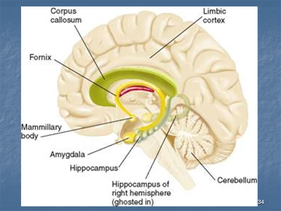


 Fig. 1.4.1.>")

 Pia mater -inner membrane, contains.>")


; Ventral (vientre means tummy); Caudal.>")
 Brain and spinal cord Peripheral Nervous System (PNS) ◦ nerves.>")