Download presentation
Presentation is loading. Please wait.

1
New Developments in Renal Dialysis WH Seto WHO CC, Hong Kong www.webbertraining.comDecember 5, 2012 Hosted by Prof. Lance Jennings University of Otago, New Zealand Sponsored by WHO Patient Safety Challenge Clean Care is Safer Care 1

3
Standards requirements Dialysis water: chemical contaminants Microbiological contaminants Concentrate Dialysis fluid: Microbiological contaminants in standard fluids Ultrapure dialysis fluid online-prepared substitution fluid Record keeping Other recommendations System design Validation of system performance: Plan Installation qualitification Operational qualification Performance qualification Routine monitoring Quality Management: Fluid quality Water treatment equipment Water storage and distribution Concentrate preparation, distribution and proportioning Microbiological control: Disinfection Microbiological monitoring Environment Personnel SUMMARY Water Fluid Machine

4
4

5
5

6
6

7
Major important infection control issues in dialysis centre New microbiological standard of fluids for dialysis and related therapies Minimize vascular access infections in hemodialysis patients Concern of hepatitis C outbreaks 7

8
Water Treatment To remove chemical, bacterial & endotoxin contaminant that could be harmful to patients Consist of : Water softener Particulate filter(s) Carbon filter(s) Deionizers, filters, Reverse osmosis (RO) Ultrafilters, UV light 8
 Carbon filter(s) Deionizers, filters, Reverse osmosis (RO) Ultrafilters, UV light 8")
9
Water softener Charcoal filters 9

10
Particulate filter (s) Reverse osmosis (RO) 10
 Reverse osmosis (RO) 10")
11
Gram-negative water bacteria Pseudomonas Flavobacterium Acinetobacter Alcaligenes Achromobacter Aeromonas Serratia Xanthomonas TYPES OF WATER MICROORGANISMS THAT HAVE BEEN FOUND IN DIALYSIS SYSTEMS (1) Endotoxin 11
 Endotoxin 11")
12
Non-tuberculous mycobacteria Mycobacterium chelonae fortuitum gordonae scrofulaceum kansasii avium intracellularis TYPES OF WATER MICROORGANISMS THAT HAVE BEEN FOUND IN DIALYSIS SYSTEMS (2) 12
 12")
13
The evolution of extracorporeal treatment of end-stage renal failure has enforced focus on the purity of dialysis fluid. 13

14
Bicarbonate dialysate are commonly used for both conventional and high- flux dialysis which a good culture medium Potential transfer of bacteria from dialysate to patient blood Bicarbonate dialysate 14

15
Conventional dialyser High-flux dialyser 15

16
High flux dialyzers have larger pores, the bacterial particles can pass more easily into the patient’s bloodstream, Patients on high flux dialysis have more frequent pyrogen reactions Adverse effect of high flux dialysis 16

17
An other major challenge of high-flux haemodialysis (HD) and haemodiafiltration relates to the necessity for ultrapure dialysis fluid and for sterile non-pyrogenic substitution fluid. 17

18
Haemodiafiltration On-line fluid replacement Ultra-filter 18

19
cfu Water <200/ml Dialysate <200/ml Dialyser disinfectant<200/ml Dialysate for infusion 1/1000 L Ultra-pure dialysate 1/10 ml AAMI 2004 Monitoring of dialysis fluid Should be done at least monthly 19

20
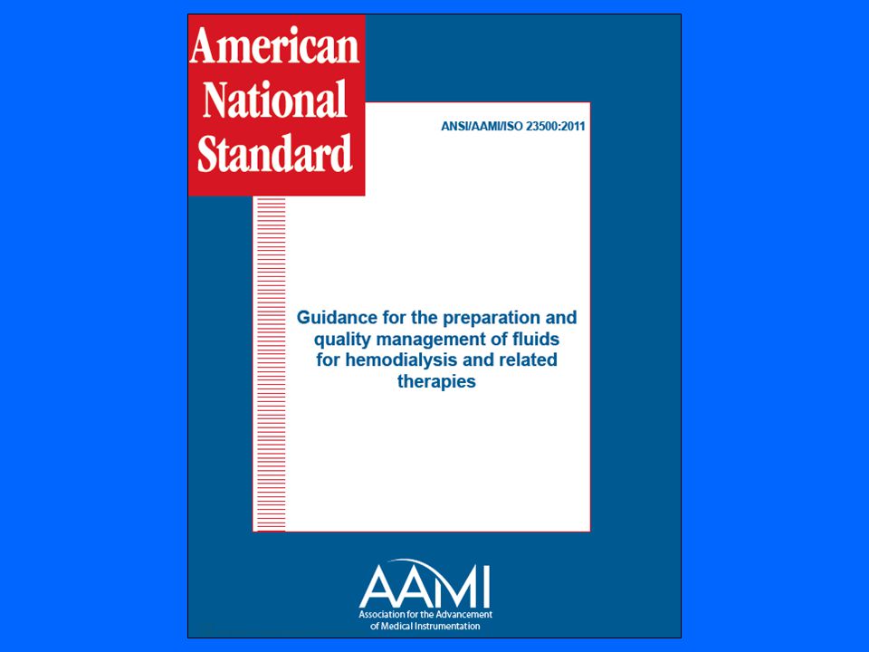
2011 New standard of fluids for hemodialysis Association for the advancement of medical instrumentation 20

21
Dialysis fluid = dialysis water and dialysate Microbial count<100 CFU/ml Endotoxin concentration <0.5 EU/ml 21

22
Dialysis water is treated water for HD, reprocess of dialysers, preparation of concentrate, fluid for on-line convective therapy

23
23

24
Dialysis water – sampling

25
Dialysis fluid

26
Test for compliance of microbiological requirement Culture agar - tryptone glucose extract agar (TGEA) Incubation T 0 - 17 0 C -23 0 C Incubation time- 168 hours (7 days) Method and sample volume -spread plate, 0.1 ml - 0.3 ml -pour plate, 0.1 ml – 1 ml Dialysis fluid routine test:
 Incubation T C C Incubation time- 168 hours (7 days) Method and sample volume -spread plate, 0.1 ml ml -pour plate, 0.1 ml – 1 ml Dialysis fluid routine test:")
27
27

28
Ultrapure dialysis fluid 1.Highly purified dialysis fluid in place of conventional 2.Feed solution infusing directly to pt’s blood Culture method by membrane filtration (10-1000ml)
")
29
“These types of samples also should be taken at least once monthly and after suspected pyrogenic reactions or changes in the water treatment system of disinfection protocols.” (pp 347) Bennett & Brachman’s 29
 Bennett & Brachman’s 29")
31
31

32
Burden of Dialysis Infections In the US, there are about 370,000 people relying on hemodialysis About 75,000 people receive hemodialysis through a central line Central lines have a higher risk of infection than a fistula or graft CDC estimates 37,000 central line- associated bloodstream infections may have occurred in U.S. hemodialysis patients in 2008 A Cause for Concern 32

33
33

34
34

35
Cuffed catheter 35

36
36

37
37

38
Prevention of vascular access infections National Kidney Foundation and CDC - USA No antibiotic prophylaxis – at insertion and use of catheter No routine change of catheter Use sterile techniques (cap, mask, sterile gown, large drape.) Limiting non-cuff catheter to 3-4 weeks Use only for HD Only trained personnel care for the catheter Replace dressing after HD or when damp, loose & soil Disinfect skin with CHG for insertion and dressing change Ensure catheter site is compatible with catheter material 38
 Limiting non-cuff catheter to 3-4 weeks Use only for HD Only trained personnel care for the catheter Replace dressing after HD or when damp, loose & soil Disinfect skin with CHG for insertion and dressing change Ensure catheter site is compatible with catheter material 38")
39
39

40
40

41
41

42
Careful infection control practices can prevent hemodialysis catheter associated bloodstream infection: Follow established guideline for access care Use proper insertion and catheter care protocol 42

43
Slide print screen of new york outbreak & Ix 162 who were being treated as of July 2008, Manhattan, NY Medical director of the dialysis center was fined $300,000 in September 2008 43

44
No of gloves for patient care No change of gloves between patient and when dirty Not using CHG for skin disinfection Did not observe aseptic technique when inserting cannula 44

45
Heparin need to be diluted with saline The dilution is done at fixed time Dilution is done in ward area Only one saline bag is used 45

46
Heparin saline prepared near clotting time test and patient care area 46

47
One-way flow of supplies Clean Dirty Medication area Patient cubicle 1 Patient cubicle 2 Patient cubicle 3 X X -No return of supplies -No transfer of supplies -No mobile cart 47

48
48

49
Multidose vials: Preservatives has no impact on HBV, HCV Medication vials 49

50
Do not store equipment with blood sampling area X 50

51
Should be prepared away from patient care area 51

52
Transducer / filters to prevent blood leak and contamination 52

53
Dedicated items for use on single patient Disposable - disposed of Reusable - disinfection before use on other patients 53

54
Infection control practices for HD patients Wear glove when caring for patient Change gloves between patient and hand hygiene Dedicated or single patient use item Designated area for admixture of medication Do not share medication vials Do not use common medication cart Do not store supplies with blood samples and patient equipment Use external transducer/filter to prevent blood leak Clean & disinfect dialysis station between patient use Cap and clamp tubing & kidney and use leak proof container when transport 54

55
Infection control issues in dialysis centre Adopt the new AAMI microbiological standard of fluids for dialysis and related therapies Eliminate vascular access infections in hemodialysis patients Enforce infection control guideline to prevent MDRO & hepatitis C outbreaks 55

56
56

57
2013 WHO Teleclasses Now available at www.webbertraining.com/schedulep1.php www.webbertraining.com 57

Similar presentations


. This treatment cleans the blood.>")


 Elizabeth Lindley St James’s University Hospital and Leeds General Infirmary Leeds, UK.>")


