Download presentation
Presentation is loading. Please wait.

2
Filariae Trichinella spiralis Experiment 2

3
To study the morphology of microfilariae and laboratory diagnostic methods. To learn the morphology of T. spiralis. To master the pathogenesis of T. spiralis through animal experiment. Objectives and Requirements

4
Filariae 丝虫

5
Wuchereria bancrofti ( 班氏吴策线虫, Wb ) Brugia malayi ( 马来布鲁线虫, Bm )
 Brugia malayi ( 马来布鲁线虫, Bm )")
6
Because these two species are similar in morphology,they will be discussed together,with notions on dissimilaritis.

7
Morphology Adult: White and thread-like. Two rings of small papillae on the head. Female: 5~10 cm in length, uterus almost occupies the body, uterus with eggs, mature eggs & larvae(microfilariae) Male: 2.5~4cm and a curved tail with two copulatory spicules.
 Male: 2.5~4cm and a curved tail with two copulatory spicules..")
8
Adult

9
Wuchereria bancrofti adult worm in a lymphatic channel adult

11
Cephalic space sheath Body nuclei Excretory pore Terminal nuclei Nervous cycle Microfilaria: * * 177~296 µm in length with sheath, * * Anterior: bluntly rounded * * End: pointed * * A large number of nuclei could be seen in the body after staining.

12
Morphological Differences of Microfilariae between W. bancroti and B. malayi _______________________________________________________________ _ Species W. bancrofti B. malayi _______________________________________________________________ _ Appearance graceful, sweeping curves irregular, stiff curves _______________________________________________________________ _ Size larger 244-296× 7 µ m smaller 177-230 × 6 µ m _______________________________________________________________ _ Cephalic space shorter(1:1 or1:2) longer(2:1) (length:width) _______________________________________________________________ _ Bodynuclei equal sized, clearly unequal sized, over lapped, defined, countable uncountable _______________________________________________________________ _ Terminal nuclei no two _______________________________________________________________ _
 longer(2:1) (length:width) _______________________________________________________________ _ Bodynuclei equal sized, clearly unequal sized, over lapped, defined, countable uncountable _______________________________________________________________ _ Terminal nuclei no two _______________________________________________________________ _.")
14
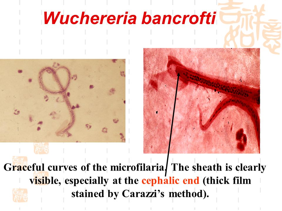
Wuchereria bancrofti Graceful curves of the microfilaria. The sheath is clearly visible, especially at the cephalic end (thick film stained by Carazzi’s method).
..")
15
Wuchereria bancrofti Caudal end of the microfilaria. No nuclei in the pointed caudal end (stained by Giemsa)
.")
16
Brugia malayi Coiled microfilaria. The nuclei are more or less distinct. The last caudal nuclei are clearly separated from the others and extends into the tip of the tail. (Giemsa stain).
..")
17
Microfilaria (unstained) Cephalic space
 Cephalic space")
18
Infective stage larvae Mouthpart of mosquito

19
A large number of larvae of a filarias emerging from the proboscis of a mosquito.

20
Diagnosis The diagnosis depends on the symptoms, signs and history of living in endemic areas, but the confirmative diagnosis depends on the demonstration of microfilariae 1. First choice method is thick blood smear, taking blood at night from 9pm-2am 2. Fresh blood smear used for mass education. 3. Immunological tests are only made reference of the diagnosis.

21
Thick blood smear

22
Trichinella spiralis 旋毛形线虫

23
Morphology Adult small & slender with slightly tapered anterior end A pair of copulatory spicules at the posterior of the male male size: 1.4-1.6 plus 0.04- 0.05 mm female size: 3-4 plus 0.06 mm pharynx is one third or half of worm body long

25
Adult reproductive system of both sex is single tract single uterus filled developing eggs, developed & hatched larva belong to ovoviviparous

26
Larvae cyst The cyst are commonly found in skeletal muscle,its size is about 0.25-0.5 plus 0.21-0.42 mm. Usually, there is 1-2 coiled larvae in a muscle fiber.

27
Trichinella spiralis encysted larva

28
Larvae of trichinella spiralis in muscle section

29
Once ingested, the larva is freed from its cyst cell, and enters the mucosa of the small intestine

30
Diagnosis Laboratory Diagnosis of Trichiniasis depends on : 1. Muscular biopsy 2. Immunodiagnosis and xenodiagnosis (1) CPT(circumlarval precipitin test) (2) ELISA (3) IHA
 CPT(circumlarval precipitin test) (2) ELISA (3) IHA.")
31
Diagnosis Muscle samples (highly active muscle like columns of the diaphragm, tongue) were squeezed between two glass plates and then microscoped at low power
 were squeezed between two glass plates and then microscoped at low power")
32
1. Label the microfilariae. 2. observe the cyst of T. spiralis Exercise

33
Homework 1.draw the two kinds of microfilariae 2.draw the cyst of T. spiralis

Similar presentations


.>")


