Download presentation
Presentation is loading. Please wait.

1
بسم الله الرحمن الرحيم

2
HEMODYNAMIC DISTURBANCES (Disorders of blood flow)
By: Dr. Gehan Mohamed

3
CIRCULATORY DISTURBANCES
1- Hyperemia 2- Congestion 3- Thrombosis 4- Embolism 5- Ischemia 6- Infarction 7- Hemorrhage 8- Edema

4
Hyperemia & Congestion
The terms hyperemia & congestion both indicate: a local increase in volume of blood in a particular tissue. Hyperemia is an active process resulting from increased arterial blood inflow because of arteriolar dilatation. - The affected tissue is reddened because of engorgement of tissues with oxygenated blood. Congestion is a passive process resulting from impaired venous outflow from a tissue. - The tissue has a red-blue color due to accumulation of deoxygenated blood.

5
HYPEREMIA Definition:
Increase in blood flow to an organ as result of active dilatation of its arterioles. It is an active process, involving change in the muscle tone of the arterioles (active hyperemia). Types: 1- Physiological: - hyperemia in skeletal muscles during exercise. - hyperemia in the gut following a meal. 2- Pathological: e.g. in acute inflammation.
. Types: 1- Physiological: - hyperemia in skeletal muscles during exercise. - hyperemia in the gut following a meal. 2- Pathological: e.g. in acute inflammation.")
7
VENOUS CONGESTION (Passive Hyperemia)
Definition: Increase in venous blood in an organ as result of obstruction of venous outflow. - the veins, venules, & capillaries in the organ become passively dilated (passive hyperemia). - although there is excess blood in the tissue, the blood flow is slow & reduced. Types: → Localized acute chronic Generalized acute
. - although there is excess blood in the tissue, the blood flow is slow & reduced. Types: → Localized acute. chronic. Generalized acute.")
8
Acute localized venous congestion
Causes: Sudden complete venous obstruction by: thrombosis, ligature, strangulation, or twisting of the pedicle of a movable organ. Effects: 1- Rapid severe distention of the veins and capillaries which may rupture → hemorrhage. 2- Edema occurs rapidly in the tissues. 3- In intestine: infarction & gangrene may occur

9
Chronic localized venous congestion
• Causes: Gradual incomplete venous obstruction by: 1- Venous compression by: a tumor, enlarged lymph node or pregnant uterus. 2- Liver cirrhosis or fibrosis → congestion in mesenteric & splenic veins. 3- Left ventricular failure → congestion of pulmonary veins • Effects: Chronic dilatation of the veins, venules and capillaries proximal to the obstruction resulting in: 1- Edema Stasis predisposes to thrombosis 3- Gradual opening of the collateral veins. 4- Development of varicoses (e.g. oesophageal varices)
")
10
Acute generalized venous congestion
Causes: - occurs in acute heart failure Effects: All viscera show acute congestion (rapid generalized congestion)
")
11
Chronic generalized venous congestion
Definition: Gradual congestion affecting the whole venous system in the body. Causes: Right sided heart failure Effects: Hypoxia & cyanosis Dyspnea (due to pulmonary Congestion) Generalised oedema Chronic venous congestion in different organs.
. 3- Generalised oedema. 4- Chronic venous congestion in different organs.")
12
Effects of chronic generalized venous congestion
Hypoxia & Cyanosis: - Hypoxia is due to defective oxygenation of blood in the congested lungs. - Cyanosis is due to increased amounts of reduced hemoglobin (due to stasis). Dyspnea: due to pulmonary congestion. Generalized edema (cardiac edema). Effects in different organs.
. Dyspnea: due to pulmonary congestion. Generalized edema (cardiac edema). Effects in different organs.")
13
Thrombosis 1- Definition 2- Causes 3- Composition 4- Types 5- Sites
6- Fate 7- N.B.

14
Definition of thrombosis
Thrombosis is: - The formation of a solid mass (compact mass) - Composed of the blood elements. In a blood vessel or heart. In circulating blood. - During life.
 - Composed of the blood elements. In a blood vessel or heart. In circulating blood. - During life.")
15
There are 3 major factors which predispose to thrombosis
Causes of thrombosis There are 3 major factors which predispose to thrombosis (Virchow’s triad) 1- Endothelial damage 2- Slowing & turbulence of blood flow 3- Changes in blood composition
 1- Endothelial damage. 2- Slowing & turbulence of blood flow. 3- Changes in blood composition.")
16
Virchow triad in thrombosis

17
Causes of thrombosis 1- Endothelial damage:
- This is the most important factor in thrombus formation. - Endothelial damage may be: Mechanical, inflammatory, or degenerative The injured endothelium becomes swollen with rough surface. 2- Staisis: There is slowing of blood flow in the heart as in mitral stenosis and in blood vessels as in varicose veins.

18
3- Changes in composition of blood:
↑ platelets e.g. after operations. ↑ fibrinogen as in pregnancy. ↑ R.B.Cs. (polythycaemia) → ↑ viscosity of blood → staisis → thrombosis. ↑ W.B.C. as in leukaemia → ↑ viscosity of blood → staisis → thrombosis.
 → ↑ viscosity of blood → staisis → thrombosis. ↑ W.B.C. as in leukaemia → ↑ viscosity of blood → staisis → thrombosis.")
19
Pathogenesis (Mechanism) of thrombosis:
- Platelets leave the blood stream, agglutinate and adhere to the damaged endothelium. They form laminae, which are arranged vertical to the blood stream and called lines of Zhan. - Soon, fibrin accumulates around them with red and white blood cells.

20
Lines of Zhan

21
Classification of thrombi
According to the color & composition of thrombi According to the site of thrombus: According to presence or absence of bacteria:

22
According to the color & composition of thrombi:
1- Pale thrombus: formed only of platelets and fibrin. 2- Red thrombus: formed mainly of red cells and fibrin. 3- Mixed thrombus: containing all blood elements.

23
According to the site of thrombus:
1- Venous thrombus (the most common): formed in veins as in varicose veins and after major abdominal operations Arterial thrombus: found in atherosclerosis and aneurysm. 3- Cardiac thrombus: found in the heart, either in the heart chambers called mural thrombus or on the heart valves called vegetations. 4- Capillary thrombi
: formed in veins as in varicose veins and after major abdominal operations. 2- Arterial thrombus: found in atherosclerosis and aneurysm. 3- Cardiac thrombus: found in the heart, either in the heart chambers called mural. thrombus or on the heart valves called vegetations. 4- Capillary thrombi.")
24
According to presence or absence of bacteria:
1- Septic thrombus: containing pyogenic bacteria Aseptic thrombi: without bacteria.

25
1- Venous thrombosis Thrombosis in veins may be either:
Thrombosis in veins is more common than other sites because of their slow blood, and thin wall. Thrombosis in veins may be either: Thrombophlebitis Phlebothrombosis

26
Thrombophlebitis ● Thrombosis is caused by inflammation of venous wall. ● Two types occur: 1- Septic thrombophlebitis: - Occurs in veins draining septic lesions. - e.g.: appendicular vein in case of acute appendicitis. - suppuration of the thrombus causes its softening - fragments of infected thrombus may break away → pyaemia 2-Aseptic thrombophlebitis: - Inflammation is caused by factors other than bacteria. - e.g.: trauma and radiations. - a small fixed aseptic thrombus occurs.

27
Phlebothrombosis 1- Thrombosis in varicose veins due to stasis.
This is thrombosis in non-inflamed veins. The thrombus may propagate and may fragment causing pulmonary embolism. Examples include: 1- Thrombosis in varicose veins due to stasis. 2- Thrombosis in calf veins (DVT) in chronic cardiac pts confined to bed (stasis & compression of calf ms). 3- Thrombosis in pelvic & femoral veins after labour or operations (↑platelets, bed recumbence, surgical injury).
 in chronic cardiac pts confined to bed (stasis & compression of calf ms). 3- Thrombosis in pelvic & femoral veins after labour or operations (↑platelets, bed recumbence, surgical injury).")
28
2- Arterial thrombosis ● Less common than venous thrombosis because of the rapid blood flow in the arteries and the thick elastic arterial wall which resists injury. ● Thrombosis occurs in arteries affected by: atherosclerosis, arteritis, & aneurysms (due to stasis, disordered blood flow & roughness of the intima). ● Arterial thrombosis → ischemia.
. ● Arterial thrombosis → ischemia.")
29
Fate of thrombi It depends upon its size & whether it is septic or aseptic. ● Septic thrombi: Fragments by proteolytic enzymes into septic emboli → pyaemic abscesses. ● Aseptic Thrombi: may undergo: - Small thrombi is dissolved and absorbed. - Large thrombus undergoes: 1- Organization (fibrosis) 2- Organization & canalization 3- Calcification 4- Fragmentation and embolism.
 2- Organization & canalization. 3- Calcification. 4- Fragmentation and embolism.")
30
Thrombus: organized & recanalized

31
Blood Clot A mass of blood elements formed by transformation of fibrinogen to fibrin, in stagnant blood. The clot is dark red with a glistening smooth surface, and is not adherent to the vessel wall. Clotting of blood may be: → Outside the CVS Inside the CVS: During life after death (e.g. in stagnant blood) (postmortem clots) red yellow
 (postmortem clots) red yellow.")
32
Difference between thrombus and clot:
1- Occurs in stagnant blood during life or after death 2- Loosely attached 3- Soft and moist 4- Red and yellow 5- No lines of Zhan 1- Occurs in circulating blood during live 2- Firmly attached 3- Friable and dry 4- Pale, pale red or red 5- May show lines of Zhan

33
EMBOLISM DEFINITION CAUSES & TYPES

34
Embolism ● Definition Embolus: An insoluble (solid, liquid or gaseous) mass circulating in the blood stream. Embolism: Is the process of impaction of the embolus in a narrow vessel.

35
Embolism ● Causes & Types: 1- Detached thrombi (thrombo-embolism)
2- Fat embolism: The fat of the bone marrow reaches the circulation after fracture of bones. 3- Air embolism: due to injury of neck & chest veins. 4- Parasitic emboli: e.g. bilharzial worms and ova. 5- Tumor emboli: groups of tumour cells penetrate the wall of blood vessels especially veins. 6- Amniotic fluid embolism.

36
1- Detached thrombi (thromboembolism)
Sites of impaction: 1- Pulmonary embolism 2- Portal embolism 3- Systemic embolism 4- Paradoxical embolism i.e.the embolus coming with venous return to be impacted in lung causing pulmonary embolism but instead of that it will pass from right side of heart to its left side through septal defect then pass to systemic circulation.

37
Effects of thromboemboli
Effects depends upon: 1- Size of the embolus. 2- Nature of the embolus (septic or aseptic). 3- State of the collateral circulation in the affected site.
. 3- State of the collateral circulation in the affected site.")
38
Effects of thromboemboli
Effects depends upon: 1- Size of the embolus. 2- Nature of the embolus (septic or aseptic). 3- State of the collateral circulation in the affected site. Effects of pulmonary embolism: Big embolus Medium sized embolus Small embolus
. 3- State of the collateral circulation in the affected site. Effects of pulmonary embolism: Big embolus Medium sized embolus Small embolus.")
39
Effects of thromboemboli
Effects depends upon: 1- Size of the embolus. 2- Nature of the embolus (septic or aseptic). 3- State of the collateral circulation in the affected site. Effects of pulmonary embolism: Big embolus Medium sized embolus Small embolus Acute Rt sided Heart failure Sudden death
. 3- State of the collateral circulation in the affected site. Effects of pulmonary embolism: Big embolus Medium sized embolus Small embolus. Acute Rt sided. Heart failure. Sudden death.")
40
Effects of thromboemboli
Effects depends upon: 1- Size of the embolus. 2- Nature of the embolus (septic or aseptic). 3- State of the collateral circulation in the affected site. Effects of pulmonary embolism: Big embolus Medium sized embolus Small embolus Acute Rt sided healthy lung Heart failure no effect Sudden death
. 3- State of the collateral circulation in the affected site. Effects of pulmonary embolism: Big embolus Medium sized embolus Small embolus. Acute Rt sided healthy lung. Heart failure. no effect. Sudden death.")
41
Effects of thromboemboli
Effects depends upon: 1- Size of the embolus. 2- Nature of the embolus (septic or aseptic). 3- State of the collateral circulation in the affected site. Effects of pulmonary embolism: Big embolus Medium sized embolus Small embolus Acute Rt sided healthy lung congested lung Heart failure no effect lung infarction Sudden death
. 3- State of the collateral circulation in the affected site. Effects of pulmonary embolism: Big embolus Medium sized embolus Small embolus. Acute Rt sided healthy lung congested lung. Heart failure. no effect lung infarction. Sudden death.")
42
Effects of thromboemboli
Effects depends upon: 1- Size of the embolus. 2- Nature of the embolus (septic or aseptic). 3- State of the collateral circulation in the affected site. Effects of pulmonary embolism: Big embolus Medium sized embolus Small embolus Acute Rt sided healthy lung congested lung no effect Heart failure no effect lung infarction Sudden death
. 3- State of the collateral circulation in the affected site. Effects of pulmonary embolism: Big embolus Medium sized embolus Small embolus. Acute Rt sided healthy lung congested lung no effect. Heart failure. no effect lung infarction. Sudden death.")
43
Air embolism Rare and may result from:
1- Injury to the large neck veins. Air is sucked by the negative pressure in the thorax. 2- During cardiothoracic surgery → air may enter veins 3- In criminal abortion → air may pass into uterine veins 4- Caisson’s disease (decompression sickness): - In deep dives, the high pressure increases the amount of gasses dissolved in the blood of the divers. - If decompression is done rapidly, gases esp. nitrogen form emboli in the blood vessels. - Small amount of air is harmless, but cc. interferes with cardiac contraction and causes acute heart failure.
: - In deep dives, the high pressure increases the amount of gasses dissolved in the blood of the divers. - If decompression is done rapidly, gases esp. nitrogen form emboli in the blood vessels. - Small amount of air is harmless, but cc. interferes with cardiac contraction and causes acute heart failure.")
44
Fat embolism Rare condition Causes include:
(1) Bone fractures and crush limb injuries. (2) Trauma to adipose tissue (infl. or burns). (3) Trauma to a grossly fatty liver. (4) Major surgery.
 Bone fractures and crush limb injuries. (2) Trauma to adipose tissue (infl. or burns). (3) Trauma to a grossly fatty liver. (4) Major surgery.")
45
Ischemia Definition: Types: Ischemia may be either:
Deficient arterial blood supply to an organ or tissue due to partial or complete occlusion of its artery. Types: Ischemia may be either: 1- Acute ischemia (complete or sudden ischemia) 2- Chronic ischemia (partial or gradual ischemia)
 2- Chronic ischemia. (partial or gradual ischemia)")
46
Acute ischemia Causes: Sudden complete arterial occlusion by:
1- Thrombosis or embolism. (most common) 2- Surgical ligature of the artery. 3- Twisting of the pedicle of a movable organ e.g intestinal loop. 4- Arterial spasm as in ergot poisoning. Effects: depends on the efficiency of collaterals: ● Sudden occlusion of end arteries or arteries with poor collaterals → infarction or gangrene. ● Sudden occlusion of arteries with efficient collaterals may not cause tissue damage.
 2- Surgical ligature of the artery. 3- Twisting of the pedicle of a movable organ e.g. intestinal loop. 4- Arterial spasm as in ergot poisoning. Effects: depends on the efficiency of collaterals: ● Sudden occlusion of end arteries or arteries with poor collaterals → infarction or gangrene. ● Sudden occlusion of arteries with efficient collaterals may not cause tissue damage.")
47
Chronic Ischemia Causes: Incomplete arterial occlusion by:
1- Atherosclerosis. 2- Pressure on the artery by enlarged lymph node, tumor ... etc. 3- End arteritis oblitrans as in chronic inflammation. Effects: depends on the efficiency of collaterals: ● With inefficient collaterals: - pain on exercise: angina pectoris, intermittent claudication.. - cellular degeneration, atrophy followed by fibrosis. ● With efficient collaterals no tissue damage occurs.

48
Infarction Definition Causes Types Pathological features Fate Examples

49
Infarction Definition
An infarct is an area of coagulative necrosis (liquefactive in the brain) caused by sudden ischemia.
 caused by sudden ischemia.")
50
Infarction Causes 1- Thrombosis that may occur inside diseased arteries. 2- Embolism. 3- Strangulation or twisting of an organ as in loops of intestine, testes or ovaries.

51
Types of infarcts: 1- Red infarcts (hemorrhagic):
- Occur in vascular organs as the lung, liver and intestine. - The red color is due to hge in the substance of the infarct. 2- Pale infarcts: - Are more common and occur in firm and less vascular organs as the kidney, heart and spleen. 3- Liquefactive infarcts: - Occur in the brain and spinal cord.

52
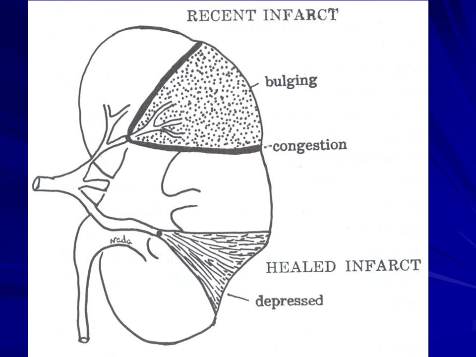
Pathological features of the infarct
● Gross picture: Shape: The infarct is wedge shaped or pyramidal. The base is directed towards the surface of the organ. Site: The infarct is subcapsular, raised when recent due to edema and depressed when healed due to fibrosis. Size: depends on the size of the occluded vessel. Color: Infarcts are of two types; red and pale. It is surrounded by a red zone of inflammatory hyperemia.

54
Infarction of the kidney

55
Infarction of the kidney

56
Infarction of the spleen

57
Infarction of the lung (hemorrhagic infarction)
")
58
Infarction of the lung (hemorrhagic infarction)
")
59
Intestinal infarction → Gangrene

60
● Microscopic picture:
Early: the cells show various post-necrotic changes. Next: structural details are lost but the outlines are preserved. Lastly: necrotic tissue appears as granular pink debris. The infarct is surrounded by a red zone of inflammatory hyperemia.

61
● General reactions: Infarcts are associated with general reactions in the form of: Fever Leucocytosis Increased sedimentation rate; ESR Elevation of certain serum enzymes as transaminase in myocardial infarction.

62
Fate of the infarct Small infarct: Large infarct:
Necrotic tissues are removed by macrophages, granulation tissue fills the defect followed by fibrosis. Large infarct: Gets surrounded by a fibrous capsule and its substance may show dystrophic calcification.

63
HAEMORRHAGE Definition Causes Types Effects Haemostasis

64
HAEMORRHAGE Definition:
Escape of blood outside the blood vessels or cardiac chambers. (loss of blood from circulation)
")
65
Causes of haemorrhage 1- Trauma: involving the heart and blood vessels. 2- Diseases of blood vessels: a) Hypertension. b) Varicose veins: as piles. c) Degeneration: as atheroma and aneurysm. d) Infection: as tuberculosis. e) Malignant cells invading blood vessels. 3- Haemorrhagic blood diseases: as haemophilia, purpura, leukaemia and scurvy.
 Hypertension. b) Varicose veins: as piles. c) Degeneration: as atheroma and aneurysm. d) Infection: as tuberculosis. e) Malignant cells invading blood vessels. 3- Haemorrhagic blood diseases: as haemophilia, purpura, leukaemia and scurvy.")
66
Types of haemorrhage External haemorrhage Internal haemorrhage
Interstitial haemorrhage

67
1- External haemorrhage
Escape of blood outside the body. 1- Epistaxis: Bleeding from the nose. 2- Hemoptysis: Coughing of blood. 3- Hematemesis: Vomiting of blood. 4- Melena: Presence of dark digested blood in stools. 5- Bleeding per rectum: passage of red blood with stool 6- Hematuria: Blood in urine. 7- Menorrhagia: Excessive or prolonged menstrual bleeding. 8- Metrorrhagia: Irregular uterine bleeding unrelated to menses 9- Bleeding from skin

68
2- Internal haemorrhage
Bleeding into body cavities. 1- Hemothorax: Hge into the pleural sac. 2- Hemopericardium: Hge. into pericardial sac. 3- Hemoperitoneum: Hge. into peritoneal sac. 4- Hematocele: Hge. into tunica vaginalis sac. 5- Hemoarthrosis: Hge. into a joint cavity.

69
3- Interstitial haemorrhage
Bleeding into interstitial tissue spaces. 1- Petechial haemorrhage: escape of small amount of blood of capillary origin → small spots of haemorrhage. 2- Ecchymosis: escape of moderate amount of blood → a bigger patch of haemorrhage. 3- Hematoma: escape of large amount of blood causing a swelling.

70
- Interstitial haemorrhage is at first dark red (arterial blood) or bluish (venous blood).
- Then, hemoglobin breaks down into biliverdin and hemosiderin. - BiIiverdin gives the area a green color but is soon absorbed in the blood. - The hemosiderin left gives the area a brown color and is gradually removed by macrophages, so the color changes to yellow and gradually fades away.

71
Effects of haemorrhage
● Small amount: No effect. ● Repeated small amounts (chronic hge): - Causes microcytic hypochromic anemia. - e.g. in piles and peptic ulcers. ● Moderate amount: (< 750 ml.) → Is compensated by: 1- reflex ↑ heart rate 2- reflex vasoconstriction in the skin, muscles & GIT 3- withdrawal of tissue fluids into the blood. 4- Proteins are added from the liver. 5- blood cells are added by the hyperplastic B.M. ● Massive amount: → hemorrhagic shock.
: - Causes microcytic hypochromic anemia. - e.g. in piles and peptic ulcers. ● Moderate amount: (< 750 ml.) → Is compensated by: 1- reflex ↑ heart rate. 2- reflex vasoconstriction in the skin, muscles & GIT. 3- withdrawal of tissue fluids into the blood. 4- Proteins are added from the liver. 5- blood cells are added by the hyperplastic B.M. ● Massive amount: → hemorrhagic shock.")
72
Edema Definition Causes Classification Pathological features

73
Edema Definition: - Edema fluid may be either transudate or exudate.
- Pathological accumulation of excess fluids in the interstitial tissue spaces and serous sacs. - Edema fluid may be either transudate or exudate.

74
Transudate Exudate Caused by conditions other than inflammation
Occurs in cases of inflammation. Low protein content (below 3 gm%). High protein content (4-8 gm%). Specific gravity below 1015. Specific gravity above 1018. Does not clot on standing. (no fibrinogen) Clots on standing (presence of fibrinogen) No inflammatory cells. Contains inflammatory cells.
. High protein content. (4-8 gm%). Specific gravity below Specific gravity above Does not clot on standing. (no fibrinogen) Clots on standing. (presence of fibrinogen) No inflammatory cells. Contains inflammatory cells.")
75
Approximately 60% of body weight is water,
two thirds of which is intracellular with the remainder is in the extracellular compartments (the interstitial tissue & plasma).
.")
76
Mechanism of edema formation
Filtration out Reabsorption in 15 mmHg HP 35 mmHg HP Mechanism of edema formation

77
Capillary hydrostatic pressure (n: 35-15 mmHg)
Rates of filtration & reabsorption across the capillary wall depend on: Capillary hydrostatic pressure (n: mmHg) Plasma osmotic pressure (n: 20 mmHg) - Normally, capillary hydrostatic & osmotic forces are balanced, so that the amount of Interstitial fluid remains constant. - At arterial end: H.P. > O.P.→ filtration of tissue fluid At venous end: H.P. < O.P.→ withdrawal of tissue fluid
 Plasma osmotic pressure (n: 20 mmHg) - Normally, capillary hydrostatic & osmotic forces are balanced, so that the amount of Interstitial fluid remains constant. - At arterial end: H.P. > O.P.→ filtration of tissue fluid. At venous end: H.P. < O.P.→ withdrawal of tissue fluid.")
78
Causes of edema 1- Increased capillary hydrostatic pressure:
occurs in cases of : - venous congestion (generalized or localized) - sodium & water retention → ↑ blood volume 2- Decreased plasma colloid osmotic pressure: occurs in cases of hypoproteinemia (fall of total plasma proteins below 2.5 gm% or fall of serum albumin below 1.5 gm%) 3- Increased capillary permeability: - Caused by toxins, hypoxia, & chemicals (e.g. histamine in acute infl.). - Escape of proteins into ISF → ↓ plasma osmotic pr. & ↑ tissue osmotic pr. → further edema.
 - sodium & water retention → ↑ blood volume. 2- Decreased plasma colloid osmotic pressure: occurs in cases of hypoproteinemia (fall of total plasma proteins below 2.5 gm% or fall of serum albumin below 1.5 gm%) 3- Increased capillary permeability: - Caused by toxins, hypoxia, & chemicals (e.g. histamine in acute infl.). - Escape of proteins into ISF → ↓ plasma osmotic pr. & ↑ tissue osmotic pr. → further edema.")
79
4- Lymphatic obstruction
- It → lymphatic edema (lymphedema). - It is caused by: 1- Lymphangitis and lymphadenitis as in Filariasis → elephantiasis. 3- Mechanical compression of lymphatics e.g. by tumors. 4- Lymphatic permeation by malignant cells. 5- Post-irradiation fibrosis in lymphatics & LNs. 6- Surgical removal of the lymph nodes.
. - It is caused by: 1- Lymphangitis and lymphadenitis as in Filariasis. → elephantiasis. 3- Mechanical compression of lymphatics e.g. by tumors. 4- Lymphatic permeation by malignant cells. 5- Post-irradiation fibrosis in lymphatics & LNs. 6- Surgical removal of the lymph nodes.")
80
Classification of edema
According to the site of edema: 1- Localized edema: 2- Generalized edema (anasarca) According to consistency of edema: 1- Pitting edema (Soft edema): 2- Non-Pitting edema (Hard edema).
 According to consistency of edema: 1- Pitting edema (Soft edema): 2- Non-Pitting edema (Hard edema).")
81
Localized edema Localized in a part of the body.
The total amount of fluids in the body is within normal but with abnormal distribution It includes: 1- Inflammatory edema: Occurs in acute inflammation. The edema fluid is an exudate. 2- Obstructive edema: Venous obstruction → ↑ hydrostatic pressure in the veins and capillaries → edema. Lymphatic obstruction

82
Generalized edema (anasarca)
The total amount of body fluids is increased. It includes: 1- Cardiac edema: Occurs in congestive heart failure. 2- Nutritional edema: Caused by hypoproteinemia due to: - Malnutrition & Malabsorption states - Chronic liver disease → ↓ formation of plasma proteins. 3- Renal edema: Occurs in renal diseases & is of two types: - Nephritic edema: Occurs in acute diffuse glomerulonephritis. - Nephrotic edema: It is caused by massive albuminuria → hypoproteinemia.

83
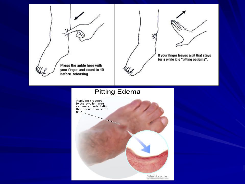
Pitting edema The accumulated fluid can be easily moved on pressing
the affected part, leaving a pit at site of pressure (it pits on pressure). This is because the edema fluid has low protein content → it is present free in the tissue spaces. Occurs when edema fluid is transutade: 1- All types of generalized edema (cardiac, renal and nutritional edema). 2- Localized edema due to venous obstruction
. This is because the edema fluid has low protein content → it is present free in the tissue spaces. Occurs when edema fluid is transutade: 1- All types of generalized edema. (cardiac, renal and nutritional edema). 2- Localized edema due to venous obstruction.")
85
Non-Pitting edema The edematous part does not pit on pressure.
This is because the edema fluid is united with the tissue elements. Occurs in cases of lymphatic edema.

86
Cardiac edema Definition: Generalized edema caused by Right side heart failur. Causes: 1- Congestion → ↑ capillary hydrostatic pressure. 2- Hypoxia → Increased capillary permeability. 3- Renal congestion → sodium and water retention. Sites: - Edema begins around the ankle (gravity effect) - Later it becomes generalized and associated with ascitis, hydrothorax and hydropericardium.
 - Later it becomes generalized and associated with ascitis, hydrothorax and hydropericardium.")
87
Thank you

88
الحمد لله رب العالمين

Similar presentations


 ~5% of total.>")


 intracellular. (1/3)extracellular (interstitial fluid) 5% blood plasma. edema = an accumulation of interstitial.>")


