Download presentation
Presentation is loading. Please wait.

2
Electrocorticography,or ECoG, is the practice of using electrodes placed directly on the exposed surface of the brain, after conducting a craniotomy. It’s goal is to record electrical activity in the production of Epileptic Seizures in search of finding the zones of epileptic origin. Two Forms: Intraoperative and Extraoperative

3
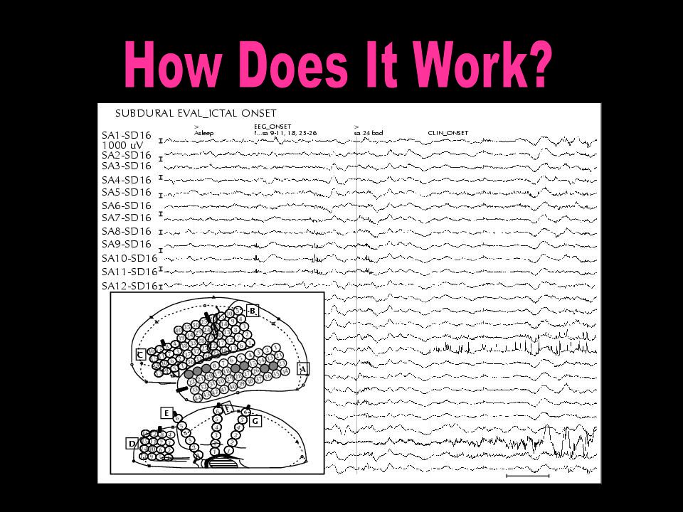
Types of ECoG electrodes: 16 disposable stainless steal electrodes placed in a ball and socket joint attached to an overlying frame. 16 disposable stainless steal electrodes placed in a ball and socket joint attached to an overlying frame. Grid Electrodes ranging from 4 to 64 electrodes. These are transparent, flexible sheets numbered at each different electrode. (Preferred method due to the ability to slip under the dura matter.) Grid Electrodes ranging from 4 to 64 electrodes. These are transparent, flexible sheets numbered at each different electrode. (Preferred method due to the ability to slip under the dura matter.)
 Grid Electrodes ranging from 4 to 64 electrodes. These are transparent, flexible sheets numbered at each different electrode. (Preferred method due to the ability to slip under the dura matter.).")
4
A seizure occurs when a brief, strong surge of an overabundance in electrical activity affects part or all of the brain lasting from a few seconds to a few minutes. It may bring about a change in sensation, awareness or behavior. They are classified as Epileptic if there is a reoccurrence typically more then twice with electrical brain activity.

6
Intraoperative ECoG: Primary Goal To localize the zone and origin point of seizure activity for mapping and resectioning of brain functions.

7
From The Beginning… 1930- Dr. Wilder Penfield developed the “Montreal Procedure” in Canada. First to detect “auras” in Epileptic patients. 1948- Rudolf M. Hess conducted first scalp EEG in a 31 year old patient in Switzerland. 1949- Hugo Krayenbuhl and Rudolf M. Hess- First attempt at ECoG surgery performed on May 18 th at University Hospital in Switzerland. Hess and Krayenbuhl worked in union, Krayenbuhl preformed the surgery; Hess monitored the electrical output of the brain. 1950- Dr. Wilder Penfield and Herbert Jasper; began using the Krayenbuhl and Hess experiment and the Montreal Procedure in combination to create further developments, being the current ECoG methods. 2002- Kuruvilla, M.D and Flink, M.D. – Uppsala University Hospital in Sweden. Began taking on the issue of Intraoperative ECoG and its reliability.

8
Spontaneous brain activity consisting of unusual spikes and sharp waves for the patient. Due to the abnormal spikes and brain activity faulty information is being recorded as the origin of epileptic activity. Seizures are rarely recorded. Requires quick decisions Limited Sampling Time Both the background brain activity and epileptic activity maybe altered by the anesthetics.

9
Schwartz, Bazil, Walczak, Chan, Pedley, and Goodman Study consisted of 29 epileptic participants. All 29 underwent standard resectioning with Intraoperative ECoG. 11 patients (38%) had residual epileptic spikes after resection. 18 (62%) had new spikes post resection. Conclusion: Spikes persisted in frequency with ill effect from resectioning with Intraoperative ECoG.
 had residual epileptic spikes after resection. 18 (62%) had new spikes post resection. Conclusion: Spikes persisted in frequency with ill effect from resectioning with Intraoperative ECoG..")
10
Spenser, Tran, Javidan, Pacia, & Marks Conducted at Yale University School of Medicine; Department of Neurology and Neurosurgery. Study consisted of 36 patients with epilepsy and present brain tumors. Two groups of 18. Pre Intraoperative ECoG: Group 1: 85% of the participants had spikes (70% over tumor bed, 63% in surrounding tissue) Group 2: 88% of the participants had spikes (55% over tumor bed, 89% in surrounding tissue) Post Intraoperative ECoG: Group 1: 60% of the participants had spikes (46% around of the resectioned area, 26% elsewhere in the brain.) Group 2: 67% of the participants had spikes (50% had spikes around the resectioned area, 67% elsewhere in the brain.) Conclusion: It was not found that Intraoperative ECoG accurately recorded active epileptic activity. The results were found to show post resection spikes and seizure recurrance.
 Group 2: 88% of the participants had spikes (55% over tumor bed, 89% in surrounding tissue) Post Intraoperative ECoG: Group 1: 60% of the participants had spikes (46% around of the resectioned area, 26% elsewhere in the brain.) Group 2: 67% of the participants had spikes (50% had spikes around the resectioned area, 67% elsewhere in the brain.) Conclusion: It was not found that Intraoperative ECoG accurately recorded active epileptic activity. The results were found to show post resection spikes and seizure recurrance..")
11
Is It Really What It Seems? Epilepsy- Intraoperative ECoG http://www.youtube.com/watch?v=ZpsMxejn xUE http://www.youtube.com/watch?v=ZpsMxejn xUE

12
Heather Jensen-Siebens “It’s trying. After surgery number two you begin to wonder if the doctors know what they are doing when you, nearly two weeks after surgery, end up having a Tonic-Clonic. You wonder if Electrocorticography is really going to work for you or anyone.” -Heather “…But only by the strength of God and my family have I been able to conquer the ups and downs that this treatment comes with.” -Heather

Similar presentations


. 1875 - Richard Canton first discovers electrical signals on the surface of animal brains 1940s - Wilder Penfield.>")


