Download presentation
Presentation is loading. Please wait.

1
Valvular Heart Disease/Myopathy/Aneurysm
By Nancy Jenkins

2
Definition Abnormal dilation of a blood vessel at a site of weakness or a tear in the vessel wall. Usually secondary to atherosclerosis. Most commonly affect the aorta

3
Bing Video: Abdominal Aortic Aneurysm Surgery

4
Aortic Aneurysms Atherosclerotic plaques deposit beneath the intima
Plaque formation is thought to cause degenerative changes in the media Leading to loss of elasticity, weakening, and aortic dilation Dilated aortic wall can become lined with thrombi than can embolize Leads to acute ischemic symptoms in distal branches Important to assess peripheral pulses

5
Aorta Largest artery Responsible for supplying oxygenated blood to essentially all vital organs **Aneurysm can occur in any artery but the aorta is most common Growth rate unpredictable **Larger the aneurysm greater risk of rupture

6
May also involve the aortic arch or the thoracic aorta,
Most (3/4) are found in abdominal aorta below renal arteries ¼ are found in the thoracic area
 are found in abdominal aorta. below renal arteries. ¼ are found in the thoracic area.")
7
Abdominal aortic aneurysms (AAA)
Studies suggest strong genetic predisposition *Male gender and smoking stronger risk factors than hypertension and diabetes Abdominal aortic aneurysms (AAA) Occur in 4.1% to 14.2% of men 0.35% to 6.2% of women over 60 Cause of 16,000 deaths per year
 Occur in 4.1% to 14.2% of men. 0.35% to 6.2% of women over 60. Cause of 16,000 deaths per year.")
8
Risk Factors- Atherosclerosis

9
Aortic Aneurysms Classification by type and location
2 basic classifications- True and False True aneurysm Wall of artery forms the aneurysm At least one vessel layer still intact Fusiform Circumferential, relatively uniform in shape Saccular Pouchlike with narrow neck connecting bulge to one side of arterial wall

11
Fusiform Most AAA are fusiform and 98% are below the renal artery

12
Aortic Aneurysms Classification
False aneurysm Also called pseudoaneurysm Not an aneurysm Disruption of all layers of arterial wall Results in bleeding contained by surrounding structures Pseudoaneurysm: an outpouching of a blood vessel, involving a defect in the two innermost layers (the tunica intima and media) with continuity of the outermost layer, the adventitia. Alternatively, all three layers are damaged and bleeding outside of the vessel is contained by a clot or by surrounding tissue.
 with continuity of the outermost layer, the adventitia. Alternatively, all three layers are damaged and bleeding outside of the vessel is contained by a clot or by surrounding tissue.")
13
False Aneurysms May result from Trauma Infection
After peripheral artery bypass graft surgery at site of anastomosis Arterial leakage after cannulae removal

14
Aortic Aneurysm Diagnostic Studies
X-rays- Most are diagnosed without symptoms on routine X-ray Chest - Demonstrate mediastinal silhouette and any abnormal widening of thoracic aorta Abdomen -May show calcification within wall of AAA ECG -to rule out MI

15
Aortic Aneurysm Diagnostic Studies
Echocardiography Assists in diagnosis of aortic valve insufficiency Related to ascending aortic dilation Ultrasonography Useful in screening for aneurysms Monitor aneurysm size

16
Aortic Aneurysm Diagnostic Studies
CT scan Most accurate test to determine Anterior to posterior length Cross-sectional diameter Presence of thrombus in aneurysm MRI Diagnose and assess the location and severity

17
Aortic Aneurysm Diagnostic Studies
Angiography Anatomic mapping of aortic system using contrast Not reliable method of determining diameter or length Can provide accurate info about involvement of intestinal, renal or distal vessels

18
Clinical Manifestations By Location- Thoracic Aortic Aneurysm
Frequently asymptomatic May have substernal, neck or back pain Coughing, due to pressure placed on the windpipe (trachea) Hoarseness Difficulty swallowing Swelling (edema) in the neck or arms Myocardial infarction, or stroke due to dissection or rupture involving the branches of the aorta
 Hoarseness. Difficulty swallowing. Swelling (edema) in the neck or arms. Myocardial infarction, or stroke due to dissection or rupture involving the branches of the aorta.")
20
Ascending Aortic Aneurysm Aortic Arch Clinical Manifestations
ASH Angina Hoarseness If presses on superior vena cava Decreased venous return can cause Distended neck veins Edema of head and arms

21
Abdominal Aortic Aneurysm Clinical Manifestations
Abdominal aortic aneurysms (AAA) Often asymptomatic Frequently detected On physical exam Pulsatile mass in periumbilical area (Grey’s Anatomy) Bruit may be auscultated When patient examined for unrelated problem (i.e., CT scan, abdominal x-ray)
 Often asymptomatic. Frequently detected. On physical exam. Pulsatile mass in periumbilical area (Grey’s Anatomy) Bruit may be auscultated. When patient examined for unrelated problem (i.e., CT scan, abdominal x-ray)")
22
Aortic Aneurysm Clinical Manifestations
AAA, con’t May mimic pain associated with abdominal or back disorders Pain correlates to the size- can be excrutiating May spontaneously embolize plaque Causing “blue toe syndrome” patchy mottling of feet/toes with presence of palpable pedal pulses It can rupture, causing shock and death in 50% of rupture cases

23
Nursing Diagnoses Risk for Ineffective Tissue Perfusion
Risk for Injury Anxiety Pain Knowledge Deficit

24
Medical Treatment of Aneurysms- if less than 5cm
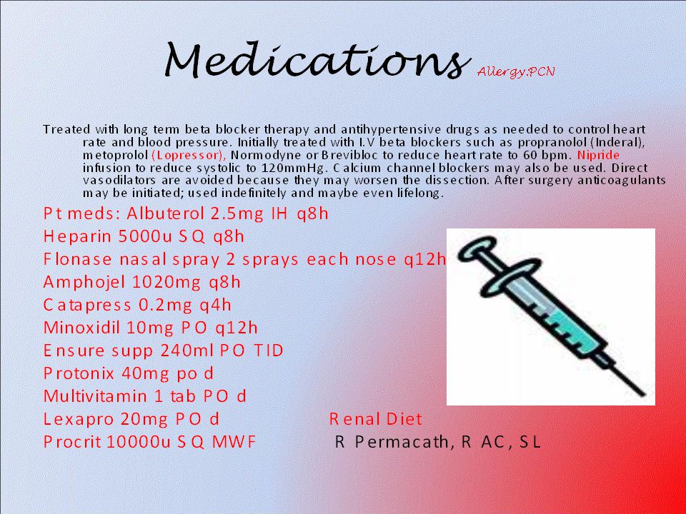
Anti-hypertensives Beta blockers, Vasodilators Calcium channel blockers Nipride Sedatives Niacin, mevocor, statins Post-op anti-coagulants

25
Complication Aortic Dissection
Blood invades or dissects the layers of the vessel wall- most often thoracic Aortic dissection - Wikipedia, the free encyclopedia

26
Dissecting aneurysms are unique and life threatening
Dissecting aneurysms are unique and life threatening. A break or tear in the tunica intima and media allows blood to invade or dissect the layers of the vessel wall. The blood is usually contained by the adventitia, forming a saccular or longitudinal aneurysm.

27
Aortic Dissection Affects men more often than women
Occurs most frequently between fourth and seventh decades of life Acute and life threatening Mortality rate 90% if not medically or surgically treated

28
Aortic Dissection Etiology and Pathophysiology
As heart contracts, each systolic pulsation ↑ pressure on damaged area Further ↑ dissection May occlude major branches of aorta Cutting off blood supply to brain, abdominal organs, kidneys, spinal cord, and extremities People with Marfan’s at risk

30
Aortic Dissection Collaborative Care
Initial goal ↓ BP and myocardial contractility to diminish pulsatile forces within aorta Drug therapy IV β-adrenergic blocker Esmolol (Brevibloc) Other hypertensive agents Calcium channel blockers Sodium Nitroprusside Angiotensin-converting enzyme
 Other hypertensive agents. Calcium channel blockers. Sodium Nitroprusside. Angiotensin-converting enzyme.")
31
Aortic Dissection Surgical therapy When drug therapy is ineffective or
When complications of aortic dissection are present Heart failure, leaking dissection, occlusion of an artery Surgery may be delayed to allow edema to decrease and permit clotting of blood. Even with prompt surgical intervention 30-day mortality of acute aortic dissections remains high (10%-28%)
")
32
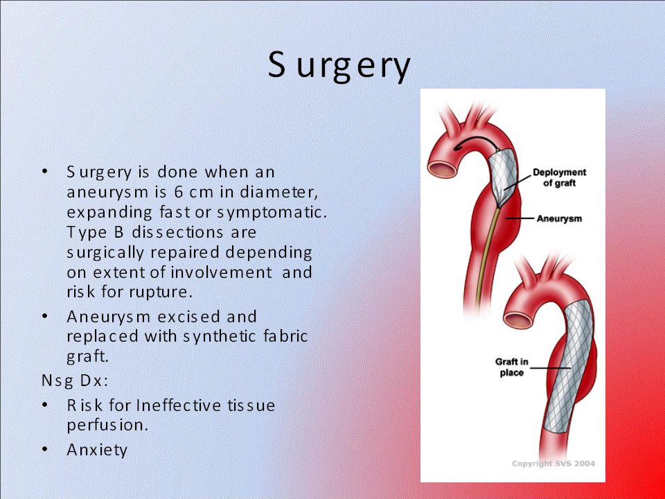
AAA-Medical Treatment - Surgery or Stent
Usually repaired if >5cm Open procedure- abd incision, cross clamp aorta,aneuysm opened and plaque removed, then graft sutured in place. Care much like after CABG (Not done as much anymore unless a rupture) Pre-op assess all peripheral pulses Post-op-check urine output and peripheral pulses hourly for 24 hours- (when to call Dr.) Endovascular stents- placed through femoral artery
 Pre-op assess all peripheral pulses. Post-op-check urine output and peripheral pulses hourly for 24 hours- (when to call Dr.) Endovascular stents- placed through femoral artery.")
33
Stent Graft Repair

34
Aortic Aneurysm Endovascular graft procedure, con’t
Approach is percutaneous femoral access Advantages Shorter operative time Shorter anesthesia time Reduction in use of general anesthesia Reduced groin complications within first 6 months

35
Open Repair of AAA Post-Op- Similar to CABG ICU monitoring
Arterial line Central venous pressure (CVP) or pulmonary artery (PA) catheter Mechanical ventilation Urinary catheter Nasogastric tube ECG Pulse oximetry Pain medication
 or pulmonary artery (PA) catheter. Mechanical ventilation. Urinary catheter. Nasogastric tube. ECG. Pulse oximetry. Pain medication.")
36
Nursing Management Nursing Implementation
Acute Intervention Postop, continued Cardiovascular status Continuous ECG monitoring Electrolyte monitoring Arterial blood gas monitoring Oxygen administration Antidysrhythmic/pain medications

37
Nursing Management Nursing Implementation
Acute Intervention Postop, continued Infection Antibiotic administration Assessment of body temperature Monitoring of WBC Adequate nutrition Observe surgical incision for signs of infection

38
Nursing Management Nursing Implementation
Acute Intervention Postop, continued Gastrointestinal status Nasogastric tube Abdominal assessment Passing of flatus is key sign of returning bowel function Watch for manifestations of bowel ischemia

39
Nursing Management Nursing Implementation
Acute Intervention Postop, continued Neurologic status Level of consciousness Pupil size and response to light Facial symmetry Speech Ability to move upper extremities Quality of hand grasps

40
Nursing Management Nursing Implementation
Acute Intervention Postop, continued Peripheral perfusion status Pulse assessment Mark pulse locations with felt-tip pen Extremity assessment (5P’s) Temperature, color, capillary refill time, sensation and movement of extremities
 Temperature, color, capillary refill time, sensation and movement of extremities.")
41
Nursing Management Nursing Implementation
Acute Intervention Postop, continued Renal perfusion status Urinary output Fluid intake Daily weight CVP/PA pressure Blood urea nitrogen/Creatinine

42
Nursing Management Ambulatory and Home Care
Encourage patient to express concerns Patient instructed to gradually increase activities No heavy lifting Educate on signs and symptoms of complications Infection Neurovascular changes

43
Prevention 1.Ultrasound is extremely effective at detecting AAAs.The U.S. Preventive Services Task Force (USPSTF) recommends that anyone aged 65 to 75 who has ever smoked undergo a one-time ultrasound screening for AAA 2.Prevent atherosclerosis 3.Treat and control hypertension 4.Diet- low cholesterol, low sodium and no stimulants 5.Careful follow-up if less than 5cm. It can grow .5cm /year
 recommends that anyone aged 65 to 75 who has ever smoked undergo a one-time ultrasound screening for AAA. 2.Prevent atherosclerosis. 3.Treat and control hypertension. 4.Diet- low cholesterol, low sodium and no stimulants. 5.Careful follow-up if less than 5cm. It can grow .5cm /year.")
44
Other Complications Rupture- signs of ecchymosis (triad) Thrombi
Back pain Hypotension Pulsating mass Thrombi Renal Failure

45
Rupture Triad Back pain Pulsating hematoma Hypotension

46
Rupture Rupture- serious complication related to untreated aneurysm
Posterior rupture Bleeding may be tamponaded by surrounding structures, thus preventing exsanguination and death Severe pain May/may not have back/flank ecchymosis Anterior rupture Massive hemorrhage Most do not survive long enough to get to the hospital WHY??

47
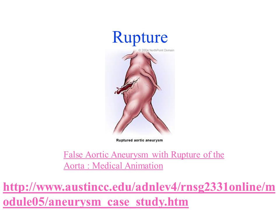
Rupture False Aortic Aneurysm with Rupture of the Aorta : Medical Animation
48
Student Case Study

Similar presentations


 Practice Group Logo here.>")


>")


 LECT7 ALI B ALHAILIY.>")

