Download presentation
Presentation is loading. Please wait.

1
CASE REPORT CASE REPORT 洪嘉蔚 醫師 / 吳維峰 主任 台北市立仁愛醫院 小兒科

2
General Data Name: 李 小弟 Name: 李 小弟 Birth day: 85/04/24 Birth day: 85/04/24 Age: 6 y/o Age: 6 y/o Chart number: 15213493 Chart number: 15213493 Admission day: 91/05/03 Admission day: 91/05/03 Discharge day: 91/05/20 Discharge day: 91/05/20 BW: 22 Kg BW: 22 Kg

3
Chief Complaint Fever off and on for 8 days Fever off and on for 8 days

4
Present Illness A 6 year-old boy suffered from fever off and on for 8 days. He also complained of cough, rhinorrhea and difficult to expectorate sputum. He was taken to LMD twice and our OPD on 91/04/30, but the symptoms persisted in spite of drugs use. So he was taken to our OPD again on 91/05/03. Physical examination revealed decreased breathing sound on right chest. CXR showed lobar pneumonia. A 6 year-old boy suffered from fever off and on for 8 days. He also complained of cough, rhinorrhea and difficult to expectorate sputum. He was taken to LMD twice and our OPD on 91/04/30, but the symptoms persisted in spite of drugs use. So he was taken to our OPD again on 91/05/03. Physical examination revealed decreased breathing sound on right chest. CXR showed lobar pneumonia.

5
Brief history Birth Hx: GA: 39 Wks, BBW:3050 gm, NSD Birth Hx: GA: 39 Wks, BBW:3050 gm, NSD Previous admission: Denied Previous admission: Denied Vaccination: As schedule Vaccination: As schedule Allergy Hx: Denied Allergy Hx: Denied Food exposure: Denied Food exposure: Denied Drug exposure: Denied Drug exposure: Denied Recent travel: Denied Recent travel: Denied Family Hx: Non-contributory Family Hx: Non-contributory

6
Physical Examination: Vital sign: BT 39.9, PR:120 bpm, RR 32/min Vital sign: BT 39.9, PR:120 bpm, RR 32/min General appearance: Acute-ill looking General appearance: Acute-ill looking HEENT: No gross anomaly HEENT: No gross anomaly Conjunctiva: not injected Conjunctiva: not injected Throat: mild injection Throat: mild injection Chest: Symmetric expansion Chest: Symmetric expansion Retraction: no Retraction: no decreased breathing sound: right lung, decreased breathing sound: right lung, fine moist rales(+) fine moist rales(+) percussion: dullness of right chest percussion: dullness of right chest
 fine moist rales(+) percussion: dullness of right chest percussion: dullness of right chest")
7
CBC/DC5/35/7 WBC910010110 Hgb11.910.6 Hct32.929.7 MCV72.675.2 PLT190000206000 Neut92.389.5 Lym3.3%5.5% Mono2.3%1.6% Eos0.3%0.5%

8
Urinalysis 5/35/4 AppearanceY-CLEARY-CLEAR SP. Gr. 1.0101.015 PH6.06.0 Protein1+1+ Glucose-- OB-- Bilirubin-- Nitrate-- RBC0-28-10 WBC4-640-50 Bacteria++

9
Biochemistry GluBUNCrASTALTNaK 5/3946.00.5117391254.5 5/4129 5/7933.00.5851791374.0

11
Blood culture 5/3 NO GROWTH 5/3 NO GROWTH 5/7 NO GROWTH 5/7 NO GROWTH

12
Urine culture 5/5 NO GROWTH 5/5 NO GROWTH

13
Serology CRP 5/4 163 mg/l 5/4 163 mg/l 5/7 126 mg/l 5/7 126 mg/l Mycoplasma Pneumoniae Antibody 5/4 NEGATIVE 5/4 NEGATIVE

14
Hospital Course (I) Initially (5/3), empiric antibiotics with Cefuroxime 500mg IV q6h and Erythromycin 250mg PO q6h were used, but intermittent high fever up to 39C was still noted. Initially (5/3), empiric antibiotics with Cefuroxime 500mg IV q6h and Erythromycin 250mg PO q6h were used, but intermittent high fever up to 39C was still noted. Gentamicin was added on 5/4 due to pyuria of urinalysis and suspected UTI Gentamicin was added on 5/4 due to pyuria of urinalysis and suspected UTI
, empiric antibiotics with Cefuroxime 500mg IV q6h and Erythromycin 250mg PO q6h were used, but intermittent high fever up to 39C was still noted. Gentamicin was added on 5/4 due to pyuria of urinalysis and suspected UTI Gentamicin was added on 5/4 due to pyuria of urinalysis and suspected UTI.")
16
WHAT’S YOUR INITIAL IMPRESSION ?

17
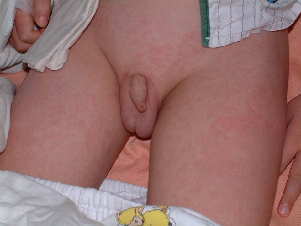
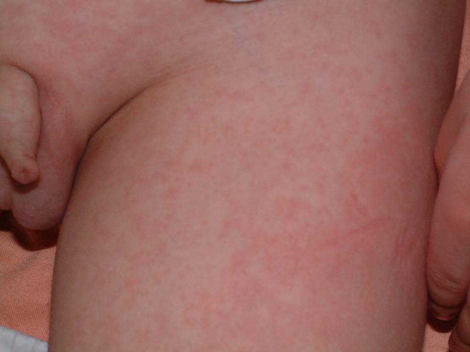
Hospital Course (II) On 5/5, multiple fine, discrete, rubella-like skin rashes developed on the face, trunk and extremities with itchy sensation. Vena infusion and Sinbaby lotion were used for symptom relief. On 5/5, multiple fine, discrete, rubella-like skin rashes developed on the face, trunk and extremities with itchy sensation. Vena infusion and Sinbaby lotion were used for symptom relief.

21
Serology 5/7 IgA 126 (70-400) 5/7 IgA 126 (70-400) IgM 105 (40-230) IgM 105 (40-230) IgG 778 (700-1600) IgG 778 (700-1600) 5/8 Measles IgM (-) 5/8 Measles IgM (-) Rubella IgM (-) Rubella IgM (-)
 5/7 IgA 126 (70-400) IgM 105 (40-230) IgM 105 (40-230) IgG 778 ( ) IgG 778 ( ) 5/8 Measles IgM (-) 5/8 Measles IgM (-) Rubella IgM (-) Rubella IgM (-)")
22
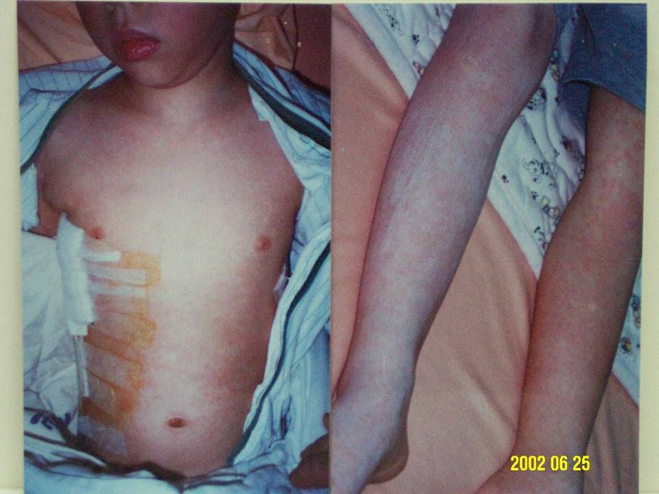
Hospital Course (III) On 5/7, followed CXR showed massive amount of pleural effusion, right lung. So we do chest CT, and erythromycin was changed to 220mg IV q6h On 5/7, followed CXR showed massive amount of pleural effusion, right lung. So we do chest CT, and erythromycin was changed to 220mg IV q6h On 5/8, thoracocentasis was done and showed exudate effusion. So we do chest tube insertion. About 200ml of yellow- reddish fluid was drained. On 5/8, thoracocentasis was done and showed exudate effusion. So we do chest tube insertion. About 200ml of yellow- reddish fluid was drained.

24
Chest CT Date 91/05/07 Date 91/05/07 Impression: Impression: Consolidation of right lower lobe and medial segment of middle lobe, pneumonia is likely. Moderate amount of right pleural effusion and scanty amount of left pleural effusion. Consolidation of right lower lobe and medial segment of middle lobe, pneumonia is likely. Moderate amount of right pleural effusion and scanty amount of left pleural effusion.

25
Abdominal echo Date 91/05/07 Date 91/05/07 Ultrasonic Impression: Ultrasonic Impression: Negative finding of abdominal ultrasonography Negative finding of abdominal ultrasonography

27
Pleural Effusion Study (I) 5/8 Pleural fluid Appearance cloudy Appearance cloudy Color reddish-yellow Color reddish-yellow Bloody (+) Bloody (+) Chylous (-) Chylous (-) Coagulation (+) Coagulation (+) Sp. Gr. 1.025 Sp. Gr. 1.025

28
Pleural Effusion Study (II) WBC 630 cumm WBC 630 cumm Polynuclear cells 55.0% Polynuclear cells 55.0% Mononuclear cells 45.0% Mononuclear cells 45.0% Abnormal cells (-) Abnormal cells (-) Pleural, Acid-Fast Stain: Not Found Pleural, Acid-Fast Stain: Not Found Pleural, Gram ’ s Stain: Not Found Pleural, Gram ’ s Stain: Not Found
 WBC 630 cumm WBC 630 cumm Polynuclear cells 55.0% Polynuclear cells 55.0% Mononuclear cells 45.0% Mononuclear cells 45.0% Abnormal cells (-) Abnormal cells (-) Pleural, Acid-Fast Stain: Not Found Pleural, Acid-Fast Stain: Not Found Pleural, Gram ’ s Stain: Not Found Pleural, Gram ’ s Stain: Not Found")
29
Pleural Effusion Study (III) Pleural Effusion Pleural Effusion Glucose 71 mg/dl Glucose 71 mg/dl LDH 3149 IU/L (H) LDH 3149 IU/L (H) Protein 3.30 g/dl (L) Protein 3.30 g/dl (L)
 Pleural Effusion Pleural Effusion Glucose 71 mg/dl Glucose 71 mg/dl LDH 3149 IU/L (H) LDH 3149 IU/L (H) Protein 3.30 g/dl (L) Protein 3.30 g/dl (L)")
30
Pleural Effusion Study (IV) Pleural effusion culture Pleural effusion culture on 5/8 no growth on 5/8 no growth on 5/13 no growth on 5/13 no growth
 Pleural effusion culture Pleural effusion culture on 5/8 no growth on 5/8 no growth on 5/13 no growth on 5/13 no growth")
31
Pleural Effusion Study (V) 5/8 Pleural effusion cytology: 5/8 Pleural effusion cytology: No evidence of malignancy No evidence of malignancy 5/14 Pleural PCR assay for mycobacteria 5/14 Pleural PCR assay for mycobacteria result: Negative result: Negative
 5/8 Pleural effusion cytology: 5/8 Pleural effusion cytology: No evidence of malignancy No evidence of malignancy 5/14 Pleural PCR assay for mycobacteria 5/14 Pleural PCR assay for mycobacteria result: Negative result: Negative")
32
Hospital Course (IV) On 5/10, followed CBC/DC showed leukocytosis with left shift (WBC 19570, Neu 92.9%). Persistent high fever was noted. So Cefuroxime was changed to Ceftriaxone 1g IV q12h On 5/10, followed CBC/DC showed leukocytosis with left shift (WBC 19570, Neu 92.9%). Persistent high fever was noted. So Cefuroxime was changed to Ceftriaxone 1g IV q12h High fever up to 40C persisted in spite of Ceftriaxone + Gentamicin + Erythromycin combined use High fever up to 40C persisted in spite of Ceftriaxone + Gentamicin + Erythromycin combined use
. Persistent high fever was noted. So Cefuroxime was changed to Ceftriaxone 1g IV q12h High fever up to 40C persisted in spite of Ceftriaxone + Gentamicin + Erythromycin combined use High fever up to 40C persisted in spite of Ceftriaxone + Gentamicin + Erythromycin combined use.")
33
CBC/DC5/35/75/105/135/15 WBC910010110195702645012270 Hgb11.910.68.88.48.9 Hct32.929.725.224.225.1 MCV72.675.285.776.274.1 PLT190000206000378000551000707000 Neut92.389.592.992.887.1 Lym3.3%5.5%4.2%3.5%6.9% Mono2.3%1.6%2.2%1.9%2.6% Eos0.3%0.5%0.4%0.8%1.8%

34
Urinalysis 5/35/45/115/14 AppearanceY-CLEARY-CLEARY-CLEARY-CLEAR SP. Gr. 1.0101.0151.0101.020 PH6.06.05.06.5 Protein1+1+-- Glucose---- OB---- Bilirubin---- Nitrate---- RBC0-28-100-20-1 WBC4-640-500-28-10 Bacteria++----

35
Biochemistry GluBUNCrASTALTNaKAlb 5/3946.00.5117391254.5 5/4129 5/7933.00.5851791374.0 5/95.00.446991315.2 5/111363.0 5/1411.20.485175 5/16711463.5

36
Blood culture 5/3 NO GROWTH 5/3 NO GROWTH 5/7 NO GROWTH 5/7 NO GROWTH 5/11 NO GROWTH 5/11 NO GROWTH

37
Urine culture 5/5 NO GROWTH 5/5 NO GROWTH 5/10 NO GROWTH 5/10 NO GROWTH 5/12 NO GROWTH 5/12 NO GROWTH

38
Serology (I) CRP 5/4 163 mg/l 5/4 163 mg/l 5/7 126 mg/l 5/7 126 mg/l 5/14 113 mg/l 5/14 113 mg/l
 CRP 5/4 163 mg/l 5/4 163 mg/l 5/7 126 mg/l 5/7 126 mg/l 5/ mg/l 5/ mg/l")
39
Serology (II) 5/13 Direct Coombs ’ test: positive Indirect Coombs ’ test: positive Indirect Coombs ’ test: positive 5/14 RA< 10.2 IU/ML (<40.0) C3 166.0 mg/dl (90.0-180.0) C3 166.0 mg/dl (90.0-180.0) C4 21.4 mg/dl (10.0- 40.0) C4 21.4 mg/dl (10.0- 40.0)
 5/13 Direct Coombs ’ test: positive Indirect Coombs ’ test: positive Indirect Coombs ’ test: positive 5/14 RA< 10.2 IU/ML (<40.0) C mg/dl ( ) C mg/dl ( ) C mg/dl ( ) C mg/dl ( )")
40
Serology (III) 5/14 Heterophil Ab: Negative 5/14 Heterophil Ab: Negative ANA Negative ANA Negative 5/14 Legionella Ab: Negative 5/14 Legionella Ab: Negative Chlamydia Ab: Negative Chlamydia Ab: Negative
 5/14 Heterophil Ab: Negative 5/14 Heterophil Ab: Negative ANA Negative ANA Negative 5/14 Legionella Ab: Negative 5/14 Legionella Ab: Negative Chlamydia Ab: Negative Chlamydia Ab: Negative")
41
Ga-67 Inflammation Survey Date 91/05/15 Date 91/05/15 A patch of abnormal tracer uptake at the right lower lung field, may be inflammatory focus. A patch of abnormal tracer uptake at the right lower lung field, may be inflammatory focus. Diffusely increase uptake of liver. This phenomenon can be found in iron deficiency anemia Diffusely increase uptake of liver. This phenomenon can be found in iron deficiency anemia

45
WHAT’S YOUR DIAGNOSIS ?

46
Serology (I) Mycoplasma Pneumoniae Antibody 5/4 Negative 5/4 Negative 5/7 160X 5/7 160X 5/14 320X 5/14 320X
 Mycoplasma Pneumoniae Antibody 5/4 Negative 5/4 Negative 5/7 160X 5/7 160X 5/14 320X 5/14 320X")
47
Pleural Effusion Study (II) 5/8 Pleural fluid for Mycoplasmal 5/8 Pleural fluid for Mycoplasmal pneumonia antibody: 80X pneumonia antibody: 80X
 5/8 Pleural fluid for Mycoplasmal 5/8 Pleural fluid for Mycoplasmal pneumonia antibody: 80X pneumonia antibody: 80X")
48
Serology (III) 5/16 Cold hemaglutination: 512 X (<32X) 5/16 Cold hemaglutination: 512 X (<32X)
 5/16 Cold hemaglutination: 512 X (<32X) 5/16 Cold hemaglutination: 512 X (<32X)")
49
Hospital Course (V) Chest tube was removed on 5/13 Chest tube was removed on 5/13 We used prednisolone (2mg/kg/day in 4 divided doses) on 5/14. Fever subsided on the night of 5/14. We used prednisolone (2mg/kg/day in 4 divided doses) on 5/14. Fever subsided on the night of 5/14. Steroid was tapered gradually Steroid was tapered gradually On 5/20, patient was discharged under stable condition. On 5/20, patient was discharged under stable condition.
 on 5/14. Fever subsided on the night of 5/14. Steroid was tapered gradually Steroid was tapered gradually On 5/20, patient was discharged under stable condition. On 5/20, patient was discharged under stable condition..")
52
CBC/DC5/35/75/105/135/155/23 WBC9100101101957026450122709780 Hgb11.910.68.88.48.910.1 Hct32.929.725.224.225.130.2 MCV72.675.285.776.274.178.9 PLT190000206000378000551000707000377000 Neut92.389.592.992.887.166.1 Lym3.3%5.5%4.2%3.5%6.9%22.3 Mono2.3%1.6%2.2%1.9%2.6%10.0 Eos0.3%0.5%0.4%0.8%1.8%1.0

53
Biochemistry GluBUNCrASTALTNaKAlb 5/3946.00.5117391254.5 5/4129 5/7933.00.5851791374.0 5/95.00.446991315.2 5/111363.0 5/1411.20.485175 5/16711463.5 5/232130

54
Serology (I) CRP 5/4 163 mg/l 5/4 163 mg/l 5/7 126 mg/l 5/7 126 mg/l 5/14 113 mg/l 5/14 113 mg/l 5/30 5.2 mg/l 5/30 5.2 mg/l
 CRP 5/4 163 mg/l 5/4 163 mg/l 5/7 126 mg/l 5/7 126 mg/l 5/ mg/l 5/ mg/l 5/ mg/l 5/ mg/l")
55
Final Diagnosis Mycoplasmal lobar pneumonia, complicated with prolonged fever, skin rashes, right lung pleural effusion, and hemolytic anemia Mycoplasmal lobar pneumonia, complicated with prolonged fever, skin rashes, right lung pleural effusion, and hemolytic anemia

56
DISCUSSION

57
Mycoplasma Pneumoniae In 1944, M. pneumoniae was reported by Monroe Eaton, originally called the Eaton agent. In 1944, M. pneumoniae was reported by Monroe Eaton, originally called the Eaton agent. Smallest free-living microorganism, belongs to the class Mollicutes. Smallest free-living microorganism, belongs to the class Mollicutes. Mycoplasmas lack a cell wall, so tend to be pleomorphic. Mycoplasmas lack a cell wall, so tend to be pleomorphic.

58
Clinical Manifestations M. pneumoniae causes approximately 20% of all cases of pneumonia. M. pneumoniae causes approximately 20% of all cases of pneumonia. Peak incidence at 6-21 years of age. Peak incidence at 6-21 years of age. Incubation period of 2-3 weeks. Incubation period of 2-3 weeks. Transmission by inhalation of infected droplet aerosols. Transmission by inhalation of infected droplet aerosols.

60
Pneumonia is the most important clinical manifestation of M. pneumoniae infection. Pneumonia is the most important clinical manifestation of M. pneumoniae infection. * Bronchopneumonia pattern mostly. * Bronchopneumonia pattern mostly. Lobar pneumonia and large amount Lobar pneumonia and large amount pleural fluid are unusual. pleural fluid are unusual. Pediat Radiol 1989;19(8):499-503 Pediat Radiol 1989;19(8):499-503 * Respiratory disease other than * Respiratory disease other than pneumonia: unspecific URI, pharyngitis, pneumonia: unspecific URI, pharyngitis, AOM, croup, sinusitis, bronchitis, AOM, croup, sinusitis, bronchitis, bronchiolitis, asthma. bronchiolitis, asthma.
: Pediat Radiol 1989;19(8): * Respiratory disease other than * Respiratory disease other than pneumonia: unspecific URI, pharyngitis, pneumonia: unspecific URI, pharyngitis, AOM, croup, sinusitis, bronchitis, AOM, croup, sinusitis, bronchitis, bronchiolitis, asthma. bronchiolitis, asthma..")
61
Cutaneous manifestations : common. Cutaneous manifestations : common. * Exanthem and enanthem of Mycoplasma * Exanthem and enanthem of Mycoplasma pneumoniae infection are observed in 5 to pneumoniae infection are observed in 5 to 24% of cases 24% of cases AAP, Report of Committee on Infectious Diseases, 1994:333-5 AAP, Report of Committee on Infectious Diseases, 1994:333-5 * Most common with an erythematous * Most common with an erythematous maculopapular rash on the trunk and back; maculopapular rash on the trunk and back; discrete (rubelliform) or confluent discrete (rubelliform) or confluent (morbilliform). (morbilliform). * Most serious presentation: Erythema * Most serious presentation: Erythema multiforme and Stevens-Johnson syndrome. multiforme and Stevens-Johnson syndrome. Clini Pediatrics 1991:30(1),42-9 Clini Pediatrics 1991:30(1),42-9
 or confluent discrete (rubelliform) or confluent (morbilliform). (morbilliform). * Most serious presentation: Erythema * Most serious presentation: Erythema multiforme and Stevens-Johnson syndrome. multiforme and Stevens-Johnson syndrome. Clini Pediatrics 1991:30(1),42-9 Clini Pediatrics 1991:30(1),42-9.")
62
Hematologic manifestations: Hematologic manifestations: * Hemolytic anemia: usually mild, * Hemolytic anemia: usually mild, however, it may become severe and however, it may become severe and result in 50% reduction in hemoglobin result in 50% reduction in hemoglobin concentration. concentration. Pediat Infec Dis J 1998;17(2):173-7 Pediat Infec Dis J 1998;17(2):173-7 * Direct Coombs test usually positive. * Direct Coombs test usually positive. * Steroid administration may be * Steroid administration may be beneficial. beneficial. South Med J 1990;83(9):1106-8 South Med J 1990;83(9):1106-8
:173-7 Pediat Infec Dis J 1998;17(2):173-7 * Direct Coombs test usually positive. * Direct Coombs test usually positive. * Steroid administration may be * Steroid administration may be beneficial. beneficial. South Med J 1990;83(9): South Med J 1990;83(9):")
63
Hemolytic anemia is presumably related to the presence of cold agglutinins in serum which at high concentration, may agglutinate erythrocytes at 37 ℃ Hemolytic anemia is presumably related to the presence of cold agglutinins in serum which at high concentration, may agglutinate erythrocytes at 37 ℃ Rev Pneumol Clin 1990,46(2),83-4 Rev Pneumol Clin 1990,46(2),83-4
,83-4 Rev Pneumol Clin 1990,46(2),83-4")
64
Gastrointestinal findings are nonspecific with nausea, vomiting, abdominal pain, and/or diarrhea. Gastrointestinal findings are nonspecific with nausea, vomiting, abdominal pain, and/or diarrhea. Neurologic disease association was reported 2.6-4.8%. Neurologic disease association was reported 2.6-4.8%. * Encephalitis, meningitis, transverse * Encephalitis, meningitis, transverse myelitis, psychosis, Bell palsy and myelitis, psychosis, Bell palsy and Guillain-Barre` syndrome. Guillain-Barre` syndrome. Arthritis in association with M.pneumoniae Arthritis in association with M.pneumoniae infection have not been established. infection have not been established.

65
Hepatitis was once thought to be unusual, but recent studies suggest that liver dysfunction may be present in up to 30% of M. pneumoniae infection. Hepatitis was once thought to be unusual, but recent studies suggest that liver dysfunction may be present in up to 30% of M. pneumoniae infection. Pediatr Pulmonol 1990;8:182-7

66
Liver dysfunction was observed more frequently in patients with pleuropneumonia than in simple pneumonia cases. Liver dysfunction was observed more frequently in patients with pleuropneumonia than in simple pneumonia cases. Pediatr Pulmonol 1990;8:182-7

67
Radiographic Manifestation (I) Interstitial infiltration was more commonly seen in pediatric than adult patients (46% vs 20%) Interstitial infiltration was more commonly seen in pediatric than adult patients (46% vs 20%) Unilateral lesions 80% Unilateral lesions 80% Single lobe lesions 77% Single lobe lesions 77% Lower lobe predominant 69% Lower lobe predominant 69% Pleural effusion 7% Pleural effusion 7% 高雄醫學科學雜誌 1993;9(4):204-11
 Interstitial infiltration was more commonly seen in pediatric than adult patients (46% vs 20%) Interstitial infiltration was more commonly seen in pediatric than adult patients (46% vs 20%) Unilateral lesions 80% Unilateral lesions 80% Single lobe lesions 77% Single lobe lesions 77% Lower lobe predominant 69% Lower lobe predominant 69% Pleural effusion 7% Pleural effusion 7% 高雄醫學科學雜誌 1993;9(4):204-11")
68
Radiographic Manifestation (II) Unilateral infiltration 84% Unilateral infiltration 84% Lower lobe predominance 60% Lower lobe predominance 60% Confluent consolidation 56% Confluent consolidation 56% Patchy consolidation 33% Patchy consolidation 33% Pleural effusion 24% Pleural effusion 24% 長庚醫學雜誌 1991;14(3):156-62
 Unilateral infiltration 84% Unilateral infiltration 84% Lower lobe predominance 60% Lower lobe predominance 60% Confluent consolidation 56% Confluent consolidation 56% Patchy consolidation 33% Patchy consolidation 33% Pleural effusion 24% Pleural effusion 24% 長庚醫學雜誌 1991;14(3):156-62")
69
Diagnosis (I) WBC, CRP, ESR are non-specific, may be normal or elevated. WBC, CRP, ESR are non-specific, may be normal or elevated. Growth of the organism takes weeks, generally only in expertise laboratories. Growth of the organism takes weeks, generally only in expertise laboratories. PCR is sensitive and specific. PCR is sensitive and specific. Serologic testing : Cold agglutinins, titer of >1:64 is suggestive of infection; Anti- mycoplasmal Ab detection, fourfold or greater rise are considered diagnostic. Serologic testing : Cold agglutinins, titer of >1:64 is suggestive of infection; Anti- mycoplasmal Ab detection, fourfold or greater rise are considered diagnostic.

70
Diagnosis (II) Imaging : Interstitial infiltrate or bronchopneumonia pattern. Lobar consolidation and pleural effusion are uncommon but may occur. Imaging : Interstitial infiltrate or bronchopneumonia pattern. Lobar consolidation and pleural effusion are uncommon but may occur.

71
Treatment Erythromycin is the drug of choice. (40-50mg/kg/24hr q6h for 10-14 days). Newer macrolides: Azithromycin (10mg/kg on day 1, and 5mg/kg/24hr on days 2-5) or Clarithromycin (15mg/kg/24hr given in two divided doses for 10 days).
 or Clarithromycin (15mg/kg/24hr given in two divided doses for 10 days)..")
72
Empiric Therapy for Lobar Pneumonia Clinically moderate to severely toxic, treat empirically for S. pneumonia, S. pyogens ( and H. influenzae type b in unimmunized children) Clinically moderate to severely toxic, treat empirically for S. pneumonia, S. pyogens ( and H. influenzae type b in unimmunized children) In toxic children, tests for Mycoplasma should be considered because focal pneumonia is a rare presentation In toxic children, tests for Mycoplasma should be considered because focal pneumonia is a rare presentation
 Clinically moderate to severely toxic, treat empirically for S. pneumonia, S. pyogens ( and H. influenzae type b in unimmunized children) In toxic children, tests for Mycoplasma should be considered because focal pneumonia is a rare presentation In toxic children, tests for Mycoplasma should be considered because focal pneumonia is a rare presentation.")
73
Cefuroxime intravenously, ceftriaxone or cefotaxime intravenously Cefuroxime intravenously, ceftriaxone or cefotaxime intravenously For anti-staphylococcal coverage, add to the above, either: nafcillin, oxacillin, or clindamycin For anti-staphylococcal coverage, add to the above, either: nafcillin, oxacillin, or clindamycin For Mycoplasma: intravenous erythromycin or azithromycin; or oral erythromycin, azithromycin, or clarithromycin. For Mycoplasma: intravenous erythromycin or azithromycin; or oral erythromycin, azithromycin, or clarithromycin.

74
Pneumonia, with pleural fluid or empyema Treat empirically for S. pneumonia, S. pyogenes, and S. aureus ( and H. influenzae type b in unimmunized children) Treat empirically for S. pneumonia, S. pyogenes, and S. aureus ( and H. influenzae type b in unimmunized children) Consider aspiration pneumonia with anaerobic oral flora as pathogens; needle or catheter aspiration of pleural fluid, with drainage, is often required for clinical cure. Consider aspiration pneumonia with anaerobic oral flora as pathogens; needle or catheter aspiration of pleural fluid, with drainage, is often required for clinical cure.
 Treat empirically for S. pneumonia, S. pyogenes, and S. aureus ( and H. influenzae type b in unimmunized children) Consider aspiration pneumonia with anaerobic oral flora as pathogens; needle or catheter aspiration of pleural fluid, with drainage, is often required for clinical cure. Consider aspiration pneumonia with anaerobic oral flora as pathogens; needle or catheter aspiration of pleural fluid, with drainage, is often required for clinical cure..")
75
Ceftriaxone or cefotaxime Ceftriaxone or cefotaxime For antistaphylococcal covarage, add to the above either: nafcillin, oxacillin, or clindamycin (also covers anaerobes found in aspiration pneumonia as well as most pneumoncocci) For antistaphylococcal covarage, add to the above either: nafcillin, oxacillin, or clindamycin (also covers anaerobes found in aspiration pneumonia as well as most pneumoncocci) Single agent therapy with meropenem, or ticarcillin/clavulanate (Timentin) both of which cover both aerobic and anaerobic pathogens Single agent therapy with meropenem, or ticarcillin/clavulanate (Timentin) both of which cover both aerobic and anaerobic pathogens
 For antistaphylococcal covarage, add to the above either: nafcillin, oxacillin, or clindamycin (also covers anaerobes found in aspiration pneumonia as well as most pneumoncocci) Single agent therapy with meropenem, or ticarcillin/clavulanate (Timentin) both of which cover both aerobic and anaerobic pathogens Single agent therapy with meropenem, or ticarcillin/clavulanate (Timentin) both of which cover both aerobic and anaerobic pathogens")
76
CONCLUSION

77
Presence of pleuropneumonia appears to be associated with more severe and prolonged course of illness Presence of pleuropneumonia appears to be associated with more severe and prolonged course of illness Pediatr Pulmonol 1990;8:182-7

78
Even in patients with clinically mild pneumonia, Mycoplasma pneumoniae may be the cause of severe anemia Even in patients with clinically mild pneumonia, Mycoplasma pneumoniae may be the cause of severe anemia Ann of Hematol 2001;80(3):180-2 Ann of Hematol 2001;80(3):180-2
:180-2 Ann of Hematol 2001;80(3):180-2")
79
Association of exanthem and pneumonia or of hemolytic anemia and pneumonia are considered to be strongly suggestive for the diagnosis of M. pneumonia infection Association of exanthem and pneumonia or of hemolytic anemia and pneumonia are considered to be strongly suggestive for the diagnosis of M. pneumonia infection Clin Infec Dis 1993;17(Suppl 1):S47-51
:S")
80
THANKS FOR YOUR ATTENTION ! THE END THE END

Similar presentations


 Admitted D6 with 1 week H/O: SOBE, Cough, minimal sputum SOBE, Cough, minimal sputum ? Fever &>")


. -Cytological tests (>")
