Download presentation
Presentation is loading. Please wait.

1
Hip and Thigh Pain Arthur Jason De Luigi, DO
Program Director, Sports Medicine Fellowship Director, Sports Medicine Director, Interventional Pain MedStar National Rehabilitation Hospital MedStar Georgetown University Hospital Medical Director and Head Team Physician US Paralympic Alpine Ski Team

2
Disclosures Nothing to Disclose

3
Overview Epidemiology Hip and Thigh Anatomy Physical Examination
Diagnostic Imaging Pathology Treatment

4
Incidence Hip and Thigh pain are very commonly the chief complaint of office visits Account for 0.61% of all visits About 1 in every 164 encounters Runners report an average yearly hip or pelvic injury rate of 2% to 11%.

5
Incidence NHANES III 14.3% of patients aged 60 years and older reported significant hip pain on most days over the previous 6 weeks. 18.4% of those who had not participated in leisure time physical activity during the previous month reported severe hip pain Opposed to 12.6% of those who did engage in physical activity

6
Common Hip Problems by Age
Newborn Congenital dislocation of hip Age 2-8 AVN of hip (Legg-Calve-Perthes), synovitis Age10-14 Slipped Cap Fem Epiphysis Age 14-25 Stress Fracture Age 20-40 Labral Tear Age >40 Osteoarthritis
, synovitis. Age Slipped Cap Fem Epiphysis. Age Stress Fracture. Age Labral Tear. Age >40. Osteoarthritis.")
7
Anatomy Bones Pelvis Ilium Ischium Pubis Sacrum Femur

8
Anatomy Ilium - anterior superior iliac spine, anterior inferior iliac spine, Iliac crest, Gluteal Line, Posterior superior iliac spine Ischium - Ramus of the ischium, Ischial spine, Ischial tuberosity, body weight rest in the sitting position Pubis - superior and inferior rami, Body of pubis, Pubic crest All three forms acetabulum –articulates with head of femur to form hip joint Three joints – SI, Pubic symphysis, Acetabulum 8

9
Anatomy

10
Anatomy

11
Anatomy

12
Anatomy Anterior Medial Posterolateral Iliopsoas Quadriceps Sartorius
Vastus Medialis Vastus Intermedius Vastus Lateralis Rectus Femoris Sartorius Medial Adductor Magnus Adductor Longus Adductor Brevis Gracilis Posterolateral Piriformis Gluteus Maximus Gluteus Medius Gluteus Minimus Tensor Fascia Lata Iliotibial Band

13
Physical Examination

14
Anterior Hip Pain Examination Inspection Palpation ROM Special Tests
Walking/Gait Pelvic position/splinting Atrophy/ecchymosis/bony deformity Palpation ROM Flexion/extension/internal/external rotation Strength Special Tests FABER FADIR Thomas test Snapping Hip Test Hernia exam

15
Range of Motion Flexion: 110 to 120 degrees
Extension: 10 to 15 degrees

16
Range of Motion Abduction: 30 to 50 degrees Adduction: 30 degrees

17
Range of Motion External rotation: 40 to 60 degrees
Internal rotation: 30 to 40 degrees

18
Special Tests modified Thomas Test hip flexor and quad flexibility

19
Special Tests Patrick’s Test (FABER) hip joint SI joint
 hip joint SI joint")
20
Special Tests Labral Injury FADIR:
Flexion, Adduction, Internal Rotation Axial Loading pain +/- click

21
Lateral Hip Pain Examination Special Tests Ober Test
Trendelenberg Test

22
Special Tests Ober Test iliotibial band flexibility

23
Posterior Hip Pain Examination ROM Leg Length Neurologic Special Tests
Reflex Strength Sensory Special Tests Piriformis FABER (Patrick) Gaenslen’s Gillet Fortin Facet Loading Straight Leg Raise Reverse SLR
 Gaenslen’s. Gillet. Fortin. Facet Loading. Straight Leg Raise. Reverse SLR.")
24
Range of Motion Lumbar Range of Motion Flexion – 80o Extension – 35o
Lat Bend – 40o Rotation – 3-18o

25
Special Tests Leg length
true leg length discrepancy congenital maldevelopment trauma functional leg length discrepancy scoliosis

26
Special Tests Leg length Measured from ASIS to medial malleolus
Functionally measured knees & hips flexed with thumbs on medial malleolus then knees and hips extended

27
Neurologic Examination

28
Special Tests Patrick’s Test (FABER) hip joint SI joint
 hip joint SI joint")
29
Special Tests Gaenslen’s Sign Pain at ipsilateral SIJ is positive test

30
Special Tests Piriformis Test Piriformis flexibility or pain
Sciatic Nerve Distribution

31
Special Tests Popliteal Angle Hamstring flexibility

32
Diagnostic Imaging

33
Diagnostic Imaging Radiographs Bone scan: stress fxs
Anterior-Posterior view Frog leg view STANDING films to r/o early OA Bone scan: stress fxs CT: subtle fractures MRI: soft tissue, stress fx Arthrogram: labral tears

34
Anterior Hip Pain Differential Dx Osteoarthritis
Inflammatory arthritis Muscle and tendon strains Tendonitis Femoral neck stress fracture Sports hernia (Occult hernia or tear of oblique aponeurosis) Obturator or ilioinguinal nerve entrapment Osteitis pubis Acetabular labral tears
 Obturator or ilioinguinal nerve entrapment. Osteitis pubis. Acetabular labral tears.")
35
Hip Ultrasound Indications for a hip examination
Include, but are not limited to: soft tissue injury tendon pathology arthritis soft tissue masses or swelling nerve entrapment effusion bone injury

36
Hip Ultrasound Specifications of a hip examination
Patient’s body habitus lower frequency transducer may be required Spatial resolution decreases with a decrease in the transducer frequency operator should use the highest possible frequency that provides adequate penetration

37
Hip Ultrasound Anterior approach Patient positioning: Planes:
supine with the hip in mild external rotation Planes: Sagittal oblique plane parallel to the long axis of the femoral neck femoral head, neck, and joint effusion Sagittal and axial planes labrum, iliopsoas tendon and bursa, femoral vessels, sartorius and rectus femoris muscles The above structures are then scanned in the axial plane, perpendicular to the original scan plane

38
—Sonograpthy of normal hip joint
Longitudinal —Hip joint aspiration using sonographic guidance. Sonography of normal hip joint. Longitudinal sonogram of normal hip joint longitudinal to femoral neck. Acetabulum is to the left, and arrowheads mark femoral head and shaft. No fluid is seen distending joint capsule. Fessell D P et al. AJR 2000;174: ©2000 by American Roentgen Ray Society

39
Hip Ultrasound Anterior Approach
Dynamic evaluation of snapping hip syndrome Anterior: iliopsoas tendon as it passes over superior pubic bone Lateral: iliotibial band crosses the greater trochanter

40
Hip Ultrasound Lateral approach Patient positioning: Planes:
lateral decubitus Planes: axial and coronal (longitudinal) greater trochanter, greater trochanteric bursa, gluteus muscles, and tensor fascia lata dynamic evaluation of iliotibial band syndrome
 greater trochanter, greater trochanteric bursa, gluteus muscles, and tensor fascia lata. dynamic evaluation of iliotibial band syndrome")
41
Hip Ultrasound Medial approach Patient positioning: Planes:
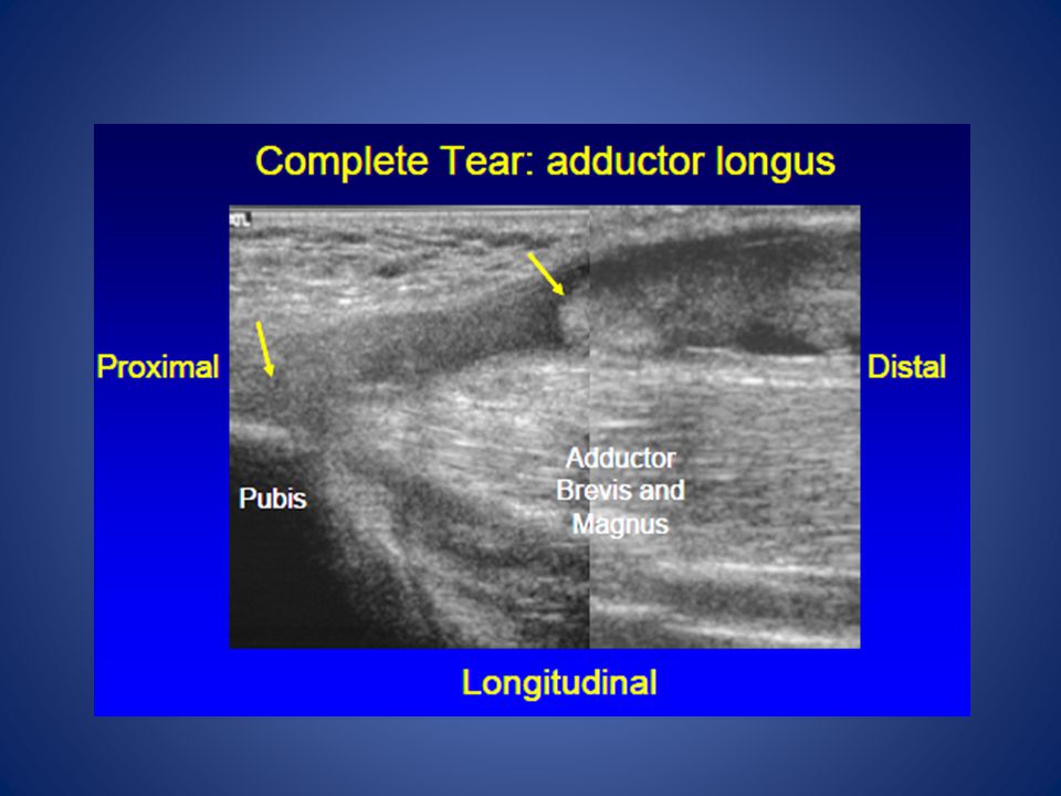
45-degree knee flexion, external rotation (frog-leg position) Planes: Sagittal oblique and axial planes (adductor muscles, pubic bone and insertion of rectus abdominis)
 Planes: Sagittal oblique and axial planes. (adductor muscles, pubic bone and insertion of rectus abdominis)")
42
Hip Pathology Snapping Hip Trochanter Athletic Pubalgia
Iliopsoas Iliotibial Band Trochanter Trochanteric Bursitis Gluteal Tendons Athletic Pubalgia Sports Hernia Direct Hernia Indirect Hernia Hip Osteoarthritis Iliopsoas Bursitis Iliopectineal Bursitis Femoroacetabular Impingement Acetabular Labral Tear Adductor Strain Tear Quadriceps Hamstrings Ischial Bursitis

43
Anterior Hip Pain Differential Dx Osteoarthritis
Inflammatory arthritis Muscle and tendon strains Tendonitis Femoral neck stress fracture Sports hernia (Occult hernia or tear of oblique aponeurosis) Obturator or ilioinguinal nerve entrapment Osteitis pubis Acetabular labral tears
 Obturator or ilioinguinal nerve entrapment. Osteitis pubis. Acetabular labral tears.")
44
Margo K, et al. Evaluation and management of hip pain: An algorithmic approach J Fam Pract. 2003, 52:8

45
Hip Pathology Snapping Hip Trochanter Femoroacetabular Impingement
Iliopsoas Iliotibial Band Trochanter Trochanteric Bursitis Gluteal Tendons Femoroacetabular Impingement Acetabular Labral Tear

46
Hip Pathology AIIS Avulsion Quadriceps Adductor Athletic Pubalgia
Strain Tear Adductor Athletic Pubalgia Sports Hernia Direct Hernia Indirect Hernia

47
Hip Anterior Recess Anterior and posterior layers
Fibrous tissue + minute layer of synovium Hyperechoic Each 2-4 mm thick Radiology 1999; 210: 499

48
Hip Effusion Separation of anterior and posterior layers (1)
Capsule distention at femoral neck > 7 mm or difference of 1 mm from opposite side (2) Extension & abduction improves visualization (3) Avoid Internal Rotation of hip during assessment: capsule thickens (1) Radiology 1999; 210: 499 (2) Scand J Rheum 1989; 18:113 (3) Acta Rad 1997; 38: 867
 Extension & abduction improves visualization (3) Avoid Internal Rotation of hip during assessment: capsule thickens. (1) Radiology 1999; 210: 499. (2) Scand J Rheum 1989; 18:113. (3) Acta Rad 1997; 38: 867.")
49
-Hip joint longitudinal -Hypoechoic hip effusion
—Hip joint aspiration using sonographic guidance. 42-year-old male IV drug abuser in whom septic hip joint was clinically suspected. Longitudinal sonogram obtained as illustrated in A shows hypoechoic hip effusion between arrowheads marking femoral cortex and corresponding arrows marking joint capsule. Culture of aspirate grew Staphylococcus aureus. Fessell D P et al. AJR 2000;174: ©2000 by American Roentgen Ray Society

50
Hip Joint

51
Hip Joint: Effusion Cannot predict infection by ultrasound
Negative power color Doppler does not exclude infection* Guided aspiration AJR 1998; 206: 731

52
Transient Hip Synovitis
Anterior Longitudinal

54
Acute Transient (“Toxic”) Synovitis
inflammatory process of hip w/ chronic irritation and excess secretion of synovial fluid within the capsule; ? cause Most common dx in limping child <10, but it’s a Dx of exclusion; r/o septic arthritis, SCFE, stress fx, etc. Xrays normal; MRI helpful ruling out other causes Labs: normal CBC, CRP S/Sx: pain w/ walking, low-grade fever Tx: relative rest, analgesics

57
Iliopsoas Bursitis Located anterior to the hip
Can be visualized in transverse plane at the level of femoral head Immediately medial to iliopsoas tendon Communicates with hip joint in up to 15% Number is increased with abnormal hip joint Bursa may be distended with simple fluid, complex fluid or synovitis Ranges from anechoic to hyperechoic May distend into abdomen Should not be confused with intra-abdominal or psoas abscess Bursitis: presence of pain with transducer pressure Increased flow on color/power Doppler Distention out of proportion to hip joint recess

58
Iliopsoas bursitis Cause: overuse of hip flexors S/Sx:
anterior hip pain, +/- snap preferred position of hip in flex/ER, TTP to deep palpation anteriorly, pain with passive hip extension Tx: relative rest, ice, brief NSAID, stretching of iliopsoas, +/- steroid injection (preferably w/ guidance)
")
60
Iliopsoas Bursitis

61
Iliopsoas Tendon

62
Osteitis Pubis Repetitive trauma to pubic symphysis due to overuse
Running/cutting, esp soccer, football, basketball Signs & Symptoms insidious onset dull anterior groin pain; may radiate TTP over Pubic Symphysis +/- pain w/ resisted Adduction or passive Abduction Treatment relative rest, brief NSAID, cross-training stretching/strength rehab consider steroid injection

63
Osteitis Pubis Radiographs

64
Athletic Pubalgia Sports “hernia”
TTP lower abd wall No palpable hernias Co-incident injuries Adductor tendinopathy Osteitis pubis Imaging: consider MRI to r/o other conditions Dynamic US helpful? Tx: relative rest, flexibility, strength surgery if refractory

68
Muscle strains Adductors, gluteals, quads, hamstring tears usually from overstretching during eccentric contraction, esp when muscle fatigued Risk factors Early in season Muscle imbalance, inflexibility, inadequate warmup S/Sx: localized pain and TTP, +/- swelling or ecchymosis , rarely palpable muscle defect, and decreased ROM Graded I, II, III similar to sprains Xrays to r/o avulsion fxs if near muscle origins; MRI if suspected complete tear Tx: PRICEMM, Rehab (ROMstrengthcardiosport-specific tng)
")
69
Quadriceps Contusions
Direct blow to muscle causes tissue damage S/Sx: localized TTP, +/-ecchymosis Grade I: knee flexion >90 Grade II: knee flexion 45-90 Grade III: knee flexion <45 Tx: PRICE; avoid NSAID 48 hrs Max knee flexion, wrap in place 24 hrs Crutches, gradual WB, rehab (ROMstrength) RTP when FROM, 90%+ strength, activity w/o pain Complications: Compartment syndrome (acute) Myositis ossificans (chronic) Slowly enlarging mass, redness, increasing pain Xrays weeks, BS/US sooner
 RTP when FROM, 90%+ strength, activity w/o pain. Complications: Compartment syndrome (acute) Myositis ossificans (chronic) Slowly enlarging mass, redness, increasing pain. Xrays weeks, BS/US sooner.")
77
Stress Fractures Caused by repetitive overuse stresses
RF’s: training errors, females, inadequate footwear, intrinsic factors Pelvic, femoral neck, femoral shaft S/Sx: insidious pain w/ activity; +/- local TTP or pain w/ hop test, +/- decreased ROM Xrays first, MRI or BS if neg but suspected Tx Femoral: immediate NWB, Ortho referral Tension sidesurgery Pelvic/femoral shaft: painless relative rest; graduated WB, strength/stretching rehab, address other RF’s

78
Hip fractures Most common through femoral neck, various traumatic causes S/Sx: pain, swelling, and loss of function Involved leg shortened and externally rotated Tx: Ortho referral, surgery

79
Hip Dislocation Femoral head usually goes posteriorly
common mechanism: knee to dashboard during traffic collision S/Sx: extreme pain, obvious deformity, unwilling to move the extremity; position typically flexion, adduction, and internal rotation (FAdIR) Tx: emergent reduction in ER under sedation (Ortho STAT!)
 Tx: emergent reduction in ER under sedation (Ortho STAT!)")
80
AVN of Femoral Head Causes:
Trauma: fxs, hip dislocation, surgery Medical conditions (numerous) S/Sx: nonspecific hip pain, may radiate to knee; exam may be relatively unremarkable, with decr IR/ER as dz advances Xrays usually diagnostic >3mo duration; MRI or BS if normal Tx: make pt NWB and refer to Ortho Conservative tx vs hip replacement depending on severity
 S/Sx: nonspecific hip pain, may radiate to knee; exam may be relatively unremarkable, with decr IR/ER as dz advances. Xrays usually diagnostic >3mo duration; MRI or BS if normal. Tx: make pt NWB and refer to Ortho. Conservative tx vs hip replacement depending on severity.")
81
Pelvic Apophysitis THE PHYSICIAN AND SPORTSMEDICINE - VOL 29 - NO. 1 - JANUARY 2001

82
Pelvic Apophysitis Cause: overuse at tendinous insertion at apophysis
Iliac crest > ASIS, AIIS, lesser troch, greater troch, ischial tuberosity S/Sx: localized pain, TTP, pain w/ passive stretch of attached muscle Xrays to r/o avulsion fxs Tx: relative rest (rare crutches), ice, brief NSAID?, cross training, strength rehab, flexibility
, ice, brief NSAID , cross training, strength rehab, flexibility.")
83
Pelvic Avulsion Fractures
Caused by violent contraction of the attaching muscle in skeletally immature athlete Sprint, jump, soccer, gymnast, dancer, football Ischial tuberosity > AIIS > ASIS > iliac crest, lesser troch, greater troch S/Sx: sudden pain +/- pop, poor ROM, local pain and TTP +/- muscle bulging away from the attachment Xrays needed to eval size/displacement Tx: PRICEMM, progressive rehab Ortho referral if displacement >2 cm

84
Slipped Capital Femoral Epiphysis (SCFE)
Slippage of femoral epiphysis laterally off femoral head Most prevalent ages 9-15, esp overweight Bilateral up to 50% S/Sx: insidious poorly localized hip/groin pain +/- radiation to knee, worse w/ activ May have limited IR Xrays usually diagnostic; MRI early if neg but dz suspected Tx: immed NWB, Ortho referral, surgery

85
Kline’s Line: tangent to superior femoral neck on AP view
Abnormal: Less or no transsection of physis Normal transsection of physis

88
Legg-Calve-Perthes Avascular necrosis of proximal femoral epiphysis
Most prevalent ages 4-9, males 4:1 Develops slowly S/Sx: intermittent deep hip pain worse w/ activity, +/- radiating to groin, ant/med thigh, knee; limping, decreased ROM, and hip flexor tightness may be noted Xrays usually diagnostic: MRI or BS early if xray neg but AVN suspected Tx: Ortho referral; crutches, pain meds

89
Lateral Hip Pain Differential Dx Greater trochanteric pain syndrome
Iliotibial band syndrome Meralgia paresthetica

91
Hip Pointer Contusion to the iliac crest
S/Sx: pain, swelling, and ecchymosis severe limit to motion +/- palpable hematoma Xrays to r/o fractures TX: rest, ice, compression, ?benefit from steroid/lido inj after acute phase, progressive ROM, strength rehab RTP: padding over area

92
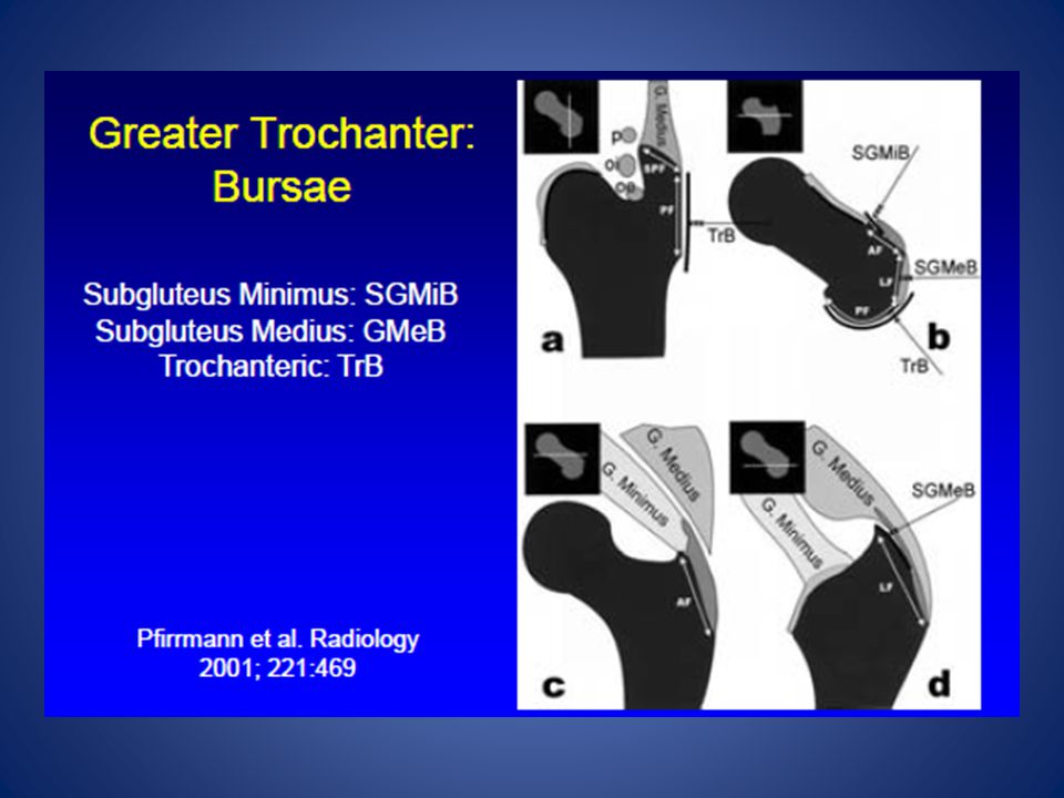
Trochanteric Bursitis
Located in posterolateral aspect of greater trochanter Located over the posterior and lateral facets of GT Deep to the gluteus maximus and Iliotibial tract Abnormal bursal distention of trochanteric bursa in lateral hip

93
Trochanteric bursitis
Causes: friction between IT band, glut medius/minimus/max and greater trochanter; common in running w/ improper biomechanics and overtraining direct blows S/Sx: local pain, tenderness over the greater trochanter Eval for leg length discrep, adductor/abductor muscle imbalance, hyperpronation Tx: relative rest, ice, brief NSAID, ITB stretching, +/- steroid injection Address biomechanical defects above

94
Trochanteric Bursitis
Trochanteric fluid seen posterolateral to GT and deep to Gluteus Maximus Best visualized if distended Distention does not indicate inflammation However is suggestive Pain with probe pressure & increased flow on color/power Doppler Increases likelihood of inflammation->Bursitis

97
Greater Trochanter Gluteus Medius and Minimus

98
Trochanteric Bursa Coronal Axial

101
Posterior Hip Pain Posterior Hip
Expand Differential to include Back Pain Evaluate for “red flags”

102
Posterior Hip Pain Differential Dx Lumbar spine disease
Degenerative disc disease Facet arthropathy Spinal stenosis Sacroiliac joint disorders Hip extensor and external rotator muscle pathology Piriformis Syndrome Aortoiliac vascular occlusive disease (rare)
")
105
Piriformis Syndrome Pain due to sciatic nerve compression at piriformis Cause: trauma, prolonged sitting, overuse; anomalies in 15-20% S/Sx: dull buttock pain +/- radiation into leg TTP over mid-buttock Pain worse with passive IR or resisted ER -Tx: relative rest, ER stretching, +/- steroid injection

106
Hip Pathology Hamstrings Ischial Bursitis Iliopsoas Bursitis Strain
Tear Ischial Bursitis Iliopsoas Bursitis

107
Ischial bursitis Cause: excessive friction over ischial tuberosity, or direct blow (hematoma, scarring) S/Sx: pain with sitting, TTP over ischial tuberosity, pain w/ passive hip flexion and active/resistive hip extension Xray to r/o fractures in traumatic hx Tx: Ice, padding, brief NSAID Prolonged: steroid injection Refractory: surgical excision

108
Lumbar and Sacroiliac Pathology
Significant Cause of Pathology referring to posterior hip and thigh Lumbar Pathology Lumbar Discogenic Pain Lumbar Facet Arthropathy Lumbar Radicular Pain Lumbar Stenosis Lumbar Spondylosis Sacroiliac Pain Sacroilitis

109
References Birrer R. and O’Connor F. Sports Medicine for the Primary Care Physician. Boca Raton: CRC Press, 2004. Greene W. Essentials of Musculoskeletal Care. Rosemont: American Academy of Orthopaedic Surgeons, 2001. Hoppenfeld S. Physical Examination of the Spine and Extremities. East Norwalk: Appleton-Century-Crofts, 1976;59-74. Lillegard W. Evaluation of Knee Injuries. In W Lillegard (ed), Handbook of Sports Medicine. Boston: Butterworth-Heinemann, 1999: Netter F. Atlas of Human Anatomy. West Caldwell: CIBA-Geigy, 1989. Tandeter H. et al. Acute Knee Injuries: Use of Decision Rules for Selective Radiograph Ordering. American Family Physician. Dec 1999; 60: (For Radiograph Images)
, Handbook of Sports Medicine. Boston: Butterworth-Heinemann, 1999: Netter F. Atlas of Human Anatomy. West Caldwell: CIBA-Geigy, Tandeter H. et al. Acute Knee Injuries: Use of Decision Rules for Selective Radiograph Ordering. American Family Physician. Dec 1999; 60: (For Radiograph Images)")
110
References ACR practice guidelines for the performance of the musculoskeletal ultrasound examination Nazarian et al.

Similar presentations