Download presentation
Presentation is loading. Please wait.

1
Abdominal wall

2
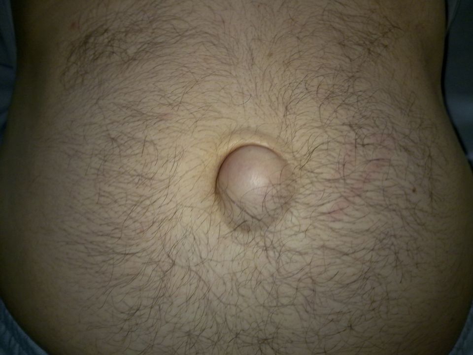
Paraumbilical hernia of adults :
(syn. supra- or inftaumbilical hernia). In adults the hernia does not occur through the umbilical scar. It is a protrusion through the linea alba just above or sometimes just below the umbilicus. As it enlarges, it becomes rounded or oval in shape with a tendency to sag downwards. Paraumbilical hernias can become very large. The neck of the sac is often remarkably narrow as compared with the size of the sac and the volume of its contents, which consist of greater omentum often accompanied by small intestine and, alternatively or in addition, a portion of the transverse colon. In long-standing cases the sac sometimes becomes loculated due to adherence of omentum to its fundus.
. In adults the hernia does not occur through the umbilical scar. It is a protrusion through the linea alba just above or sometimes just below the umbilicus. As it enlarges, it becomes rounded or oval in shape with a tendency to sag downwards. Paraumbilical hernias can become very large. The neck of the sac is often remarkably narrow as compared with the size of the sac and the volume of its contents, which consist of greater omentum often accompanied by small intestine and, alternatively or in addition, a portion of the transverse colon. In long-standing cases the sac sometimes becomes loculated due to adherence of omentum to its fundus.")
3
Clinical features : Women are affected five times more frequently than men. The patient is usually between the ages of 35 and 50. Increasing obesity, with flabbiness of the abdominal muscles, and repeated pregnancy’ are important antecedents. These hernias soon become irreducible because of omental adhesions within the sac. A large umbilical hernia causes a local dragging pain by its weight. Gastrointestinal symptoms are common and are probably due to traction on the stomach or transverse colon. Often there are transient attacks of intestinal colic due to subacute intestinal obstruction. In long-standing cases, intertrigo of the adjacent surfaces of the skin is a troublesome complication.

9
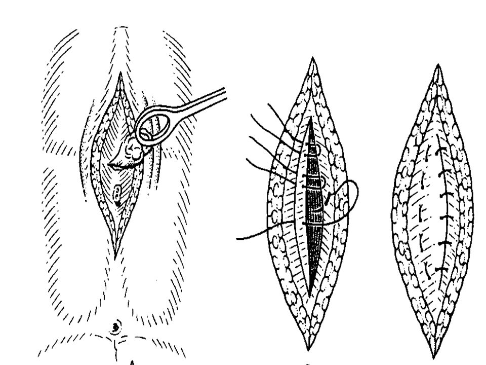
Treatment : Untreated, the hernia increases in size, and more and more of its contents become irreducible. Eventually, strangulation may occur. Therefore without undue delay operation should be advised in nearly all cases. When small, the defect can be closed by a simple repair using interrupted unabsorbable sutures: for larger hernias, a Mayo technique is advisable. Or mesh repair can be done

11
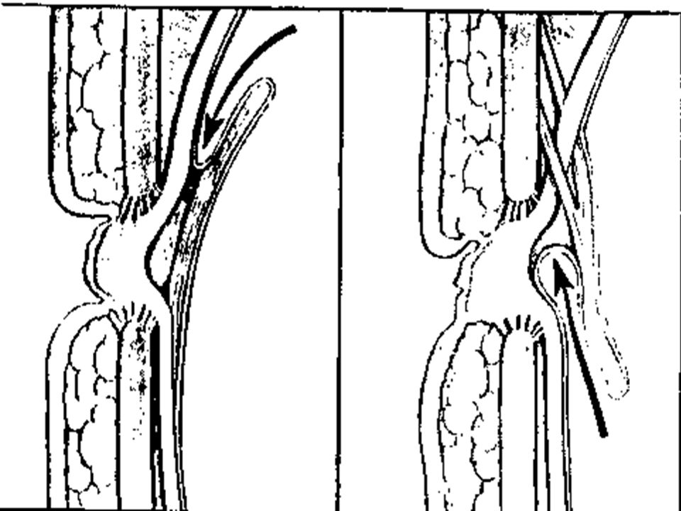
Epigastric hernia A midline epigastric hernia (syn. fatty hernia of the linea alba) occurs through the linea alba anywhere between the xiphoid process and the umbilicus, usually midway between these structures. Such a hernia commences as a protrusion of extraperitoneal fat through the linea alba, where it is pierced by a small blood vessel. More than one hernia may be present and recurrence can happen due to failure of identification of other defect at time of original repair. A swelling the size of a pea consists of a protrusion of extraperitoneal fat only (fatty hernia of the linea alba). If the protrusion enlarges, it drags a pouch of peritoneurn after it, and so becomes a true epigastric hernia. The mouth of the hernia is rarely large enough to permit a portion of hollow viscus to enter it; consequently, either the sac is empty or it contains a small portion of greater omentum.
 occurs through the linea alba anywhere between the xiphoid process and the umbilicus, usually midway between these structures. Such a hernia commences as a protrusion of extraperitoneal fat through the linea alba, where it is pierced by a small blood vessel. More than one hernia may be present and recurrence can happen due to failure of identification of other defect at time of original repair. A swelling the size of a pea consists of a protrusion of extraperitoneal fat only (fatty hernia of the linea alba). If the protrusion enlarges, it drags a pouch of peritoneurn after it, and so becomes a true epigastric hernia. The mouth of the hernia is rarely large enough to permit a portion of hollow viscus to enter it; consequently, either the sac is empty or it contains a small portion of greater omentum.")
15
Clinical features. • Symptomless. A small fatty hernia of the linea alba can be felt better than it can be seen, and may be symptomless, being discovered only in the course of routine abdominal palpation. • Painfnl. Sometimes such a hernia gives rise to attacks of local pain (worse on physical exertion) and also tenderness to touch and tight clothing; possibly because the fatty contents become nipped sufficiently to produce partial strangulation. •Referred pain (dyspeptic cases). It is not uncommon to find that the patient, who may not have noticed the hernia, complains of pain relating to digestion. Which may simulate features of peptic ulcers. Treatment. If the hernia is giving rise to symptoms, operation should be undertaken. It is essential to mark the hernia before the anaesthesia is given as it may be impossible to locate the defect if the fatty protrusion retracts into the abdomen.
 and also tenderness to touch and tight clothing; possibly because the fatty contents become nipped sufficiently to produce partial strangulation. •Referred pain (dyspeptic cases). It is not uncommon to find that the patient, who may not have noticed the hernia, complains of pain relating to digestion. Which may simulate features of peptic ulcers. Treatment. If the hernia is giving rise to symptoms, operation should be undertaken. It is essential to mark the hernia before the anaesthesia is given as it may be impossible to locate the defect if the fatty protrusion retracts into the abdomen.")
17
BURST ABDOMEN AND INCISIONAL HERNIA
ABDOMINAL WALL BURST ABDOMEN AND INCISIONAL HERNIA Factors relating to the incidence of burst abdomen and incisionsal hernia. Technique of wound closure: • choice ~ suture materials — catgut leads to a higher incidence of bursts than the use of non-absorbable monofilament polypropylene. Polyamide.

18
• method ~ closure — interrupted suturing has a low incidence
• method ~ closure — interrupted suturing has a low incidence. Thru’ and Thru’ suturing is good for the obstructed case. A one-layer closure has low incidence but it is higher than that following a two-layered closure. Interrupted ‘far and near’ sutures are a recommended technique for single layer mass closures. When continuous suturing of layers (one or two) is performed a particular fault is the use of a short length of material, pulled tightly, for in an anaesthetised relaxed patient the incision is shortened thereby, and made taut so that the material will act as if it were a cheese wire cutter when the patient is conscious and coughing. • drainage directly through a wound leads to a higher incidence of ‘bursts’ than employing drainage through a separate (stab) incision.
 is performed a particular fault is the use of a short length of material, pulled tightly, for in an anaesthetised relaxed patient the incision is shortened thereby, and made taut so that the material will act as if it were a cheese wire cutter when the patient is conscious and coughing. • drainage directly through a wound leads to a higher incidence of ‘bursts’ than employing drainage through a separate (stab) incision.")
19
Factors relating to incisions.
Midline and vertical incisions have a tendency to burst which is higher than those which are transverse. Reason for operation Infected case: deep wound infection has a notorious reputation for causing burst abdomen and/or late incisional hernia. Operations on the pancreas, with leakage of enzymes, and on obstructed cases are other reasons for disruption. Coughing,; vomiting; distension. At the completion of an operation any violent coughing set off by the removal of an endotracheal tube and suction of the laryngopharynx strains the sutures. Likewise cough, vomiting and distension (e.g. due to ileus) in the early postoperative period. Overvigorous postoperative ventilation in sedated patients can lead to wound disruption.
 in the early postoperative period. Overvigorous postoperative ventilation in sedated patients can lead to wound disruption.")
20
Causes of burst abdomen
■ Poor closure technique ■ Deep wound infection ■ Coughing or vomiting ■ Poor metabolic state of patient

21
General condition of the patient.
Obesity, jaundice, malignant disease, hypoproteinaemia. anaemia are all factors conducive to disruption of a laparotomy wound Abdominal wounds in pregnancy are notorious for a high risk of disruption.

22
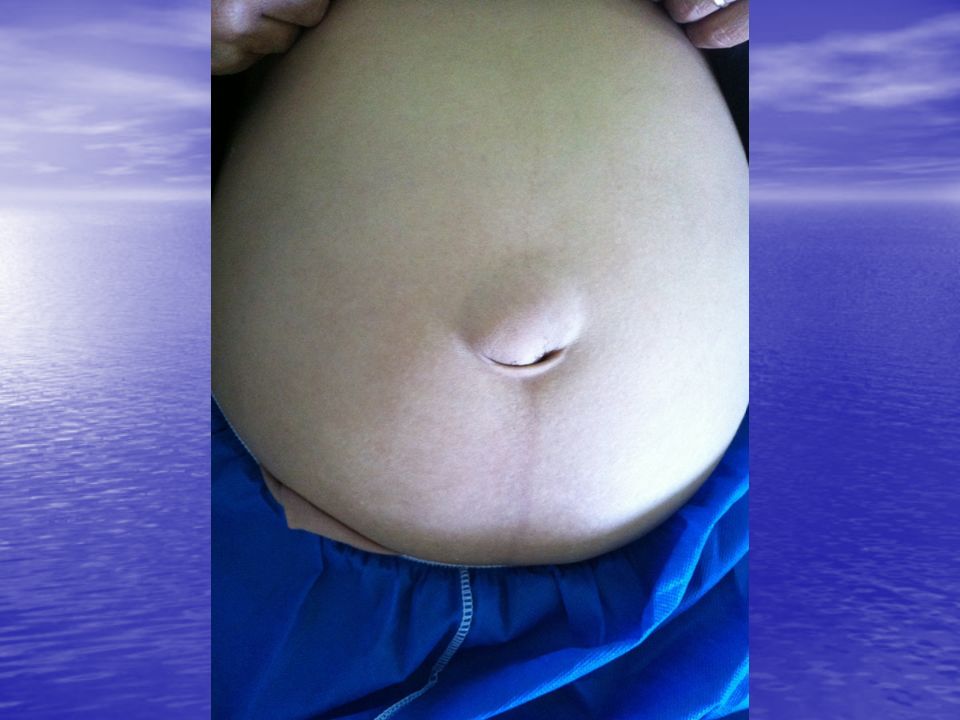
Burst abdomen and incisional hernia
In 1–2% of cases, mostly between the sixth and eighth day after operation, an abdominal wound bursts open and viscera are extruded. The disruption of the wound tends to occur a few days Before hand when the sutures apposing the deep layers (peritoneum,posterior rectus sheath) tear through or even become untied. An incisional hernia usually starts as a symptomless partial disruption of the deeper layers during the immediate or early postoperative period, the event passing unnoticed if the skin wound remains intact after the skin sutures have been removed.
 tear through or even become untied. An incisional hernia usually starts as a symptomless partial disruption of the deeper layers during the immediate or early postoperative period, the event passing unnoticed if the skin wound remains intact after the skin sutures have been removed.")
23
Burst abdomen (syn. abdominal dehiscence)
Clinical features. A serosanguineous (pink) discharge from the wound is a forerunner of disruption in fully 50 per cent of cases. It is the most pathognomonic sign of impending wound disruption, and it signifies that intraperitoneal contents are lying extraperitoneally. Patients often volunteer the information that they ‘felt something give way’. If skin sutures have been removed, omentum or coils of intestine may be forced through the wound and will be found lying on the skin. Pain and shock are often absent. It is important to note that there may be symptoms and signs of intestinal obstruction. Treatment. An emergency operation is required to replace the bowel, relieve any obstruction, and to resuture the wound. While awaiting operation, reassure the patient and cover the wound with a sterile towel. The stomach is emptied by a gastric tube and intravenous fluid therapy commenced.
 discharge from the wound is a forerunner of disruption in fully 50 per cent of cases. It is the most pathognomonic sign of impending wound disruption, and it signifies that intraperitoneal contents are lying extraperitoneally. Patients often volunteer the information that they ‘felt something give way’. If skin sutures have been removed, omentum or coils of intestine may be forced through the wound and will be found lying on the skin. Pain and shock are often absent. It is important to note that there may be symptoms and signs of intestinal obstruction. Treatment. An emergency operation is required to replace the bowel, relieve any obstruction, and to resuture the wound. While awaiting operation, reassure the patient and cover the wound with a sterile towel. The stomach is emptied by a gastric tube and intravenous fluid therapy commenced.")
24
Incisional hernia (syn. ventral hernia; postoperative hernia) Aetiology. Incisional hernia occurs most often in obese individuals, and a persistent postoperative cough and postoperative abdominal distension are its precursors. There is a high incidence of incisional hernia following operations for peritonitis because, as a rule, the wound becomes infected. The placing of a drainage tube through a separate stab incision, as opposed to bringing such a tube through the laparotomy wound, reduces the frequency. An incisional hernia usually starts as a symptomless partial disruption of the deeper layers of a laparotomy wound during the immediate or very early postoperative period. Often the event passes unnoticed if the skin wound remains intact after the stitches have been removed. A serosanguineous discharge is often the signal of dehiscence, and resuture of the deeper disrupted layers of the incision obviates the more difficult repair of an established and much larger hernia later on

25
Clinical features. There are great variations in the degree of herniation. The hernia may occur through a small portion of the scar, often the lower end. More frequently there is a diffuse bulging of the whole length of the incision. A postoperative hernia, especially one through a lower abdominal scar, usually increases steadily in size, and more and more of its contents become irreducible. Sometimes the skin overlying it is so thin and atrophic that normal peristalsis can be seen in the underlying coils of intestine. Attacks of subacute intestinal obstruction are common, and strangulation is liable to occur at the neck of a small sac or in a loculus of a large one, Nevertheless, most cases of incisional hernia are asymptomatic and broad-necked and do not need treatment.

33
Treatment. Palliative. An abdominal belt is sometimes satisfactory, especially in cases of a hernia through an upper abdominal incision. Operation. Many procedures are advocated, which is testimony to the facts that the repairs may be difficult to accomplish and no single procedure is dearly superior to the rest.

34
THANK YOU

Similar presentations