Download presentation
Presentation is loading. Please wait.

1
Nephrogenic Systemic Fibrosis: An Update
Neil M. Rofsky, MD BETH ISRAEL DEACONESS MEDICAL CENTER Harvard Medical School

2
MR Contrast Agents (Magnetopharmacuticals)
Signal Augmentation Differential distribution Safety

3
MR Contrast Agents: Principles
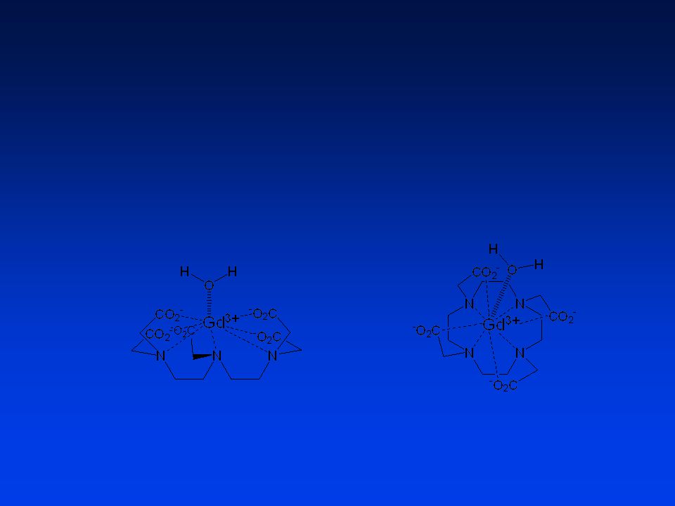
Unpaired electrons alter magnetic environment A trait of certain metal ions Indirectly affects the local H20 Naked metal ions are toxic! Ligands for safety (metal + ligand = chelate) Gd3+ Gd-DTPA-BMA (Omniscan) Gd-DOTA (Dotarem)
 Gd3+ Gd-DTPA-BMA. (Omniscan) Gd-DOTA. (Dotarem)")
4
Nephrogenic Systemic Fibrosis (NSF): The Basics
Originally known as Nephrogenic Fibrosing Dermopathy (NFD) Systemic proliferation of connective tissue (NSF) “Over 215 cases reported worldwide..” from 1997-present Strong epidemiologic association with Gd Appears to be a class issue (Omniscan >>> Magnevist, OptiMARK) Almost all renal insufficiency at exposure (most ESRD, on dialysis) Proinflammatory events in many11 (e.g., vascular surgery, sepsis, thrombosis) Some data suggests 3-5% incidence w/ Gd in setting of renal failure11 So far no co-factor identified (dialysate, ACEIs, EPO, etc.) Theories of pathogenesis: Liberation of Gd ion from carrier molecule10 Cutaneous deposition of free Gd ion10 Gd target attracts circulating fibrocytes (CFs) CFs differentiate in the dermis to resemble dermal fibroblasts Since it appears not to have existed prior to 1997, has been strong suspicion that a recently introduced agent or shift in the std of care might be the culprit Investigators have examined: dialysate (and contaminants thereof), EPO, iron, coag abnormalities, ACE inhibitors, vascular surgery, induced anti-phospholipid antibodies Investigation by dermatopathologists and the CDC has so far failed to identify a single causative medication Male ≈ female Middle-aged are most affected Variety of ethnic backgrounds are represented
 Systemic proliferation of connective tissue (NSF) Over 215 cases reported worldwide.. from 1997-present. Strong epidemiologic association with Gd. Appears to be a class issue (Omniscan >>> Magnevist, OptiMARK) Almost all renal insufficiency at exposure (most ESRD, on dialysis) Proinflammatory events in many11 (e.g., vascular surgery, sepsis, thrombosis) Some data suggests 3-5% incidence w/ Gd in setting of renal failure11. So far no co-factor identified (dialysate, ACEIs, EPO, etc.) Theories of pathogenesis: Liberation of Gd ion from carrier molecule10. Cutaneous deposition of free Gd ion10. Gd target attracts circulating fibrocytes (CFs) CFs differentiate in the dermis to resemble dermal fibroblasts. Since it appears not to have existed prior to 1997, has been strong suspicion that a recently introduced agent or shift in the std of care might be the culprit. Investigators have examined: dialysate (and contaminants thereof), EPO, iron, coag abnormalities, ACE inhibitors, vascular surgery, induced anti-phospholipid antibodies. Investigation by dermatopathologists and the CDC has so far failed to identify a single causative medication. Male ≈ female. Middle-aged are most affected. Variety of ethnic backgrounds are represented.")
5
Patient Safety a Decade Later…
First recognized case of “NFD” Gd “trigger” proposed for NSF (Grobner and Markmann) FDA issues Public Health Advisory FDA revises Public Health Advisory Literature reports Gd in NSF skin biopsies7 1997 2000 Apr 2006 May 2006 Jun 2006 Dec 2006 Jan 2007 Detected in 1997 Described in 2000 by Dr. Shawn Cowper, a dermatopathologist at Yale Popular press=MSNBC Gd detected in 4 of 13 specimens from 7 NSF patients -> detected in 4 of 7 patients First description of NSF in the literature Danish Medicines Agency reports 25 cases of Gd-associated NSF Press reports FDA warning to “kidney patients” Editorial in Radiology
 FDA issues Public Health Advisory. FDA revises Public Health Advisory. Literature reports Gd in NSF skin biopsies Apr May Jun Dec Jan Detected in Described in 2000 by Dr. Shawn Cowper, a dermatopathologist at Yale. Popular press=MSNBC. Gd detected in 4 of 13 specimens from 7 NSF patients -> detected in 4 of 7 patients. First description of NSF in the literature. Danish Medicines Agency reports 25 cases of Gd-associated NSF. Press reports FDA warning to kidney patients Editorial in Radiology.")
6
Gadolinium Agents in Review
Generic Brand Mfr FDA Approval Market Share (Doses) Gadopentetate dimeglumine Magnevist Berlex 1988 (70 mil) ~50% Gadodiamide Omniscan GE 1993 35% Gadoversetamide OptiMARK Tyco / Mallinckrodt 1999 5% Gadoteridol ProHance Bracco 1992 10% Gadobenate dimeglumine MultiHance 2004 The relative distribution of Omniscan related cases is out of proportion to the market share. } NSF: A Class Issue??
 Gadopentetate dimeglumine. Magnevist. Berlex (70 mil) ~50% Gadodiamide. Omniscan. GE % Gadoversetamide. OptiMARK. Tyco / Mallinckrodt % Gadoteridol. ProHance. Bracco % Gadobenate dimeglumine. MultiHance The relative distribution of Omniscan related cases is out of proportion to the market share. } NSF: A Class Issue")
7
Uses of High Dose Gd (+/- renal insufficiency):
MRA Peripheral Renal Neuro-onc (local practice patterns) X-ray use (k-edge of Gd is inefficient) CT Conventional Angio In clinical practice, where is high dose Gd being utilized???
 X-ray use (k-edge of Gd is inefficient) CT. Conventional Angio. In clinical practice, where is high dose Gd being utilized")
8
Nephrogenic Systemic Fibrosis (NSF): Diagnosis
Most prominent and visible effects in the skin Discoloration & texture changes Tightening, thickening, swelling → joint immobility Burning, itching, sharp pain Skin changes can be insidious -> confused w/ peripheral edema Resembles scleroderma and eosinophilic fasciitis Absent: monoclonal gammopathies9, Raynaud phenomenon and autoantibodies2 Yellowish scleral plaques Fibrotic changes can be widespread (liver, lungs, heart) Biopsy Findings Skin biopsy: thickened collagen bundles with surrounding clefts, mucin deposition, ↑ fibroblasts, ↑ CD34+ dendrocytes2 Muscle biopsy: ↑ myofibroblasts2
 Biopsy Findings. Skin biopsy: thickened collagen bundles with surrounding clefts, mucin deposition, ↑ fibroblasts, ↑ CD34+ dendrocytes2. Muscle biopsy: ↑ myofibroblasts2.")
9
Nephrogenic Systemic Fibrosis (NSF): Clinical Appearance
Reddened or darkened areas Texture changes “woody” or peau d’orange Cowper SE. Nephrogenic Fibrosing Dermopathy [NFD/NSF Website] Available at Accessed 02/01/2007.

10
Nephrogenic Systemic Fibrosis (NSF): Prognosis and Treatment
Course is chronic, progressive, variable May be severely debilitating Contractures - musculoskeletal Wheelchair requirement in some Complications may be fatal Falls, fractures Immobility, pneumonia No consistently successful treatment Symptoms may improve if renal function improves Limited evidence for kidney transplantation, extracorporeal photopheresis (ECP) Also in the literature: oral steroids, plasmapheresis Sometimes stabilizes, but rarely spontaneously remits No consistently effective therapy, but rapid correction in renal function may halt progression or reverse symptoms
 Also in the literature: oral steroids, plasmapheresis. Sometimes stabilizes, but rarely spontaneously remits. No consistently effective therapy, but rapid correction in renal function may halt progression or reverse symptoms.")
11
What we know about Gd and NSF
Causation not established; data are suspicious, but have limitations Retrospective studies Info is limited (e.g., Creatinine at time of Gd exposure, contemporaneous administration) Markedly prolonged half-life in renal failure All cases had renal dysfunction at time of Gd exposure Relationship between risk and level of dysfunction Relationship between risk and cumulative dose? Theoretical risk with any Gd contrast agent Risk different across agents (e.g., due to excess chelate)? Cases typically develop in days to few months after Gd exposure Appears that cases in NSF registry were acidotic at the time of Gd exposure; Cumulative dose is a frequent admins in short time interval. Roughly 90% of NSF cases associated w/ Omniscan, rate out of proportion to 35-40% market share Documented cases developed within 2 days to 18 months after Gd exposure; most within a few months Half-life of gadodiamide (hours)4 Normal renal function 1.3 h End-stage renal failure 34.3 h Hemodialysis (HD) 2.6 h Peritoneal dialysis (PD) 52.7 h
 Markedly prolonged half-life in renal failure. All cases had renal dysfunction at time of Gd exposure. Relationship between risk and level of dysfunction. Relationship between risk and cumulative dose Theoretical risk with any Gd contrast agent. Risk different across agents (e.g., due to excess chelate) Cases typically develop in days to few months after Gd exposure. Appears that cases in NSF registry were acidotic at the time of Gd exposure; Cumulative dose is a frequent admins in short time interval. Roughly 90% of NSF cases associated w/ Omniscan, rate out of proportion to 35-40% market share. Documented cases developed within 2 days to 18 months after Gd exposure; most within a few months. Half-life of gadodiamide (hours)4. Normal renal function. 1.3 h. End-stage renal failure h. Hemodialysis (HD) 2.6 h. Peritoneal dialysis (PD) 52.7 h.")
12
Amine backbone structure
Brand Name, Chemical Name Amine backbone structure log Kst (Stability constant) OptiMark, GdDTPA-BMEA Linear 16.8 Omniscan, GdDTPA-BMA Magnevist, GdDTPA 22.2 MultiHance GdBOPTA 22.6 Gadovist GdDO3A-butrol Macrocyclic 21.0 ProHance, GdHPDO3A 23.8 Dotarem GdDOTA 25.6 Thermodynamic stability
 OptiMark, GdDTPA-BMEA. Linear Omniscan, GdDTPA-BMA. Magnevist, GdDTPA MultiHance. GdBOPTA Gadovist. GdDO3A-butrol. Macrocyclic ProHance, GdHPDO3A Dotarem. GdDOTA Thermodynamic. stability.")
13
Gd-DTPA (Magnevist) Gd-DTPA-BMA (Omniscan) Gd-BOPTA (MultiHANCE) (Optimark) Gd-DOTA (Dotarem) Gd-EOB-DTPA (Primovist) Gd-BT-DO3A (Gadovist) Gd-HP-DO3A (ProHANCE)
 Gd-BT-DO3A (Gadovist) Gd-HP-DO3A (ProHANCE)")
14
ML M + L KD = [ M ] [ L ] [ ML ] 10 -23 = x x 500 (mM)
OR, 5 x 10 –21 = x2 OR, 7 x 10 –10 = x = [Gd 3+ ]
![ML M + L KD = [ M ] [ L ] [ ML ] = x x 500 (mM)](http://slideplayer.com/slide/2489931/9/images/14/ML+%EF%82%AE+M+%2B+L+KD+%3D+%5B+M+%5D+%5B+L+%5D+%5B+ML+%5D+%3D+x+%EF%80%AA+x+500+%28mM%29.jpg "OR, 5 x 10 –21 = x2. OR, 7 x 10 –10 = x = [Gd 3+ ]")
15
Brand Name, Chemical Name Amine backbone structure log Kst
(Stability constant) Dissociation rate in 0.1M HCl ( sec-1) OptiMark, GdDTPA-BMEA Linear 16.8 >2.2x10⁻² Omniscan, GdDTPA-BMA >2x10⁻² Magnevist, GdDTPA 22.2 1.2x10⁻³ MultiHance GdBOPTA 22.6 -not reported- Gadovist GdDO3A-butrol Macrocyclic 21.0 2.8x10⁻⁶ (estimated from data) ProHance, GdHPDO3A 23.8 6.4x10⁻⁵ Dotarem GdDOTA 25.6 8.4x10⁻⁷
 Dissociation rate in 0.1M HCl ( sec-1) OptiMark, GdDTPA-BMEA. Linear >2.2x10⁻². Omniscan, GdDTPA-BMA. >2x10⁻². Magnevist, GdDTPA x10-3. MultiHance. GdBOPTA not reported- Gadovist. GdDO3A-butrol. Macrocyclic x10-6. (estimated from data) ProHance, GdHPDO3A x10-5. Dotarem. GdDOTA x10-7.")
16
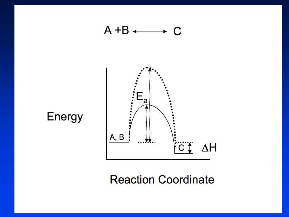
The themodynamic stability constant determines the concentrations
the themodynamic stability constant determines the concentrations of the Gd-chelate, free chelate, and free gadolinium at equilibrium, while rates of formation and dissociation, dictated by Ea , determine how rapidly these compounds reach equilibrium. In principle it is possible to have a chelate with a relatively low stability constant and high value of Ea, resulting in a ”kinetically trapped” chelate that does not dissociate on any relevant time scale. The themodynamic stability constant determines the concentrations of the Gd-chelate, free chelate, and free gadolinium at equilibrium; The rates of formation and dissociation, dictated by Ea , determine how rapidly these compounds reach equilibrium.

17
Zn Gd L ↕ Gd + L Relaxivity ’s in solution pH 7.0 PO4
Compound R1 at 3 days Gd-DTPA ↓50% Gd-DTPA-BMA ↓90% Gd –BOPTA ↓60% Gd-HP-DO NO SIG PO4 PO4 PO4 PbO4 Laurent S, et al.. Contrast Media Mol Imaging 2006;1:

18
Wedeking, Kumar and Tweedle
a very strong correlation between the dissociation rates of chelates in acid and the long-term deposition of Gd3+ in rat tissues such as liver and bone (femur). a very strong correlation between the dissociation rates of chelates in acid and the long-term deposition of Gd3+ in rat tissues such as liver and bone (femur). Wedeking, Kumar and Tweedle
. a very strong correlation between the dissociation rates of chelates in acid and the long-term deposition of Gd3+ in rat tissues such as liver and bone (femur). Wedeking, Kumar and Tweedle.")
19
“Acid dissociation rate constants were the most accurate parameters linking in vitro and in vivo dissociation. “ “Gd(HP-D03A) and Gd(DOTA)-, had the lowest residual Gd3+ in whole animals.” “No evidence of free Gd3+ could be detected for Gd(HP-D03A) using the free Gd3+ target tissues (liver and femur) at long residence times.” Wedeking, Kumar and Tweedle, Mag Res Imag 1992

20
Algorithm for Gd-Enhanced MRI
Gd-MRI in last 7 days? Consider delay to allow 7 days between Gd doses START YES NO Proceeding Inpatient Outpatient / EU “NO” to all Response to Choyke questions eGFR w/in 3 days If worsening trend, day of exam “YES” to any eGFR within 4 weeks DIALYSIS eGFR < 30 On PD eGFR < 30 On HD eGFR < 30 No dialysis eGFR 30-60 eGFR > 60 We have recently come across a questionarre that will obviate the need to obtain a serum creatinine in all patients. Discussion w/ referrer Discussion w/ referrer Discussion w/ referrer Limit Gd to mmol/kg*; Consider hydration Proceeding Proceeding Proceeding Obtain central venous access HD x 2 w/in 2 hrs and 24 hrs Informed consent ProHance™ or MultiHance™: No > 0.1 mmol/kg OK to proceed *Except for run-offs, which are permitted up to 0.2 mmol/kg after risk:benefit discussion w/ referrer.

21
Algorithm for Gd-Enhanced MRI
Gd-MRI in last 7 days? Consider delay to allow 7 days between Gd doses START YES NO Proceeding Outpatient / EU “NO” to all Response to Choyke questions Point of service query We have recently come across a questionarre that will obviate the need to obtain a serum creatinine in all patients. OK to proceed

22
Minimizing the Risk of NSF
Risk : benefit analysis Reduce use of Gd in renal disease FDA recommends avoiding for eGFR < 30 Consider non-contrast protocols Consider alternate modality (e.g., CT, conventional angiogram) Minimize dose if Gd is deemed imperative Consider alternative agents Gd-BOPTA (MultiHANCE®) No reports (yet…) Can reduce dose (has higher R1) HOWEVER…clearance kinetics less favorable (binds protein) ProHANCE Hemodialyze patients with ESRD asap ?? * Gd excretory rates 78%, 96%, 99% from 1st to 3rd HD session5 When using Gd, maximize pt condition*: Hold drugs that decrease renal function (e.g., diuretics, NSAIDs) Hydrate (consider bicarb – ? role of metabolic acidosis in NSF) Informed consent eGFR formula less accurate at 60, so may disqualify many people who can safely take Gd Since 96% of Gd is cleared by end of 2nd HD session, consider 2 sessions, w/in 3 hours and w/in 24 hours There are chemistry theories that predict dissociation of the Gd from its chelate in acidic environments. *(not evidence based!!)
 Minimize dose if Gd is deemed imperative. Consider alternative agents. Gd-BOPTA (MultiHANCE®) No reports (yet…) Can reduce dose (has higher R1) HOWEVER…clearance kinetics less favorable (binds protein) ProHANCE. Hemodialyze patients with ESRD asap * Gd excretory rates 78%, 96%, 99% from 1st to 3rd HD session5. When using Gd, maximize pt condition*: Hold drugs that decrease renal function (e.g., diuretics, NSAIDs) Hydrate (consider bicarb – role of metabolic acidosis in NSF) Informed consent. eGFR formula less accurate at 60, so may disqualify many people who can safely take Gd. Since 96% of Gd is cleared by end of 2nd HD session, consider 2 sessions, w/in 3 hours and w/in 24 hours. There are chemistry theories that predict dissociation of the Gd from its chelate in acidic environments. *(not evidence based!!)")
23
How do we screen for risk?
Choyke Questionnaire Serum Creatinine Point of Service Devices??

24
The Choyke Questionnaire
Pre-existing renal disease (OR 13.6) Proteinuria (OR 8.7) Prior kidney surgery (OR 8.1) Hypertension (OR 5.4) Gout (OR 4.6) Diabetes (OR 3.2) Completely negative responses: 450 (67%) of 673 446/450 (99%) Cr values 1.7 mg/dL 424/450 (94%) - normal Cr values Choyke, et al. Tech Urol 1998; 4: 65.
 Proteinuria (OR 8.7) Prior kidney surgery (OR 8.1) Hypertension (OR 5.4) Gout (OR 4.6) Diabetes (OR 3.2) Completely negative responses: 450 (67%) of /450 (99%) Cr values 1.7 mg/dL. 424/450 (94%) - normal Cr values. Choyke, et al. Tech Urol 1998; 4: 65.")
25
Algorithm for Gd-Enhanced MRI
Gd-MRI in last 7 days? Consider delay to allow 7 days between Gd doses START YES NO Proceeding Inpatient Outpatient / EU “NO” to all Response to Choyke questions eGFR w/in 3 days If worsening trend, day of exam “YES” to any eGFR within 4 weeks DIALYSIS eGFR < 30 On PD eGFR < 30 On HD eGFR < 30 No dialysis eGFR 30-60 eGFR > 60 We have recently come across a questionarre that will obviate the need to obtain a serum creatinine in all patients. Discussion w/ referrer Discussion w/ referrer Discussion w/ referrer Limit Gd to mmol/kg*; Consider hydration Proceeding Proceeding Proceeding Obtain central venous access HD x 2 w/in 2 hrs and 24 hrs Informed consent ProHance™ or MultiHance™: No > 0.1 mmol/kg OK to proceed *Except for run-offs, which are permitted up to 0.2 mmol/kg after risk:benefit discussion w/ referrer.

26
Algorithm for Gd-Enhanced MRI
Gd-MRI in last 7 days? Consider delay to allow 7 days between Gd doses START YES NO Proceeding Inpatient Outpatient / EU Response to Choyke questions eGFR w/in 3 days If worsening trend, day of exam “YES” to any Point of service (stat creat)! eGFR within 4 weeks eGFR < 30 No dialysis eGFR 30-60 eGFR > 60 We have recently come across a questionarre that will obviate the need to obtain a serum creatinine in all patients. Discussion w/ referrer Limit Gd to mmol/kg*; Consider hydration Proceeding Informed consent ProHance™ or MultiHance™: No > 0.1 mmol/kg OK to proceed *Except for run-offs, which are permitted up to 0.2 mmol/kg after risk:benefit discussion w/ referrer.
! eGFR within 4 weeks. eGFR < 30. No dialysis. eGFR eGFR > 60. We have recently come across a questionarre that will obviate the need to obtain a serum creatinine in all patients. Discussion w/ referrer. Limit Gd to 0.1 mmol/kg*; Consider hydration. Proceeding. Informed consent. ProHance™ or MultiHance™: No > 0.1 mmol/kg. OK to proceed. *Except for run-offs, which are permitted up to 0.2 mmol/kg after risk:benefit discussion w/ referrer.")
27
Algorithm for Gd-Enhanced MRI
Gd-MRI in last 7 days? Consider delay to allow 7 days between Gd doses START YES NO Proceeding Inpatient eGFR w/in 3 days If worsening trend, day of exam DIALYSIS eGFR < 30 On PD eGFR < 30 On HD We have recently come across a questionarre that will obviate the need to obtain a serum creatinine in all patients. Discussion w/ referrer Discussion w/ referrer Proceeding Proceeding Obtain central venous access HD x 2 w/in 2 hrs and 24 hrs Informed consent ProHance™ or MultiHance™: No > 0.1 mmol/kg *Except for run-offs, which are permitted up to 0.2 mmol/kg after risk:benefit discussion w/ referrer.

28
For Dialysis Patients

29
Hydration & HCO3 ???? Oral hydration Intravenous hydration Bicarb
1 Liter of H20 by mouth pre- and post- injection of contrast Intravenous hydration Contact the ordering physician or house staff for orders Bicarb 150mEq of NaHCO3 (e.g. dilute in 1L D5W) Pre: 1 hr prior to contrast administration @ 3cc/kg/hr and for Post: 6 hrs after contrast administration @ 1cc/kg/hr Modifications possible for pts with renal failure/CHF) Encourage oral fluid intake if not on fluid restrictions
 Pre: 1 hr prior to contrast 3cc/kg/hr and for. Post: 6 hrs after contrast 1cc/kg/hr. Modifications possible for pts with renal failure/CHF) Encourage oral fluid intake if not on fluid restrictions.")
30
Perspective – Iodinated Contrast
Risk for severe adverse reactions 0.147% HI-ICM 0.031% NI-ICM (3/10,000) Death ~ 1/100,000 either high or low osmolality. Non Ionic Iodinated contast media Caro AJR 1991 Apr; 156(4):
 Death. ~ 1/100,000 either high or low osmolality. Non Ionic Iodinated contast media. Caro AJR 1991 Apr; 156(4):")
31
Conclusions Gd is associated with NSF in pts with substantial renal insufficiency Role of acidity seems likely Risk:Benefit assessment is vital What is the risk of not giving CE-MRI? Guidelines should be submitted for institutional approval Education is essential Keep reading, keeping conversing! Consider using Gadoteridol in high risk situations

32
References Cowper SE. Nephrogenic Fibrosing Dermopathy [NFD/NSF Website] Available at Accessed 01/17/2007. Cowper SE. Nephrogenic fibrosing dermopathy: the first 6 years. Curr Opin Rheumatol 2003; 15:785. Grobner T: Gadolinium – a specific trigger for the development of nephrogenic fibrosing dermopathy and nephrogenic systemic fibrosis? Nephrology Dialysis Transplantation 21(4): , April 2006. Joffe P, Thomsen HS, Meusel M: Pharmacokinetics of gadodiamide injection in patients with severe renal insufficiency and patients undergoing hemodialysis or continuous ambulatory peritoneal dialysis. Acad Radiol 5: , 1998. Okada S et al. Safety of gadolinium contrast agent in hemodialysis patient. Acta Radiologica, 2001, 42(3): Rofsky N et al. Renal lesion characterization with gadolinium-enhanced MR imaging: Efficacy and safety in patients with renal insufficiency. Radiology, July 1991, 180: High WA, Ayers RA, Chandler J, Zito G, and Cowper SE. Gadolinium is detectable within the tissue of patients with nephrogenic systemic fibrosis. J Am Acad Dermatol 2007;56(1):21-26. Stenver DI. Investigation of the safety of MRI contrast medium Omniscan. Danish Medicines Agency. Published May 29, Accessed February 6, 2007. Boyd AS, Zic JA, and Abraham JL. Gadolinium deposition in nephrogenic fibrosing dermopathy. J Am Acad Dermatol. January Marckmann P, Skov L, Rossen K, Dupont A, Damholt MB, Heaf JG, et al. Nephrogenic systemic fibrosis: suspected causative role of gadodiamide used for contrast-enhanced magnetic resonance imaging. J Am Soc Nephrol 2006;17: Sadowski EA, Bennett LK, Chan MR, Wentland AL, Garrett AL, Garrett RW, et al. Nephrogenic systemic fibrosis: Risk factors and incidence estimation. Radiology Published January 31, Accessed February 1, 2007. Gadolinium-based MR Contrast Agents and Nephrogenic Systemic Fibrosis1 Phillip H. Kuo, MD, PhD, Emanuel Kanal, MD, Ali K. Abu-Alfa, MD and Shawn E. Cowper, MD Published online before print January 9, 2007 (Radiology 2007, /radiol ) This Article Submit a response Alert me when this article is cited Alert me when eLetters are posted Alert me if a correction is posted Services this article to a friend Similar articles in this journal Similar articles in PubMed Alert me to new issues of the journal Download to citation manager Google Scholar Articles by Kuo, P. H. Articles by Cowper, S. E. PubMed PubMed Citation © RSNA, 2007
![References Cowper SE. Nephrogenic Fibrosing Dermopathy [NFD/NSF Website] Available at Accessed 01/17/2007.](http://slideplayer.com/slide/2489931/9/images/32/References+Cowper+SE.+Nephrogenic+Fibrosing+Dermopathy+%5BNFD%2FNSF+Website%5D+Available+at+++Accessed+01%2F17%2F2007..jpg "Cowper SE. Nephrogenic fibrosing dermopathy: the first 6 years. Curr Opin Rheumatol 2003; 15:785. Grobner T: Gadolinium – a specific trigger for the development of nephrogenic fibrosing dermopathy and nephrogenic systemic fibrosis Nephrology Dialysis Transplantation 21(4): , April Joffe P, Thomsen HS, Meusel M: Pharmacokinetics of gadodiamide injection in patients with severe renal insufficiency and patients undergoing hemodialysis or continuous ambulatory peritoneal dialysis. Acad Radiol 5: , Okada S et al. Safety of gadolinium contrast agent in hemodialysis patient. Acta Radiologica, 2001, 42(3): Rofsky N et al. Renal lesion characterization with gadolinium-enhanced MR imaging: Efficacy and safety in patients with renal insufficiency. Radiology, July 1991, 180: High WA, Ayers RA, Chandler J, Zito G, and Cowper SE. Gadolinium is detectable within the tissue of patients with nephrogenic systemic fibrosis. J Am Acad Dermatol 2007;56(1): Stenver DI. Investigation of the safety of MRI contrast medium Omniscan. Danish Medicines Agency. asrtikelID=8931. Published May 29, Accessed February 6, Boyd AS, Zic JA, and Abraham JL. Gadolinium deposition in nephrogenic fibrosing dermopathy. J Am Acad Dermatol. January Marckmann P, Skov L, Rossen K, Dupont A, Damholt MB, Heaf JG, et al. Nephrogenic systemic fibrosis: suspected causative role of gadodiamide used for contrast-enhanced magnetic resonance imaging. J Am Soc Nephrol 2006;17: Sadowski EA, Bennett LK, Chan MR, Wentland AL, Garrett AL, Garrett RW, et al. Nephrogenic systemic fibrosis: Risk factors and incidence estimation. Radiology Published January 31, Accessed February 1, Gadolinium-based MR Contrast Agents and Nephrogenic Systemic Fibrosis1. Phillip H. Kuo, MD, PhD, Emanuel Kanal, MD, Ali K. Abu-Alfa, MD and Shawn E. Cowper, MD Published online before print January 9, 2007 (Radiology 2007, /radiol ) This Article. Submit a response. Alert me when this article is cited. Alert me when eLetters are posted. Alert me if a correction is posted. Services. this article to a friend. Similar articles in this journal Similar articles in PubMed. Alert me to new issues of the journal. Download to citation manager. Google Scholar. Articles by Kuo, P. H. Articles by Cowper, S. E. PubMed. PubMed Citation. © RSNA,")
34
For Non-Dialysis Patients (Conservative)
Creatinine / eGFR value OR Choyke Questionnaire We have recently come across a questionarre that will obviate the need to obtain a serum creatinine in all patients. *diuretics, NSAIDs, and ACE inhibitors (the latter when used with a diuretic) **Intravenous hydration with sodium bicarbonate for 1hr prior and 6hrs post Gd.
 **Intravenous hydration with sodium bicarbonate for 1hr prior and 6hrs post Gd.")
35
“Acid dissociation rate constants were the most accurate parameters linking in vitro and in vivo dissociation. “ “The most stable and least dissociation labile macrocycles, Gd(HP-D03A) and Gd(DOTA)-, had the lowest residual Gd3+ in whole animals.” “No evidence of free Gd3+ could be detected for Gd(HP-D03A) using the free Gd3+ target tissues (liver and femur) at long residence times.”
 and Gd(DOTA)-, had the lowest residual Gd3+ in whole animals. No evidence of free Gd3+ could be detected for Gd(HP-D03A) using the free Gd3+ target tissues (liver and femur) at long residence times. .")
36
Gadolinium Chelates MR contrast agents (metal + ligand = chelate)
Gd Brightening on T1-WI’s Gd is toxic! Chelate ‘shields’ the free metal while preserving its relaxation effect. Gd Agents differ by how they are enclosed by the agents

37
Gadolinium Chelates MR contrast agents
Brightening on T1-weighted images Gd is toxic! Chelate used to ‘shield’ the free metal while preserving its relaxation effect. Gd3+ Gd-DTPA-BMA (Omniscan) Gd-DOTA (Dotarem) Agents differ by how they are enclosed by the agents
 Gd-DOTA. (Dotarem) Agents differ by how they are enclosed by the agents.")
38
Nephrogenic Systemic Fibrosis (NSF): The Basics
Originally known as Nephrogenic Fibrosing Dermopathy (NFD) Systemic proliferation of connective tissue (NSF) ~ 215 cases reported worldwide from Strong epidemiologic association with Gd All FDA-reviewed cases had prior Gd exposure Appears to be a class issue (Omniscan >>> Magnevist, OptiMARK) All had renal insufficiency at exposure (most ESRD, on dialysis) Proinflammatory events in many11 (e.g., vascular surgery, sepsis, thrombosis) Early data suggests 3-5% incidence w/ Gd in setting of renal failure11 So far no co-factor identified (dialysate, ACEIs, EPO, etc.) Theories of pathogenesis: Liberation of Gd ion from carrier molecule10 Cutaneous deposition of free Gd ion10 Gd target attracts circulating fibrocytes (CFs) CFs differentiate in the dermis to resemble dermal fibroblasts Since it appears not to have existed prior to 1997, has been strong suspicion that a recently introduced agent or shift in the std of care might be the culprit Investigators have examined: dialysate (and contaminants thereof), EPO, iron, coag abnormalities, ACE inhibitors, vascular surgery, induced anti-phospholipid antibodies Investigation by dermatopathologists and the CDC has so far failed to identify a single causative medication Male ≈ female Middle-aged are most affected Variety of ethnic backgrounds are represented
 Systemic proliferation of connective tissue (NSF) ~ 215 cases reported worldwide from Strong epidemiologic association with Gd. All FDA-reviewed cases had prior Gd exposure. Appears to be a class issue (Omniscan >>> Magnevist, OptiMARK) All had renal insufficiency at exposure (most ESRD, on dialysis) Proinflammatory events in many11 (e.g., vascular surgery, sepsis, thrombosis) Early data suggests 3-5% incidence w/ Gd in setting of renal failure11. So far no co-factor identified (dialysate, ACEIs, EPO, etc.) Theories of pathogenesis: Liberation of Gd ion from carrier molecule10. Cutaneous deposition of free Gd ion10. Gd target attracts circulating fibrocytes (CFs) CFs differentiate in the dermis to resemble dermal fibroblasts. Since it appears not to have existed prior to 1997, has been strong suspicion that a recently introduced agent or shift in the std of care might be the culprit. Investigators have examined: dialysate (and contaminants thereof), EPO, iron, coag abnormalities, ACE inhibitors, vascular surgery, induced anti-phospholipid antibodies. Investigation by dermatopathologists and the CDC has so far failed to identify a single causative medication. Male ≈ female. Middle-aged are most affected. Variety of ethnic backgrounds are represented.")
40
G=S G=-RT(LogKeq) k=Ae-Ea/RT
 k=Ae-Ea/RT")
43
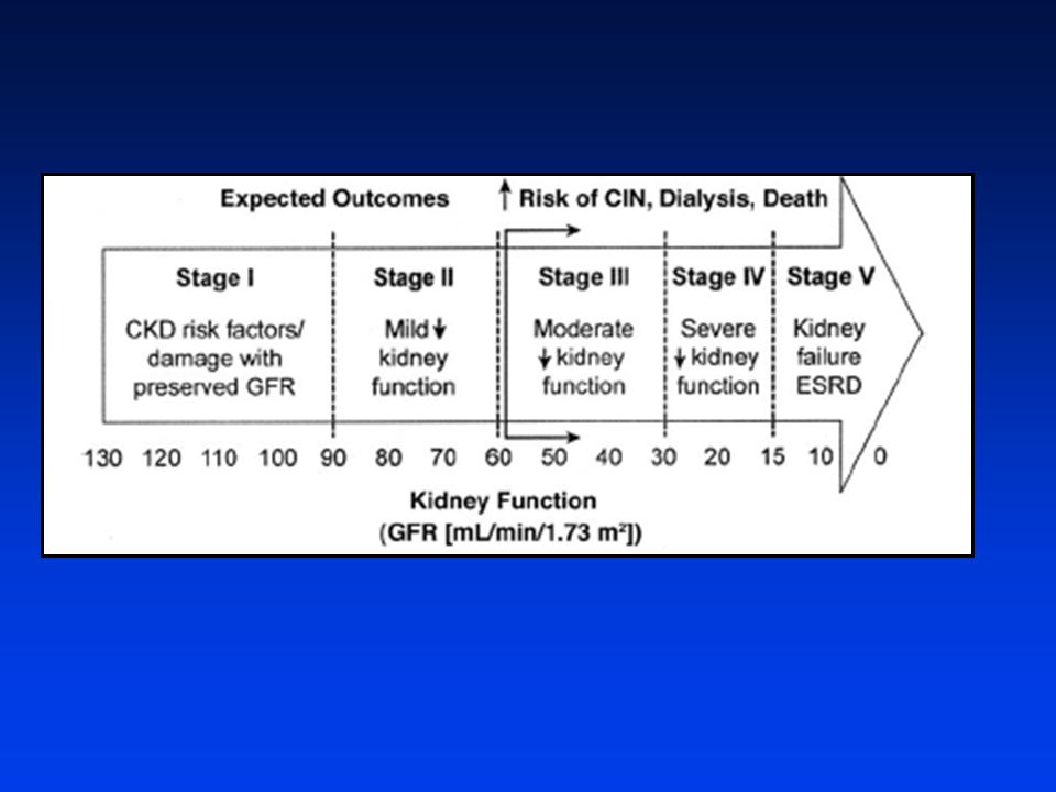
Staging of Chronic Kidney Dz
Death Expected Outcomes ↑Risk of CIN, Dialysis, Death ↑Risk of NSF Stage 1 Stage II Stage III Stage IV Stage V CKD risk factors/ Mild ↓ Moderate ↓ Severe ↓ Kidney damage w/ kidney kidney kidney failure preserved GFR fxn fxn fxn ESRD Kidney Fxn (GFR [ml/min/1.73 m2]) Modified from: Am J Kidney Dis 2002;39(suppl):S1-266.
 Modified from: Am J Kidney Dis 2002;39(suppl):S")
45
Algorithm for Choyke Screen; Outpt Efficacy Study
Outpatient / EU Response to Choyke questions Pre- service Creat/ eGFR “Yes” to any “NO” to all Point of service query “Yes”, “Yes” “Yes”, “No” “No”, “Yes” “No”, “No” Point of service Creat/ eGFR Point of service Creat/ eGFR Exam w/o Creat No F/U Creat/ eGFR F/U Creat/ eGFR; date St 1 St 2 St 3 St 4 St 5

46
Algorithm for Gd-Enhanced MRI
Gd-MRI in last 7 days? Consider delay to allow 7 days between Gd doses START YES NO Proceeding Inpatient Outpatient / EU Response to Choyke questions eGFR w/in 3 days If worsening trend, day of exam “YES” to any Point of service (stat creat)! eGFR within 4 weeks eGFR < 30 No dialysis eGFR 30-60 eGFR > 60 We have recently come across a questionarre that will obviate the need to obtain a serum creatinine in all patients. Discussion w/ referrer Limit Gd to mmol/kg*; Consider hydration Proceeding Informed consent ProHance™ or MultiHance™: No > 0.1 mmol/kg OK to proceed *Except for run-offs, which are permitted up to 0.2 mmol/kg after risk:benefit discussion w/ referrer.
! eGFR within 4 weeks. eGFR < 30. No dialysis. eGFR eGFR > 60. We have recently come across a questionarre that will obviate the need to obtain a serum creatinine in all patients. Discussion w/ referrer. Limit Gd to 0.1 mmol/kg*; Consider hydration. Proceeding. Informed consent. ProHance™ or MultiHance™: No > 0.1 mmol/kg. OK to proceed. *Except for run-offs, which are permitted up to 0.2 mmol/kg after risk:benefit discussion w/ referrer.")
47
What we know about Gd and NSF
Causation not established; data are suspicious, but have limitations Retrospective studies Info is limited (e.g., Creatinine at time of Gd exposure, contemporaneous administration) Markedly prolonged half-life in renal failure All cases had renal dysfunction at time of Gd exposure Relationship between risk and level of dysfunction Relationship between risk and cumulative dose? Theoretical risk with any Gd contrast agent Documented cases with Omniscan >>Magnevist, OptiMARK Risk different across agents (e.g., due to excess chelate)? Cases typically develop in days to few months after Gd exposure Appears that cases in NSF registry were acidotic at the time of Gd exposure; Cumulative dose is a frequent admins in short time interval. Roughly 90% of NSF cases associated w/ Omniscan, rate out of proportion to 35-40% market share Documented cases developed within 2 days to 18 months after Gd exposure; most within a few months Half-life of gadodiamide (hours)4 Normal renal function 1.3 h End-stage renal failure 34.3 h Hemodialysis (HD) 2.6 h Peritoneal dialysis (PD) 52.7 h
 Markedly prolonged half-life in renal failure. All cases had renal dysfunction at time of Gd exposure. Relationship between risk and level of dysfunction. Relationship between risk and cumulative dose Theoretical risk with any Gd contrast agent. Documented cases with Omniscan >>Magnevist, OptiMARK. Risk different across agents (e.g., due to excess chelate) Cases typically develop in days to few months after Gd exposure. Appears that cases in NSF registry were acidotic at the time of Gd exposure; Cumulative dose is a frequent admins in short time interval. Roughly 90% of NSF cases associated w/ Omniscan, rate out of proportion to 35-40% market share. Documented cases developed within 2 days to 18 months after Gd exposure; most within a few months. Half-life of gadodiamide (hours)4. Normal renal function. 1.3 h. End-stage renal failure h. Hemodialysis (HD) 2.6 h. Peritoneal dialysis (PD) 52.7 h.")
Similar presentations


>")


?>")


