Download presentation
Presentation is loading. Please wait.

1
Altered Cerebral Function & Increased Intracranial Pressure
RNSG 2432 Enhanced Concepts of Adult Health Lisa Randall, RN, MSN, ACNS-BC

2
Objectives Define and discuss altered cerebral function and increased ICP Analyze etiology and pathophysiology of altered cerebral function Discuss/illustrate signs and symptoms, diagnostics, and treatment Formulate nursing diagnoses that address physical, psychosocial, and learning needs Prioritize and evaluate nursing interventions

3
Definitions Cerebral function Mental status Speech Eyes Cranial nerves
Motor Sensory Reflexes

4
Definitions Consciousness Lethargy Obtundation Stupor Coma Arousal
Awareness Lethargy < alertness < awareness < thought process Obtundation << A/A Clouding Stupor Deep sleeplike state Vigorous stimulation Coma Unresponsiveness PVS MCS

5
Comatose State Unarousability Absence of sleep/wake cycles
Inability to interact with the environment GCS =/< 8

6
Persistent Vegetative State
Intermittent wakefulness Sleep-wake cycles No awareness of self or environment

7
Minimally Conscious State
Altered consciousness Evidence of self or environmental awareness is demonstrated

8
Anatomy

9
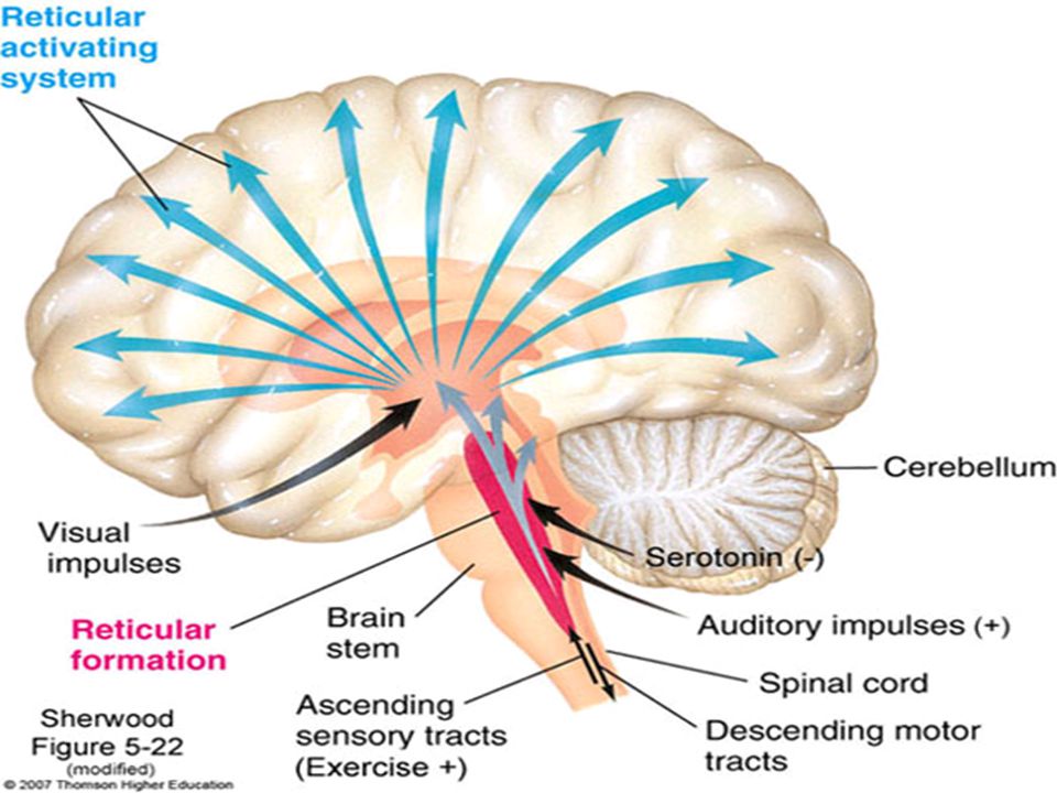
Pathophysiology Reticular Activating System (RAS) Reticular Formation
Gray cells within brainstem extends into thalamus Wakefulness Arousal Alertness

11
Etiology Altered Cerebral Dysfunction
Lesion/injury to the RAS or cerebral cortex Metabolic disorders Anoxic injury Drugs Seizures

12
Assessment LOC Health history Physical exam Vital signs
drugs/head injury/metabolic Physical exam Vital signs Temperature Cushing’s reflex/triad Neuro Vital Signs LOC, Pupils, Strength/Movement, Sensation Glasgow coma scale NIH Stroke Scale

13
Cushing Triad Edema Increased intracranial pressure
Increased systolic BP Widening pulse pressure Normal = 40 mmHg Decreased pulse rate Irregular respirations

14
GLASGOW COMA SCALE Eyes Spontaneous opening 4 Open to speech 3
Open to pain 2 Do not open 1 Verbal Response Oriented 5 Confused Inappropriate Incomprehensible None GLASGOW COMA SCALE

15
GLASGOW COMA SCALE Motor Response Obeys commands 6 Localizes to pain
Pushes your hand away 5 Withdraws from pain 4 Decorticate/flexion 3 Decerebrate/extension 2 None 1 GLASGOW COMA SCALE Range of possible scores = A score of 13 to 14 indicates mild deficit. A score between 9 and 12 points to moderate deficit, and a score of 8 or less indicates severe coma.

16
Decorticate posturing- abnormal flexion Decerebrate posturing- abnormal extension

17
Assessment Mental status General appearance/behavior
State of conciousness Mood and affect Thought content Intellectual capacity

19
Cranial Nerves

20
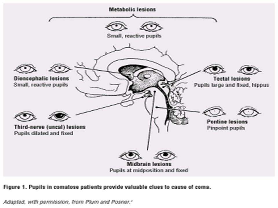
Assessment of arousal/cognition Vision & Pupillary light reflex
Sensory: CN II - Optic Visual acuity Motor: CN III - Oculomotor PERRL Direct/consensual EOMs (CN IV/VI) Compare pupil size, shape, movement, and reactivity. CN III compressed pupil on that/ipsilateral side dilates. Pin point pupils – pons or meds. Fixed pupil unresponsive to light – IICP, nerve injury, previous surgery, or mydriatic eye gtts.
 Compare pupil size, shape, movement, and reactivity. CN III compressed pupil on that/ipsilateral side dilates. Pin point pupils – pons or meds. Fixed pupil unresponsive to light – IICP, nerve injury, previous surgery, or mydriatic eye gtts. v=cuZXz92hd8g&feature=relate.")
22
Assessment Arosual/cognition EOM’S & Brain stem function
Eye movement CN III,IV,VI Oculocephalic reflex Doll’s eyes Sensory CN VIII Motor CN III,IV,VI Dolls eyes (+) opposite direction intact brain stem (-) no movement Eye movement 3,4,& 6 also tests brain stem function Corneal reflex – CN 5 & 7
 opposite direction. intact brain stem. (-) no movement. Eye movement 3,4,& 6 also tests brain stem function. Corneal reflex – CN 5 & 7.")
23
Cranial Nerve Assessment
Trigeminal (V) Corneal reflex Sensory mastication Facial (VII) Expression Taste Acoustic (VIII) Glossopharyngeal (IX) Gag/swallow Vagus (X) Gag/Swallow Spinal Accessory (XI) Shoulder shrug Hypoglossal (XII) TML
 Corneal reflex. Sensory. mastication. Facial (VII) Expression. Taste. Acoustic (VIII) Glossopharyngeal (IX) Gag/swallow. Vagus (X) Gag/Swallow. Spinal Accessory (XI) Shoulder shrug. Hypoglossal (XII) TML.")
24
Motor Ability to move, strength, and symmetry Coordination
Grips, arm strength, & drift Planter flexion, dorsiflexion, & leg strength Coordination Finger to nose, heel up and down shin Planter Reflex- Babinski testing Meningeal signs- Brudzinski & Kernig’s sign Motor- squeeze nurse’s hands, palmar drift, raise foot off bed Resistance to movement during ROM or movement to painful stimuli also indicate ability to move and strength

25
Planter Reflex and Babinski testing
Babinski's reflex (+) great toe flexes and the other toes fan out Abnormal after the age of 2.
 great toe flexes and the other toes fan out. Abnormal after the age of 2.")
26
Meningeal signs- Brudzinski, nuchal rigidity
Hips and knees flex when the neck is flexed

27
Meningitis signs- Kernig’s sign
Stiffness of the hamstrings causes an inability to straighten the leg when the hip is flexed to 90 degrees.

28
Neuro assessment - Sensation
Visual fields Dull vs. sharp Sensation same or different with eyes closed Face Hands Arms Abdomen Feet Legs

29
Homonculus

30
Assessment – Respiratory Changes
Brainstem compression Yawning & sighing Cheyne-Stokes Central neurogenic hyperventilation Apneustic breathing Cluster breathing Ataxic respirations

31
Assessment Ec

32
Question A patient with an intracranial problem does not open his eyes to any stimulus, has no verbal response except muttering when stimulated, and flexes his arm in response to painful stimuli. The nurse records the patient’s GCS score as A. 6 B. 8 C. 9 D. 11

33
Question The nurse recognizes the presence of Cushing’s triad in the patient with A. increased pulse, irregular respiration, increased BP B. decreased pulse, irregular respiration, increased pulse pressure C. Increased pulse, decreased respiration, increased pulse pressure D. decreased pulse, increased respiration, decreased systolic BP

34
Question CN III originating in the midbrain is assessed by the nurse for an early indication of pressure on the brainstem by A. assessing for nystagmus B. testing the corneal reflex C. testing pupillary reaction to light D. testing for oculocephalic (doll’s eyes) reflex
 reflex.")
35
Question An unconscious patient with increased ICP is on ventilatory support. The nurse notifies the healthcare provider when arterial blood gas (ABG) measurement results reveal a A. pH of 7.43 B. SaO2 of 94% C. PaO2 of 50mm Hg D. PaCO2 of 30mm Hg
 measurement results reveal a. A. pH of B. SaO2 of 94% C. PaO2 of 50mm Hg. D. PaCO2 of 30mm Hg.")
36
Diagnostics R/O and identify cause of LOC
BG Electrolytes/Osmolali ty ABGs CBC Liver function Kidney function Toxicology CT MRI EEG Cerebral angiogram TCD LP

38
Increased Intracranial Pressure

39
ICP Concepts Monro-Kellie hypothesis Autoregulation 80/10/10 rule
Cerebral arterioles MAP (Mean arterial pressure) Perfusion depends on B/P and chemical (CO2) Normal MAP is 70 to 100 < 60 - peripheral organs not perfused < 50 – brain not perfused Critical to maintain normal MAP with Increased ICP
 Perfusion depends on B/P and chemical (CO2) Normal MAP is 70 to 100. < 60 - peripheral organs not perfused. < 50 – brain not perfused. Critical to maintain normal MAP with Increased ICP.")
40
Compensatory Mechanisms
Vasoconstriction Decreased CSF CSF shunting Increased CSF reabsorption

41
Compliance Brain’s ability to tolerate an increase in volume without an increase in pressure Indications of decreased compliance: Sustained increase in ICP in response to stimuli Greater increases to non-noxious stimuli

42
Normal Pressure v Compensated v Uncompensated
NP Compensated Uncompensated 10mmHg 15mmHg 30mmHg Blood 10%, CSF 10% Blood 5%, CSF 5% Blood 4%, CSF 4% Stable Stiff ICP increases

43
“Rules” of Compensation
A slowly expanding mass is tolerated better that a rapidly expanding mass Brain tissue is compressible, but functional impairment, possibly irreversible does occur Location matters

44
Cerebral Perfusion Pressure
Pressure needed for adequate blood flow to brain CPP = MAP – ICP Need higher MAP if ICP increased mmHg <50 mmHg = ischemia <30 mmHg = death

45
MAP – ICP = CPP Autoregulation Danger of CPP < 50 mmHg
Arterial Blood Pressure - Brain & CS Fluid Compression = Actual Cerebral Blood Flow CPP 70 to 100 mmHg Danger of CPP < 50 mmHg MAP 50 to 150 mmHg Autoregulation Edema, CS Fluid, Tumor Increased ICP > 20 mmHg Normal ICP 0 to 15 mmHg Increased MAP needed to perfuse brain

46
Pathophysiology Changes in contents of cranial vault

47
Causes of Increased ICP
Mass effect Tumor Blood clot Edema Increased CBF Increased blood flow Increased PaCO2 Decreased PaO2 Vasodilators Increased intrathoracic pressure Coughing Straining Suctioning Peep Impairment of cerebral venous drainage Positioning

48
ICP indicators Changes in LOC Worsening headache Cognitive deficits
Pupillary changes Increasing B/P with widening pulse pressure Irregular respiratory patterns Bradycardia Seizures Aphasia Dysconjugate gaze Hemiparesis or hemiplegia

49
Assessment Health history- assess brain involvement PE
Altered cerebral function assessment Frequency depends on potential IICP Early sign- change in LOC 3rd Cranial nerve compression Papilledema Projectile vomiting Vision changes Seizures Late sign- Cushing VS changes

50
Pertinent Nursing Problems and Interventions
Ineffective tissue perfusion: cerebral Assess/report sign IICP Adequate airway Promote venous drainage Control environment stimuli Plan nursing care – avoid clustering care Avoid Valsalva’s maneuver If bone flap out post op- assess & position Assess external shunts/drains

51
Medical Management Concurrent Nursing Care
Maintenance of airway and ventilation Endotracheal intubation Oxygenation Mechanical ventilation Fluid balance/Euvolemia Medications

52
Medications Sedation, analgesia, neuromuscular blockade
Barbiturate coma Prophylactic anticonvulsant Mannitol/3% NaCl Lasix Atracrium Vasopressors Tylenol

53
Medical Management Concurrent Nursing Care
Temperature control Electrolyte balance Proper positioning Adequate nutrition Ventriculostomy Paralytics Hypothermia Pentobarbital coma Craniectomy

54
Surgical Intervention

55
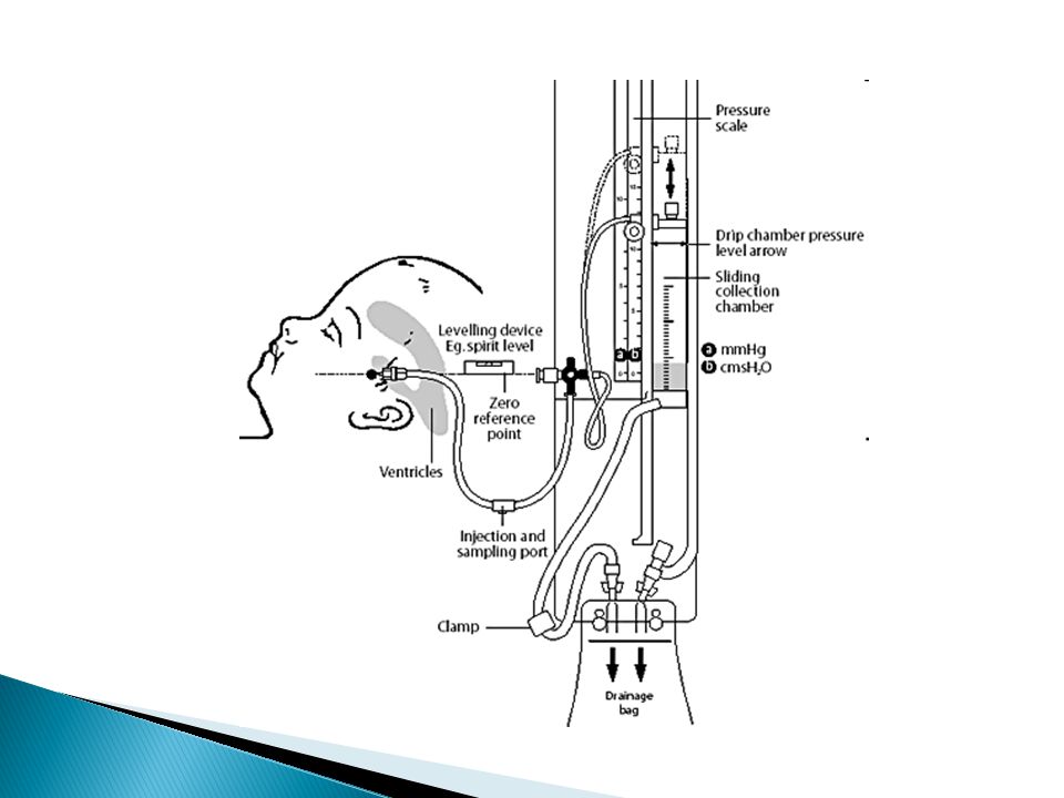
Intracranial monitoring
LICOX PbtO2 Normal mmHg Jugular venous bulb cath SjvO2 Normal SjvO2 is 60% to 80% <50 to 55% of O2 in venous blood indicates impairment of flow and brain taking out more O2 than normal ICP Waveforms (P1, P2, & P3) P1 arterial pulse wave should be highest P2 is intracranial compliance – if higher than P1 compliance is compromised P3 is the venous pulsation and should be the lowest P1 P2 P3
 P1 arterial pulse wave should be highest. P2 is intracranial compliance – if higher than P1 compliance is compromised. P3 is the venous pulsation and should be the lowest. P1 P2 P3.")
56
Intraventricular and subarachnoid monitoring devices for IICP

57
Ventriculostomy (EVD)
")
58
Standing Orders Per hospital policy

59
Deterioration/Complications
Neurological Meningitis Seizures Cerebral salt wasting (CSW) Syndrome of inappropriate antidiuretic hormone (SIADH) Hydrocephalus Cerebral edema/Increased ICP
 Syndrome of inappropriate antidiuretic hormone (SIADH) Hydrocephalus. Cerebral edema/Increased ICP.")
60
Syndrome of Inappropriate ADH (SIADH)
Increased secretion of ADH from abnormal stimuli Results in water retention Hyponatremia Na+ excreted in urine

61
Signs & Symtoms (SAIDH)
Decreased UOP Increased urine specific gravity Low serum osmo Hyponatremia Hypervolemia

62
Management Fluid restrictiion Replace sodium Diuretics Democlocycline
Fludrocortisone Hypertonic saline Oral salt Diuretics

63
Cerebral Salt Wasting Controversial Hyponatremia
Failure of CNS to regulate Na+ reabsorption Increase in circulating atrial natriuretic peptide (ANP)
")
64
Signs & Symptoms (CSW) Increased UOP Hyponatremia
Normal to increased osmo Hypovolemia Increased urine specific gravity

65
Management Volume replacement Sodium replacement
Reducing renal Na+ excretion Fludrocortisone Urea

66
SIADH v CSW Parameter SIADH CSW Serum Na+ Decreased Serum osmolarity
Urine Na+ Increased Normal-increased Urine OP Volume Normo/hypervolemic Hypovolemic Body weight

67
Cerebral Edema Hydrocephalus
Vasogenic Cytotoxic interstitial Hydrocephalus Noncommunicating Communicating ICP Production – choroid plexus; Absorption – arachniod villi

68
Hydrocephalus Normal MRI Brain MRI Hydrocephalus

70
Ventriculoperitneal Shunt

71
Manifestations/Complications
Irreversible coma Persistent vegetative state Locked-in Syndrome (not true coma) Functioning RAS & cortex; pons level interference Aware, communicate with eyes Brain death Loss of all brain function- flat EEG, no blood flow
 Functioning RAS & cortex; pons level interference. Aware, communicate with eyes. Brain death. Loss of all brain function- flat EEG, no blood flow.")
72
Brain Herniation A. Cingulate B. Uncal C. Central D. Extracranial
E. Tonsillar

73
Cingulated Herniation (a)
Cingulate gyrus slips under falx cerebri Usually caused tumor or bleed Non life threatening

74
Uncal or Lateral Herniation (b)
Uncus of temporal lobe slips through notch of tentorium and compresses the ipsilateral CN 3, brainstem, & vital centers Life threatening

75
Central or Transtentorial Herniation (c)
Downward pressure General cerebral edema Brainstem compression Compresses RAS & vital centers Abnormal heart rhythms, disturbances or cessation of breathing, cardiac arrest, and death Life threatening

76
Infratentorial (subtentorial or Tonsillar) Herniation (e)
Downward displacement of infratentorial structures through the foramen magnum Life threatening

77
Extracranial Herniation (d)
Occurs with displacement of brain through a cranial defect. Usually Non-life threatening

78
Surgical Decompression (Craniectomy)

79
CT s/p craniectomy

80
Question A patient has ICP monitoring with an intraventricular catheter. A priority nursing intervention for the patient is A. aseptic technique to prevent infection B. constant monitoring of ICP waveforms C. removal of CSF to maintain normal ICP D. sampling CSF to determine abnormalities

81
Question A patient has a nursing diagnosis of altered cerebral tissue perfusion related to cerebral edema. An appropriate nursing intervention for the patient is A. avoiding positioning the patient with neck and hip flexion B. maintaning hyperventilation to a PaCO2 of mm Hg C. clustering nursing activities to provide periods of uniterrupted rest D. routine suctioning to prevent accumulation of respiratory secretions

82
Question The earliest signs of increased ICP the nurse should assess for include A. Cushing’s triad B. unexpected vomiting C. decreasing level of consciousness (LOC) D. dilated pupil with sluggish response to light
 D. dilated pupil with sluggish response to light.")
83
Nursing Evaluation VS/NVS ICP CPP MAP PbtO2 PaCO2 CVP Labs Imaging

84
Legal/Ethical Considerations
Category status Advanced directives Prognosis Withdraw of care Palliative care End of life specialists SW/Chaplain

85
Prognosis Varies according to underlying cause and pathologic process
GCS GOS Physical/mental disability

86
Comic relief

87
More comic relief

88
Case Study Day 2 22 yo female Harvard law student Day 3
ICP Hypothermia Tracrium Day 3 Flexion 22 yo female Harvard law student Horseback riding GCS 7 Localized

89
References AANN Core Curriculum for Neuroscience Louis, MO. Nursing, 4th Ed Saunders. St. Davis, F.A. (2001). Taber’s Cyclopedic Medical Dictionary. F.A. Davis, Philadelphia. Greenberg, Mark. (2006). Handbook of Neurosurgery. Greenberg Graphics, Tampa, Florida. Lewis, S., Heitkemper, M., O’Brien, P., Bucher, L. (2007). Medical-Surgical Nursign. Assessment of Management of Medical Problems. Mosby Elsevier, St. Louis, Missouri Silvestri, Linda. (2008). Comprehensive review for the NCLEX-RN Examination. Saunders Elsevier, St. Louis, Missouri.
. Taber’s Cyclopedic Medical Dictionary. F.A. Davis, Philadelphia. Greenberg, Mark. (2006). Handbook of Neurosurgery. Greenberg Graphics, Tampa, Florida. Lewis, S., Heitkemper, M., O’Brien, P., Bucher, L. (2007). Medical-Surgical Nursign. Assessment of Management of Medical Problems. Mosby Elsevier, St. Louis, Missouri. Silvestri, Linda. (2008). Comprehensive review for the NCLEX-RN Examination. Saunders Elsevier, St. Louis, Missouri.")
Similar presentations


 Marnie Quick, RN, MSN, CNRN.>")


