Download presentation
Presentation is loading. Please wait.

1
The hypercoagulability panel in stroke: Which tests should be done?
Michael Rippee, MD Assistant Professor Department of Neurology University of Kansas Medical Center

2
A. Yes B. No Case #1: JS 38 y/o woman
Sudden onset dizzness, right sided HA, slurred speech, diplopia, left arm weakness, right sided facial droop H/o headaches MRI reveals right thalamic infarct Hypercoagulable panel? A. Yes B. No

3
Case #1: JS AT3 126 (H) (nl = 80-120) Cardiolipin
IgG 3.6 IgM 14.9 (H) (nl = <12.5) Activated Protein C Activity nl Protein C & S nl DRVVT nl Hex lupus anticoagulant not done Factor 2 gene mutation neg ANA neg Beta2 glycoprotein neg Homocysteine nl
 (nl = <12.5) Activated Protein C Activity nl. Protein C & S nl. DRVVT nl. Hex lupus anticoagulant not done. Factor 2 gene mutation neg. ANA neg. Beta2 glycoprotein neg. Homocysteine nl.")
4
Stroke Causes in Young Which is most common? A. Hypercoagulable State
B. Cardioembolism C. Atherosclerotic factors D. Other determined causes (e.g. dissection)
")
5
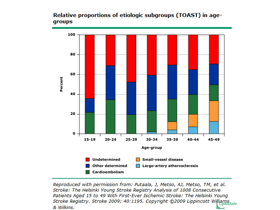
Risk factors: Young Adult
Most frequent risk factors: Dyslipidemia 60% Smoking 44% Hypertension 39% Most common etiologies: Cardioembolism 20% Cervicocephalic arterial dissection 15% Proportions of small vessel disease (14%) and large artery atherosclerosis (8%) increased beginning at age 30 to 35 Frequency of undetermined etiology (33%) decreased with age
 and large artery atherosclerosis (8%) increased beginning at age 30 to 35. Frequency of undetermined etiology (33%) decreased with age.")
7
A. Yes B. No Case #2: TW 34 y/o man
Nausea, vision loss (quadrantanopia), HA MRI revealed right occipito-temporal infarct No fam hx Otherwise healthy LDL 163 Hypercoagulable panel? A. Yes B. No
, HA. MRI revealed right occipito-temporal infarct. No fam hx. Otherwise healthy. LDL 163. Hypercoagulable panel A. Yes B. No.")
8
Case #2: TW AT3 nl Factor 8 nl Cardiolipin Abs nl
Activated Protein C nl Protein C & S nl DRVVT nl Hex lupus anticoagulant not done Factor 2 gene mutation neg Homocysteine nl ANA neg MTHFR neg Angiogram confirmed vertebral artery dissection

9
Hypercoagulabilty & Stroke
The role of hypercoagulable states in strokes is controversial Blood disorders implicated 5-10% In patients without other traditional risk factors and etiologies for stroke, hypercoagulable state should be considered Abnormal findings on routine screening coagulation tests (aPTT) should also raise a red flag Hypercoagulable state may be more important in the younger patient with stroke
 should also raise a red flag. Hypercoagulable state may be more important in the younger patient with stroke.")
10
Hypercoagulabilty & Stroke
Hypercoagulability should be suspected in patients with ischemic stroke who have the following characteristics: Younger than 50 years with no obvious cause of stroke History of multiple unexplained strokes Previous history of venous thrombosis Family history of thrombosis Abnormalities on routine screening coagulation tests

11
Thrombosis Virchow’s triad for venous thrombosis

12
Hypercoagulable State
Defined as a group of inherited or acquired conditions associated with a predisposition to: Venous thrombosis including: Upper and lower extremity deep venous thrombosis with or without pulmonary embolism Cerebral venous thrombosis Intra-abdominal venous thrombosis Arterial thrombosis including: Myocardial infarction Stroke Acute limb ischemia Splanchnic ischemia Venous thromboembolic disease is the most common Most inherited conditions appear to increase only the risk of venous thrombosis Some of the acquired conditions have been associated with both venous and arterial thrombosis Age of onset for initial thrombotic event is typically before age 45

13
Hypercoagulable State
Recent data support the role of more than one prothrombotic mutation or additional acquired conditions to be necessary for clinically apparent clotting Immobilization Surgery Cancer Pregnancy Use of hormone therapy or oral contraceptive medication Clues to the diagnosis: History of recurrent fetal loss Thrombocytopenia Livedo reticularis or Sneddon syndrome Skin necrosis during initiation of oral anticoagulant therapy Protein C and/or S deficiency

15
Hypercoagulable States
Inherited Acquired Factor V Leiden Prothrombin gene mutation Anti-thrombin deficiency Protein C & S deficiencies Elevated homocysteine Dysfibrinogenemia Elevated Factor VIII levels Abnormal fibrinolytic system Sickle Cell disease Antiphospholipid antibody syndrome Supplemental estrogen use HIT Cancer Medications Central venous catheter Obesity Pregnancy

16
Hypercoagulable workup
PT and PTT Protein C Protein S Antithrombin III activity Prothrombin gene mutations Factor V Leiden gene mutation Activated Protein C resistance Anticardiolipin antibodies (IgG and IgM) Beta2-glycoprotein I antibodies (IgG and IgM) Lupus anticoagulant tests dilute Russell viper venom time dilute activated PTT hexagonal phospholipid Homocysteine Factor VIII activity D-dimer Lipoprotein (a) MTHFR
 Beta2-glycoprotein I antibodies (IgG and IgM) Lupus anticoagulant tests. dilute Russell viper venom time. dilute activated PTT. hexagonal phospholipid. Homocysteine. Factor VIII activity. D-dimer. Lipoprotein (a) MTHFR.")
17
Hypercoagulable workup
How much does it cost? A. $450 B. $900 C. $2200 D. $5000

18
Copyright © 2012 American Medical Association. All rights reserved.
From: Hypercoagulability Syndromes Arch Intern Med. 2001;161(20): doi: /pubs.Arch Intern Med.-ISSN ira00051 Figure Legend: $ Costs of Hypercoagulable Workup at the University of Miami Date of download: 9/25/2012 Copyright © 2012 American Medical Association. All rights reserved.
: doi: /pubs.Arch Intern Med.-ISSN ira Figure Legend: $ Costs of Hypercoagulable Workup at the University of Miami. Date of download: 9/25/2012. Copyright © 2012 American Medical Association. All rights reserved.")
19
Hypercoagulable workup: Cost
A lot of facilities conduct these as send-out tests Quest $1300-$2500 (avg $2000) Pts bill $2200-$3000 Safe estimates are $2-4K Generally the panel is repeated at least once!
 Pts bill $2200-$3000. Safe estimates are $2-4K. Generally the panel is repeated at least once!")
20
So which test to order?

21
Case #3: TS 35 y/o woman Severe, unrelenting HA
Venous sinus thrombosis On OCPs No Fam Hx

22
What tests to order? A. Routine stroke, but no hypercoagulable B. Full hypercoagulable panel C. Anti-cardiolipin, lupus anticoagulant panels only D. Protein C & S, AT3, Factor V Leiden, Prothrombin mutation only E. None

23
Case #3: TS Cardiolipin nl Protein C&S nl DRVVT nl
Hex lupus anticoagulant 11 (H) (nl <8) Factor 2 mutation neg ANA neg RF neg Beta2 glycoprotein nl 9 mos later Hex lupus 6.1 DRVVT 47.6
 (nl <8) Factor 2 mutation neg. ANA neg. RF neg. Beta2 glycoprotein nl. 9 mos later. Hex lupus 6.1. DRVVT")
24
Case #4: JR 45 y/o man Left hemiparesis & facial droop
MRI showed right pontine infarct LDL 129, quit smoking 3 weeks prior to stroke No Fam Hx

25
What tests to order? A. Routine stroke, but no hypercoagulable B. Full hypercoagulable panel C. Anti-cardiolipin, lupus anticoagulant panels only D. Protein C & S, AT3, Factor V Leiden, Prothrombin mutation only E. None

26
Case #4: JR Antiphospholipid Ab Syndrome October Cardiolipin
IgG 6.8 IgM 13.0 (H) (nl = <12.5) Protein C & S nL November AT3 nl DRVVT nl Homocysteine 18.6 (H) (nl <15) Beta2 glycoprotein IgG 16 (H) (nl <15) IgM nl IgA nl Alphagalactosidase (Fabry’s) ordered but not done due to cost April Cardiolipin tests normalized Factor 8 nL Activated Protein C nl MTHFR neg Hex Lupus Anticoagulant nl Factor 2 mutation neg Factor V Leiden ordered but not done Antiphospholipid Ab Syndrome
 (nl = <12.5) Protein C & S nL. November. AT3 nl. DRVVT nl. Homocysteine 18.6 (H) (nl <15) Beta2 glycoprotein IgG 16 (H) (nl <15) IgM nl. IgA nl. Alphagalactosidase (Fabry’s) ordered but not done due to cost. April. Cardiolipin tests normalized. Factor 8 nL. Activated Protein C nl. MTHFR neg. Hex Lupus Anticoagulant nl. Factor 2 mutation neg. Factor V Leiden ordered but not done. Antiphospholipid Ab Syndrome.")
27
Which tests to order? Keep in mind whether the results will influence therapy and/or patient outcome Not advocated to screen all stroke patients for a “hypercoagulable workup” Typically, will have a prior history of one or more unexplained thromboembolic events Yield for diagnosing a hypercoagulable state is typically greatest for: Young stroke patients Family history of thrombosis No other explanations for their stroke (cryptogenic stroke) Assaying for specific prothrombotic states has limitations based on the assay and the timing of the test Levine SR. Hypercoagulable States & Stroke: A Selective Review. CNS Spectr. 2005;10(7):
 Assaying for specific prothrombotic states has limitations based on the assay and the timing of the test. Levine SR. Hypercoagulable States & Stroke: A Selective Review. CNS Spectr. 2005;10(7):")
28
Which tests to order? Blood work to diagnose a hypercoagulable state does not preclude the routine work up of any stroke patient: Neuroimaging (brain CT and MRI) Carotid ultrasound Echocardiogram Basic blood tests including a CBC, prothrombin time (PT), partial thromboplastin time (aPTT), and a fasting lipid profile Important points to be noted before ordering a work-up for hypercoagulable state: Use of anticoagulation can affect results of aCL, LA, protein C, protein S, and antithrombin III Results should be repeated in 4–8 weeks to exclude false positives that may be related to an acute phase reaction Vaishnav, AG. (2008). “Hypercoagulable States and Stroke.” In D. Alway, J.W. Cole (Eds.). Stroke Essentials for Primary Care. Human Press.
 Carotid ultrasound. Echocardiogram. Basic blood tests including a CBC, prothrombin time (PT), partial thromboplastin time (aPTT), and a fasting lipid profile. Important points to be noted before ordering a work-up for hypercoagulable state: Use of anticoagulation can affect results of aCL, LA, protein C, protein S, and antithrombin III. Results should be repeated in 4–8 weeks to exclude false positives that may be related to an acute phase reaction. Vaishnav, AG. (2008). Hypercoagulable States and Stroke. In D. Alway, J.W. Cole (Eds.). Stroke Essentials for Primary Care. Human Press.")
29
Which disorders associated with stroke?
Prothrombotic states implicated in ischemic stroke: Deficiencies of factors inhibiting coagulation Antithrombin III, protein S, and protein C Increased levels of factors promoting coagulation Factors V and VII Decreased activity in the fibrinolytic pathway Plasminogen or plasminogen activator deficiencies

30
Association with stroke
Inherited thrombophilias (eg, protein C, protein S, or antithrombin III deficiency; factor V Leiden; prothrombin G20210A mutation), and MTHFR Rarely contribute to adult stroke May play a larger role in pediatric stroke Studies in younger patients (<55 years of age) have shown an association between prothrombotic genetic variants and ischemic stroke Remains controversial in an older population with vascular risk factors Even in the young, results have been inconsistent Small study of cryptogenic stroke patients <50 years of age Increased risk associated with the PT G20210A mutation No significant association with FVL 2 other studies of young (<50 years) patients Found no association between ischemic stroke and the FVL, PT G20210A, or MTHFR The association between APL antibodies and stroke is strongest for young adults (<50 years of age) Furie KL, et al. Guidelines for the Prevention of Stroke in Patients with Stroke or Transient Ischemic Attack: A Guideline for Healthcare Professionals. Stroke. 2011;42:
, and MTHFR. Rarely contribute to adult stroke. May play a larger role in pediatric stroke. Studies in younger patients (<55 years of age) have shown an association between prothrombotic genetic variants and ischemic stroke. Remains controversial in an older population with vascular risk factors. Even in the young, results have been inconsistent. Small study of cryptogenic stroke patients <50 years of age. Increased risk associated with the PT G20210A mutation. No significant association with FVL. 2 other studies of young (<50 years) patients. Found no association between ischemic stroke and the FVL, PT G20210A, or MTHFR. The association between APL antibodies and stroke is strongest for young adults (<50 years of age) Furie KL, et al. Guidelines for the Prevention of Stroke in Patients with Stroke or Transient Ischemic Attack: A Guideline for Healthcare Professionals. Stroke. 2011;42:")
31
Hereditary Hypercoagulable Disorders
Factor V Leiden mutation Most common hereditary hypercoagulable disorder associated with cerebral venous thrombosis Scant evidence of its association with arterial strokes Caused by a mutation that makes Factor V resistant to inactivation by activated Protein C (APC resistance) APC resistance can also be induced by pregnancy and estrogen Homozygous forms are much more prone to thrombosis than a heterozygous mutation Vaishnav, AG. (2008). “Hypercoagulable States and Stroke.” In D. Alway, J.W. Cole (Eds.). Stroke Essentials for Primary Care. Human Press.
 APC resistance can also be induced by pregnancy and estrogen. Homozygous forms are much more prone to thrombosis than a heterozygous mutation. Vaishnav, AG. (2008). Hypercoagulable States and Stroke. In D. Alway, J.W. Cole (Eds.). Stroke Essentials for Primary Care. Human Press.")
32
Hereditary Hypercoagulable Disorders
Antithrombin III, Protein C, and Protein S deficiency: These conditions are relatively rare More potent cause of cerebral venous thrombosis than Factor V Leiden No evidence of their association with arterial strokes Prothrombin gene mutation (G20210A) Occurs in approximately 2–5% of individuals and in itself is a weak procoagulant in its action
 Occurs in approximately 2–5% of individuals and in itself is a weak procoagulant in its action.")
33
Hereditary Hypercoagulable Disorders
Protein C Used to screen for a primary protein C deficiency or to diagnose protein C deficiency secondary to dysproteinemia To confirm protein C deficiency, and to differentiate it from dysproteinemia, the protein C antigen is measured Protein S Activity is measured by a functional assay Both the total Protein S and free Protein S functional assays are performed because the free assay is a more reliable marker for hypercoagulability To confirm protein S deficiency, and to differentiate it from dysproteinemia, the protein S antigen is measured Antithrombin III It is recommended to repeat the level in 4–6 weeks if a deficiency was initially found in the setting of an acute thrombotic event, pregnancy, or warfarin use Vaishnav, AG. (2008). “Hypercoagulable States and Stroke.” In D. Alway, J.W. Cole (Eds.). Stroke Essentials for Primary Care. Human Press.
. Hypercoagulable States and Stroke. In D. Alway, J.W. Cole (Eds.). Stroke Essentials for Primary Care. Human Press.")
34
Antiphospholipid Antibody (aPL) Syndrome
Antiphospholipids have been associated with both arterial and venous strokes The two major types of clinically relevant aPLs: Anticardiolipin antibodies (aCLs) Require the presence of serum cofactor beta-2 glycoprotein for binding Lupus anticoagulant (LA) May not require the presence of beta-2 glycoprotein About 70% of patients with aPS have both aCL and LA. Antiphospholipid antibody syndrome (APS) is defined as both: Thrombosis or recurrent, unexplained fetal loss AND aCLs (IgG or IgM) of medium to high titres or LA on at least two occasions at least 8 weeks apart Patients with primary APS do not have systemic lupus erythematosus (SLE) or any other underlying autoimmune disorders Patients with aPLs suffer from both venous and arterial strokes Vaishnav, AG. (2008). “Hypercoagulable States and Stroke.” In D. Alway, J.W. Cole (Eds.). Stroke Essentials for Primary Care. Human Press.
 Require the presence of serum cofactor beta-2 glycoprotein for binding. Lupus anticoagulant (LA) May not require the presence of beta-2 glycoprotein. About 70% of patients with aPS have both aCL and LA. Antiphospholipid antibody syndrome (APS) is defined as both: Thrombosis or recurrent, unexplained fetal loss AND. aCLs (IgG or IgM) of medium to high titres or LA on at least two occasions at least 8 weeks apart. Patients with primary APS do not have systemic lupus erythematosus (SLE) or any other underlying autoimmune disorders. Patients with aPLs suffer from both venous and arterial strokes. Vaishnav, AG. (2008). Hypercoagulable States and Stroke. In D. Alway, J.W. Cole (Eds.). Stroke Essentials for Primary Care. Human Press.")
35
Antiphospholipid Antibody (aPL) Syndrome
 Syndrome")
36
Cerebral Venous Sinus Thrombosis
Routine blood studies: CBC, chemistry panel, PT and aPTT should be performed Screening for potential prothrombotic conditions that may predispose a person to CVT Use of contraceptives, underlying inflammatory disease, infectious process Testing for prothrombotic conditions Testing for protein C, protein S, and antithrombin deficiency is generally indicated 2 to 4 weeks after completion of anticoagulation There is a very limited value of testing in the acute setting or in patients taking warfarin Saposnik G, et al. Diagnosis & Management of Cerebral Venous Thrombosis. Stroke. 2011;42:

37
Strategies for testing

38
Which tests to order?

39
Use of screening tests Screening Test Confirmatory Test
Activated Protein C Resistance Factor V Leiden PCR Antithrombin, Protein C & S activity (functional) levels Antigenic assays DRVVT Lupus anticoagulant
 levels. Antigenic assays. DRVVT. Lupus anticoagulant.")
40
Selecting tests Use of pre-test probablity, appropriate selection of patients, and sensitivity/specifity yield higher post-test probabilities

41
Bushnell CB, Goldstein LB
Bushnell CB, Goldstein LB. Screening for Hypercoagulable Syndromes Following Stroke. Current Atherosclerosis Reports. 2003, 5:

42
Common mistakes Ordering too many tests Testing in the acute phase
Specifically ones with a low pre-test probability Duplicate tests (APC and FVL) Testing in the acute phase Testing while on warfarin or heparin
 Testing in the acute phase. Testing while on warfarin or heparin.")
43
Summary Common causes of venous thrombosis are unlikely to cause stroke Activated Protein C resistance/Factor V Leiden Protein C, Protein S, Antithrombin Prothrombin mutation This “typical” hypercoagulable panel is low yeild in arterial stroke These tests are more high yield in CVT In young patients without known etiology/risk factors anticardiolipin and lupus anticoagulant are high yield Should also order beta-2 glycoproteins MTHFR and homocysteine are not helpful Testing in the acute phase can be misleading Testing should be done off of heparin or warfarin

44
Questions

45
JM 58 y/o man H/o RA and SLE HLD and tobacco
Driving back from vacation Developed left sided weakness, facial droop MRI revealed right hemisphere ischemic stroke No Fam Hx

46
What tests to order? A. Routine stroke, but no hypercoagulable B. Full hypercoagulable panel C. Anti-cardiolipin, lupus anticoagulant panels only D. Protein C & S, AT3, Factor V Leiden, Prothrombin mutation only E. None

47
Hex Lupus Anticoagulant 31 (H) (nl = <8) Cardiolipin
Prolonged aPTT DRVVT 1.6 (H) (nl = <1.2) Hex Lupus Anticoagulant 31 (H) (nl = <8) Cardiolipin IgG 9.616.2 IgM 20.615.2 Homocysteine 10.7 Anti-phospholipid Ab Syndrome
 (nl = <1.2) Hex Lupus Anticoagulant 31 (H) (nl = <8) Cardiolipin. IgG 9.616.2. IgM 20.615.2. Homocysteine Anti-phospholipid Ab Syndrome.")
48
TP 44 y/o woman Admitted with prolonged, severe headache
Found to have extensive venous sinus thrombosis On OCP, also on HCG diet (shown to increase potential for thrombus) No Fam Hx
 No Fam Hx.")
49
What tests to order? A. Routine stroke, but no hypercoagulable B. Full hypercoagulable panel C. Anti-cardiolipin, lupus anticoagulant panels only D. Protein C & S, AT3, Factor V Leiden, Prothrombin mutation only E. None

50
Activated PC resistance nL Hex Lupus Anticoagulant nL DRVVT nL
Factor 8: 140 (nl ) Cardiolipin Ab IgG 4.0 IgM 8.2 Activated PC resistance nL Hex Lupus Anticoagulant nL DRVVT nL Protein C nL Protein S nL Factor 2 mutation neg AT3 130 (H) (nl = ) Beta-2 glycoprotein neg
 Cardiolipin Ab. IgG 4.0. IgM 8.2. Activated PC resistance nL. Hex Lupus Anticoagulant nL. DRVVT nL. Protein C nL. Protein S nL. Factor 2 mutation neg. AT3 130 (H) (nl = ) Beta-2 glycoprotein neg.")
51
GM 62 y/o man Sudden onset homonymous hemianopia, dizziness/imbalance
MRI confirmed left PCA infarct H/o hypertension & HLD

52
What tests to order? A. Routine stroke, but no hypercoagulable B. Full hypercoagulable panel C. Anti-cardiolipin, lupus anticoagulant panels only D. Protein C & S, AT3, Factor V Leiden, Prothrombin mutation only E. None

53
AT3 nl Prothrombin gene mutation neg Homocysteine nl Cardiolipin Abs nl Activated protein C nl

54
LP 29 y/o woman Stroke in 2005, found to have PFOclosed
Had sudden vision loss on right side and difficulty forming words MRI negative Fam Hx of dysrhythmia but not stroke or blood clots

Similar presentations


 Is it a stroke? (2) What part of the brain is affected? (3) What caused this stroke? Is it a haemorrhage or an infarct? Can.>")
 SIGNS Intense, griping pain in the left chest, neck to jaw through the shoulder blades Pale, cool.>")
 Stroke - Overview Third leading cause of death in industrialized countries. Total cost of strokes in the U.S. is roughly.>")


 neurological defecit of sudden onset and lasting> 24h (or leading to death), and of presumed vascular origin 5-10 per.>")


