Download presentation
Presentation is loading. Please wait.

1
Mobile Radiography Course
Instructor :TAGELDIN ABUGROON

2
Overview RAD 323 Course Mobile Equipment features.
Mobile Image Intensifier Units (C arm) Work with Mobile Equipment in the Ward. Sterilization. Tomography. Introduction to Digital Radiography.
 Work with Mobile Equipment in the Ward. Sterilization. Tomography. Introduction to Digital Radiography.")
3
Mobile Equipment Feature

4
Introduction What is mobile x-ray equipment?
It is equipment which can be moved from one place to another and used at the patient’s bedside. What is used for? It can be used to provide facilities for the following cases: (1) In-patients who could not leave their beds, especially those who cannot be moved away from their ward. (2) Surgeons who required X-ray control guidance during the course of their work in the operation theatre.
 In-patients who could not leave their beds, especially those who cannot be moved away from their ward. (2) Surgeons who required X-ray control guidance during the course of their work in the operation theatre.")
5
These days bedridden patients who are in need of radiography are now transferred safely from their ward to the imaging department, where they can be examined with all the efficiency that the department can offer. These facilities include full radiation protection: for the patient, of course, but also for other personnel who otherwise, in the hospital ward, could have been at risk. Therefore, their use should be restricted. Despite these points, the service of mobile X-ray equipment can be in demand all day especially in a busy hospital and with complicated situation patient. Study of the principal features of their construction and operation is necessary, as always, for correct and safe use.

7
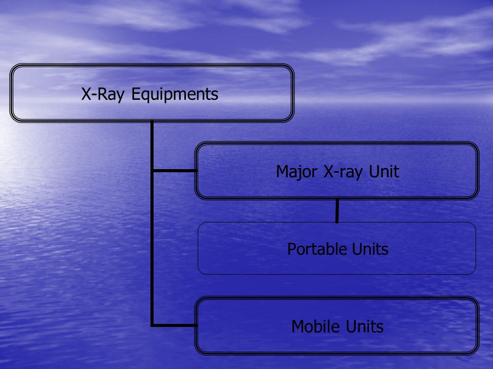
What are the differences between mobile & portable unit?
The broad distinction between the two being the difference in power output and the ability to transfer equipment. Portable units are small and light in weight, which normally can be dismantled and carried, usually by one person, and have relatively low radiation output. While Mobile x-ray units are bigger and heavier than ‘Portable’ units which is movable but because of its weight and size can only be wheeled along flat surfaces to place where required, they are usually have higher radiation output.

8
Portable unit which can be dismantled & carried by one person

9
P=I.E Portable units features Advantages: 1- Light weight.
2- Standard power supply. Disadvantages 1- Low output. 2- Lack of stability. 3- Lack of precision. The output of the machine can be determined by The following equation: Where: P= Power (Watt) I= Current (Amper) E= Voltage (Volt) P=I.E
 I= Current (Amper) E= Voltage (Volt) P=I.E.")
10
Example: Exposure factors: 100 kVp, 50 mA. Supply: 240 V
P= _50_ A × 100,000 = 5000VA 1000 I= 5000 = 21A 240 if we change the Volts to 480V, what would be happened? I= 5000 =10.5 A 480 As we increase the line voltage, the current flowing is reduced. Therefore the output of the machine will be decreased. Remember that changing the exposure factors change the current requirements. Therefore you need to know the current required and the voltage of the power supply before you do exposure.

11
Components of Portable units:
1- Oil-Filled lead lined tank: In order to make the equipment simpler, lighter, less expensive and easy to carry, it is usually constructed with: (A) X-ray tube: Self-rectified, Stationary anode. (B) High-tension transformer. (HTT) (C) Filament transformer. This is described as a tank construction and the whole enclosure is called the tube head. 2- A small control unit: (A) Rheostats: to vary the mA and kV. (B) Timer (S) (C) Exposure switches. Output: The maximum output is usually 80 kVp and 20 mA.
 X-ray tube: Self-rectified, Stationary anode. (B) High-tension transformer. (HTT) (C) Filament transformer. This is described as a tank construction and the whole enclosure is called the tube head. 2- A small control unit: (A) Rheostats: to vary the mA and kV. (B) Timer (S). (C) Exposure switches. Output: The maximum output is usually 80 kVp and 20 mA.")
12
Mobile units feature: Capable of being moved either manually or motorized drive. Advantages 1. Great stability High output. 3. Wide range of KV & mA selections. 4. The control panel and the high tension generator (with full-wave rectification) all are carried by strong machine base, away from the tube which have an advantage to greatly increased the x-ray output. 5. It has a rotating anode. 6.Flexible tube movement.
 all are carried by strong machine base, away from the tube which have an advantage to greatly increased the x-ray output. 5. It has a rotating anode. 6.Flexible tube movement.")
13
Disadvantages: 1. It is heavy in weight. 2. It needs especial maintenance. Output Up to 300 mA and maximum of 125 kVp.

14
Mobile Unit in

15
Mobile units

16
7 6 5 8 4 3 2 9 10 11 1 12

17
Construction of Mobile Unit:
1.High generation tank containing the HT transformer 2. Handle for pushing the unit. 3. Control unit Lock Tube Column. 6. Bowden cable allowing vertical movement of the cross arm. 7. Movement of the cross arm. 8. Hinge joint to allow extension of the cross arm. 9. HT cables. 10. Column base allowing rotation of the cross arm around column. 11. Base plate. 12. Large wheels for easy movement.

18
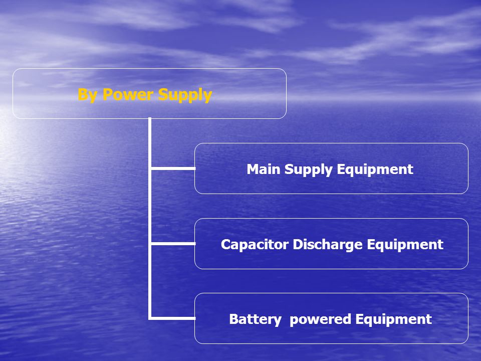
Mobile units Classification
They are classified by two ways: output & Power Supply. (A) By output: 1-Low Power Mobiles : These operate at max. x-ray current (mA) of 10 to 30 with a range of 40 to 90 kVp. Example: Dental, war x-ray equipment. 2- Average Power Mobiles : These operate at a range x-ray current (mA) of 50 to 60, 100 to 150 mA with a range of 40 to 90 kVp, 95 kVp. Example: for skull, limbs. 3-High power mobiles : These operate at max. of 300 mA, at a max. 125 kVp. Example: for CXR , abdomen, skull, Pelvimetry, IVU, pediatrics.
 By output: 1-Low Power Mobiles : These operate at max. x-ray current (mA) of 10 to 30 with a range of 40 to 90 kVp. Example: Dental, war x-ray equipment. 2- Average Power Mobiles : These operate at a range x-ray current (mA) of 50 to 60, 100 to 150 mA with a range of 40 to 90 kVp, 95 kVp. Example: for skull, limbs. 3-High power mobiles : These operate at max. of 300 mA, at a max. 125 kVp. Example: for CXR , abdomen, skull, Pelvimetry, IVU, pediatrics.")
20
Main Supply Equipment Advantages & disadvantages:
Energy may be drawn for X-ray exposure directly from the mains voltage supply. This is convenience if there is no need for the energy to be carried around within the mobile unit. But cable resistance and supply variation at different locations where the unit is used can raise difficulties affecting image quality.

21
Special Features This kind of equipment must have a robust connection cable. This encloses 3 low resistance conductors, each with a relatively large cross-sectional area. Two of the conductors carry the current, which the generator draws when an exposure is made. The third provides a safe, reliable connection to earth. The cable must be long enough (2 meter) for equipment to be used in most locations, provided there is a convenient mains outlet socket, but not so long that its resistance becomes significant. The cable should be manufactured to tolerate physical damage.
 for equipment to be used in most locations, provided there is a convenient mains outlet socket, but not so long that its resistance becomes significant. The cable should be manufactured to tolerate physical damage.")
22
Capacitor Discharge Equipment
Advantages & disadvantages 1.Small in size and light in weight, because there is no H.T.T. 2.This unit has the advantage of storing energy in the form of electric charge for later use for X-ray exposures. Therefore, it can be used in places where there is no electricity, or no a convenient mains outlet socket, or there is electrical power but not adequate (not enough to operate the X-ray unit), or suffers ‘drops’ in its values. 3.The unit has the advantage of providing high X-ray output (usually high mA). The given kVp is from 30 or 40 up to 100 to 125 kVp and 500 mAs.
, or suffers ‘drops’ in its values. 3.The unit has the advantage of providing high X-ray output (usually high mA). The given kVp is from 30 or 40 up to 100 to 125 kVp and 500 mAs.")
23
4.Shorter exposure time. By using grid-controller, this grid plate is exist between the anode and the cathode in the tube. This grid allow the electrons to pass through it and reach the anode (by carrying zero charge or no charge on it), this grid can stop the electrons passage after a specific time by carrying -ve charge on it which will reverse action and stop the exposure. 5.It has no timing control device since only the kVp has to be selected, so the mAs depends on the kVp. As the kVp in the capacitor discharge falls the mAs does so. 6.Simple in operation, it can be supplied by any voltage from 60V to 220V (so we shouldn’t worry from voltage fluctuation).
, this grid can stop the electrons passage after a specific time by carrying -ve charge on it which will reverse action and stop the exposure. 5.It has no timing control device since only the kVp has to be selected, so the mAs depends on the kVp. As the kVp in the capacitor discharge falls the mAs does so. 6.Simple in operation, it can be supplied by any voltage from 60V to 220V (so we shouldn’t worry from voltage fluctuation).")
24
7.Provides consistently reliable output and results (uniform wave), which has the advantage of less absorbed dose to the patient and good image quality. This consistency happened because the main voltage and resistance variations only have their effects upon the act of charging the capacitor, which takes place just before the X-ray exposure is made. While in the mobile unit that use the conventional generator provide the X-ray tube with a pulsating waveform voltage which varies from peak to zero then go back to peak which effect the image quality and patient protection. 8.Capacitor discharge is usually limited so that the drop in kVp is only 35% on heavy exposure. 9.The disadvantage that it has the limitation in exposures. Few times of exposure needs capacitor charge.

27
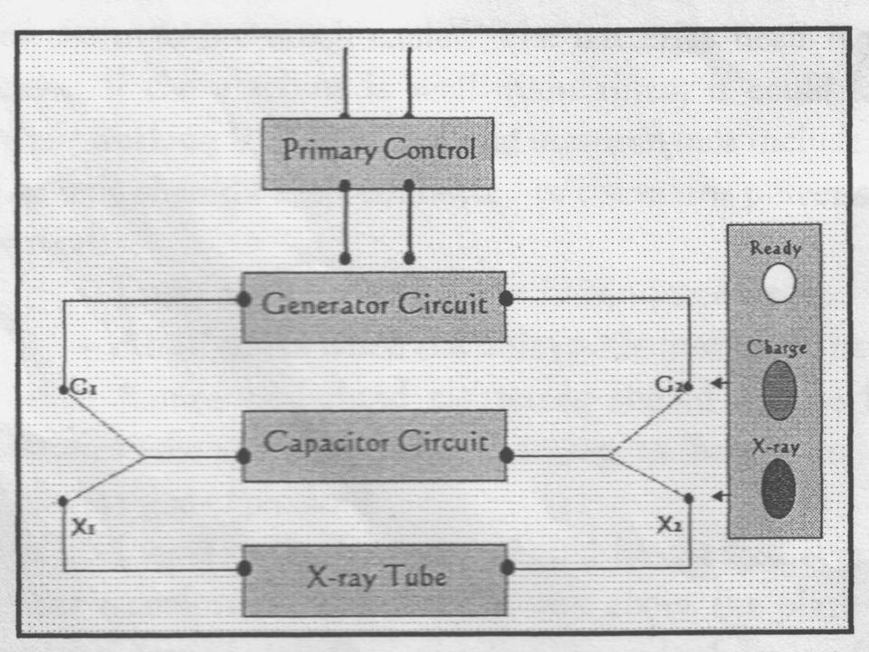
Working of the Capacitor
Charging Stage After the radiological Technologist connects the unit to the mains and selects a kVp value, the switch operated to charge the capacitor to the required kVp (G1 & G2 will be connected), via the high tension generator, then the capacitor acts as a store or source of energy. Discharging-exposure stage By pressing the exposure switch the X1 & X2 will be connected then the capacitor will discharges electrons through the X-ray tube and produce X-ray exposure.
, via the high tension generator, then the capacitor acts as a store or source of energy. Discharging-exposure stage. By pressing the exposure switch the X1 & X2 will be connected then the capacitor will discharges electrons through the X-ray tube and produce X-ray exposure.")
29
Special Features 1.Charging circuit contains solid-state rectifiers (selenium types) and capacitors these two acts as energy storing elements and as voltage multipliers (raise voltage to higher kV values, instead of the high-tension transformer). 2.The size of the capacitor determines the maximum output (mAs); this is called Capacitance Value (C.V.). For example; if the typical C.V. for charging capacitor is 1.0 the typical mAs are between mAs. After every exposure we do the mAs will be reduced. 3.Its tube has rotating anode and grid-controller.
 and capacitors these two acts as energy storing elements and as voltage multipliers (raise voltage to higher kV values, instead of the high-tension transformer). 2.The size of the capacitor determines the maximum output (mAs); this is called Capacitance Value (C.V.). For example; if the typical C.V. for charging capacitor is 1.0 the typical mAs are between mAs. After every exposure we do the mAs will be reduced. 3.Its tube has rotating anode and grid-controller.")
30
Battery Powered Generator
This is called also cordless mobile unit (mains-independent). This unit use batteries as a source of energy for X-ray exposure. Advantages: (1) This machine can be used freely, provides wide range of satisfactory kVp & mAs. (2) This machine could be recharging less frequently that the capacitor one. Some machine allows you to take up to 500 exposures without re-charging. Disadvantages: It needs special care and maintenance.
. This unit use batteries as a source of energy for X-ray exposure. Advantages: (1) This machine can be used freely, provides wide range of satisfactory kVp & mAs. (2) This machine could be recharging less frequently that the capacitor one. Some machine allows you to take up to 500 exposures without re-charging. Disadvantages: It needs special care and maintenance.")
31
Special Features: This kind of machine usually uses 2 batteries of car kind 12V (24v d.c.). All batteries are sealed for safety. Charging is achieved by connecting the generator to the mains at times when it is not required for radiography. These batteries also used as a motor for the unit wheels that makes the driving of the machine much easier. The batteries need regular care and maintenance. If the machine is well maintained, it could last up to two years of their working life.

32
Care & Maintenance of Battery powered Generator
This care and maintenance includes: (A) The unit should be left connected to the mains power supply of (200v or 115v): – Every night – During weekends. – At all times when the unit is idle (not being used). (B) Naked flames or lighted cigarettes should not be held near the batteries when they are being charging up (because of the risk of the hydrogen gas explosion). (C) The acid level in the batteries should be checked every weeks. Its proper recommended level is 6.0 mm above the plate separation. If the level drops below this, distilled water (only) must be added.
 The unit should be left connected to the mains power supply of (200v or 115v): – Every night. – During weekends. – At all times when the unit is idle (not being used). (B) Naked flames or lighted cigarettes should not be held near the batteries when they are being charging up (because of the risk of the hydrogen gas explosion). (C) The acid level in the batteries should be checked every 2 weeks. Its proper recommended level is 6.0 mm above the plate separation. If the level drops below this, distilled water (only) must be added.")
Similar presentations