Download presentation
Presentation is loading. Please wait.

1
BROWN MELANOTIC LESIONS

2
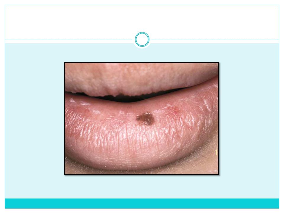
Mucosal Melanotic Macule
Etiology Most idiopathic, some postinflammatory, some drug-induced Multiple lesions suggest syndrome association, as follows: Peutz-Jeghers syndrome Laugier-Hunziker phenomenon Carney’s syndrome LEOPARD syndrome

3
Clinical Presentation
Most in adulthood (fourth decade and beyond) Most are solitary and well circumscribed Lower lip vermilion border most common site, mostly in young women (labial melanotic macule) Buccal mucosa, palate, and attached gingiva also involved (mucosal melanotic macule) Usually brown, uniformly pigmented, round to ovoid shape with slightly irregular border Usually < 5 mm in diameter
 Most are solitary and well circumscribed. Lower lip vermilion border most common site, mostly in young women (labial melanotic macule) Buccal mucosa, palate, and attached gingiva also involved (mucosal melanotic macule) Usually brown, uniformly pigmented, round to ovoid shape with slightly irregular border. Usually < 5 mm in diameter.")
5
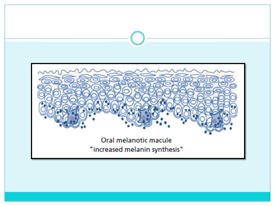
Microscopic Findings Normal melanocyte density and morphology
Increased melanin in basal cells and subjacent macrophages (mucosal melanotic macule) Increased melanin in basal cells with elongated rete pegs (ephelides)
 Increased melanin in basal cells with elongated rete pegs (ephelides)")
7
Differential Diagnosis
Biopsy Differential Diagnosis Melanoacanthoma Mucosal melanotic macule Congenital syndromes (Carney’s, Peutz-Jeghers, LEOPARD, Laugier-Hunziker)
")
8
Treatment Prognosis Observation Biopsy for esthetics
If increase in size or development of atypical signs occurs, macule should be removed to rule out malignant melanoma, particularly if on palate or alveolar mucosa. Prognosis Excellent

9
Nevus Etiology Clinical Presentation Unknown
Lesion of melanocytic origin within mucosa and skin Clinical Presentation Usually elevated, symmetric papule Pigmentation usually uniformly distributed Common on skin; unusual intraorally Palate and gingiva most often involved

10
Microscopic Findings Most are intramucosal (“dermal”)
Blue nevi are deeply situated and are composed of spindled nevus cells. Other variants are rare; junctional and compound nevi (no dysplastic nevi occur orally) Nevus cells are oval/round and are found in unencapsulated nests (theques). Melanin production is variable.
 Nevus cells are oval/round and are found in unencapsulated nests (theques). Melanin production is variable.")
11
When nevus cells are located in the epithelium connective tissue junction, the lesion is called a junctional nevus

12
When nevus cells are located in connective tissue, the lesion is called an intradermal nevus or intramucosal nevus

13
When nevus cells are located in a combination of zones, the lesion is called a compound nevus.

14
A fourth type of nevus, in which cells arc spindle shaped and found deep in the connective tissue, is known as blue nevus.

16
Differential Diagnosis
Clinical features Biopsy Differential Diagnosis Melanoma Varix Amalgam tattoo/foreign body Mucosal melanotic macule Kaposi’s sarcoma Ecchymosis Melanoacanthoma

17
Treatment Excision of all pigmented oral lesions to rule out malignant melanoma is advised. Malignant transformation of oral nevi probably does not occur. Prognosis Excellent

18
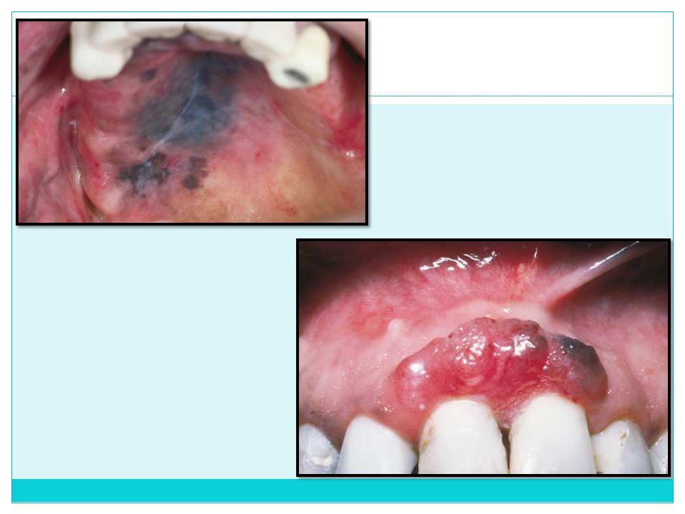
Malignant Melanoma Etiology Unknown
Cutaneous malignant melanoma with relation to sun exposure or familial-dysplastic melanocytic lesions

19
Clinical Presentation
Rare in oral cavity (< 1% of all melanomas) and sinonasal tract Most cases occur in those older than 30 years of age. Usually arises on maxillary gingiva and hard palate May exhibit early in situ phase: a macular, pigmented patch with irregular borders Progression to deeply pigmented, nodular quality with ulceration May arise de novo as a pigmented or amelanotic nodule Rarely may be metastatic to the oral cavity as a nodular, usually pigmented mass
 and sinonasal tract. Most cases occur in those older than 30 years of age. Usually arises on maxillary gingiva and hard palate. May exhibit early in situ phase: a macular, pigmented patch with irregular borders. Progression to deeply pigmented, nodular quality with ulceration. May arise de novo as a pigmented or amelanotic nodule. Rarely may be metastatic to the oral cavity as a nodular, usually pigmented mass.")
20
Microscopic Findings Early stage: atypical melanocytes at epithelial–connective tissue interface, occasionally with intraepithelial spread Later infiltration into lamina propria and muscle Strict correlation to cutaneous malignant melanoma is not well established, although, as in skin, a similar horizontal or in situ growth phase often precedes the vertical invasive phase. Amelanotic forms may require use of immunohistochemical identification: S-100 protein, HMB-45, Melan-A expression

22
Differential Diagnosis
Biopsy High index of suspicion Differential Diagnosis Mucosal nevus Extrinsic pigmentation Melanoacanthoma Kaposi’s sarcoma Vascular malformation Amalgam tattoo Mucosal melanotic macule

23
Treatment Prognosis Surgical excision
Marginal parameters related to depth of invasion and presence of lateral growth Wide surgical margins; resection (including maxillectomy) for large, deeper lesions Neck dissection in cases of deep invasion (< 1.25 mm) Prognosis Generally poor for most oral malignant melanomas Less than 20% survival at 5 years in most studies
 for large, deeper lesions. Neck dissection in cases of deep invasion (< 1.25 mm) Prognosis. Generally poor for most oral malignant melanomas. Less than 20% survival at 5 years in most studies.")
24
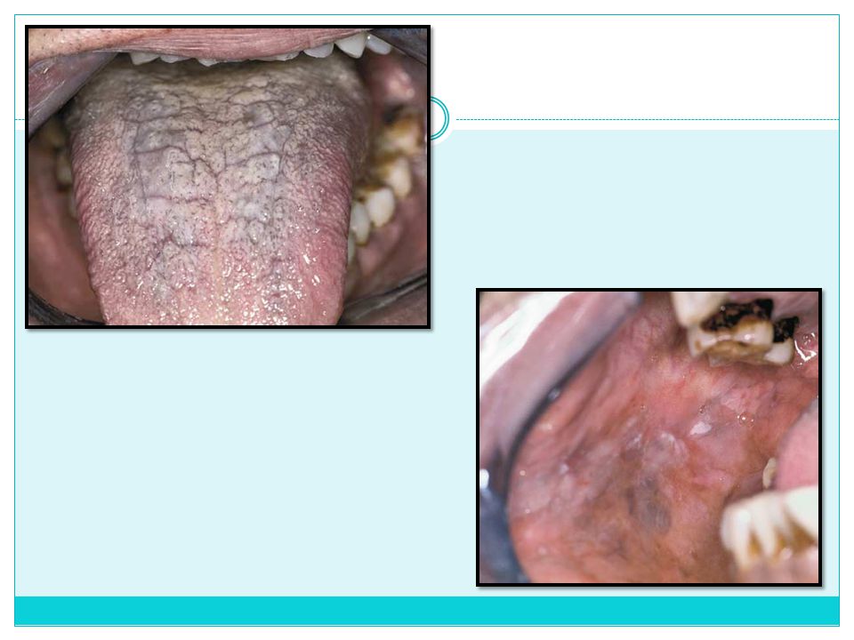
Drug-Induced Melanosis
Etiology Occupational exposure—metals vapors (lead, mercury) Therapeutic—metal salt deposits (bismuth, cis-platinum, silver, gold); also nonmetal agents, such as chloroquine, minocycline, zidovudine, chlorpromazine, phenolphthalein, clofazimine, and others
 Therapeutic—metal salt deposits (bismuth, cis-platinum, silver, gold); also nonmetal agents, such as chloroquine, minocycline, zidovudine, chlorpromazine, phenolphthalein, clofazimine, and others.")
25
Clinical Presentation
Focal to diffuse areas of pigmentary change If heavy metals are the cause, a typical gray to black color is seen along the gingival margin or areas of inflammation. Palatal changes characteristic with antimalarial drugs and minocycline Most medications cause color alteration of buccal-labial mucosa and attached gingiva. Darkened alveolar bone with minocycline therapy (10% at 1 year, 20% at 4 years of therapy)
")
27
Differential Diagnosis
History of exposure to, or ingestion of, heavy metals or drugs Differentiation from melanocyte-related pigmentation by biopsy if necessary Differential Diagnosis When localized: amalgam tattoo, mucosal melanotic macule, melanoacanthoma, mucosal nevus, ephelides, Kaposi’s sarcoma, purpura, malignant melanoma, ecchymosis When generalized: ethnic pigmentation, Addison’s disease If asymmetric, in situ melanoma must be ruled out by biopsy.

28
Treatment Prognosis Investigation of cause and elimination if possible
Excellent

29
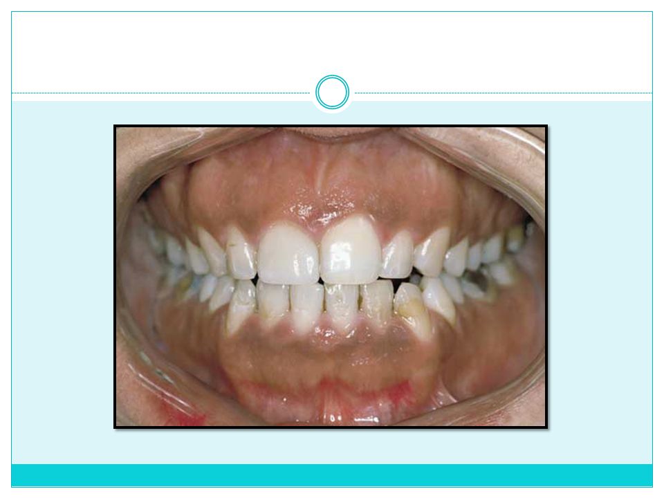
Physiologic Pigmentation
Etiology Normal melanocyte activity Clinical Presentation Seen in all ages Symmetric distribution over many sites, gingiva most commonly Surface architecture, texture unchanged

31
Prognosis Diagnosis Differential Diagnosis Treatment History
Distribution Differential Diagnosis Mucosal melanotic macule Smoking-associated melanosis Superficial malignant melanoma Treatment None Prognosis Excellent

32
Smoker’s Melanosis Etiology
Melanin pigmentation of oral mucosa in heavy smokers May occur in up to 1 of 5 smokers, especially females taking birth control pills or hormone replacement Melanocytes stimulated by a component in tobacco smoke

33
Clinical Presentation
Brownish discoloration of alveolar and attached labial gingiva, buccal mucosa Pigmentation is diffuse and uniformly distributed; symmetric gingival pigmentation occurs most often. Degree of pigmentation is positively influenced by female hormones (birth control pills, hormone replacement therapy).
.")
35
Microscopic Findings Increased melanin in basal cell layer
Increased melanin production by normal numbers of melanocytes Melanin incontinence

36
Differential Diagnosis
History of chronic, heavy smoking Biopsy Clinical appearance Differential Diagnosis Physiologic pigmentation Addison’s disease Medication-related pigmentation (drug-induced pigmentation by chloroquine, clofazimine, mepacrine, chlorpromazine, quinidine, or zidovudine) Malignant melanoma
 Malignant melanoma.")
37
Treatment Prognosis None Reversible, if smoking is discontinued
Good, with smoking cessation

38
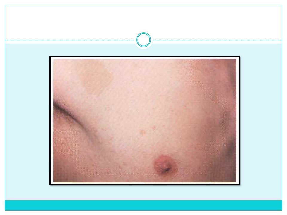
Cafe-au-lait macules Café-au-lait macules are discrete melanin-pigmented patches of skin that have irregular margins and a brown coloration. They are noted at birth or soon thereafter and may also be seen in normal children. No treatment is required, but they may be indicative of a syndrome of greater significance

39
Individuals with six or more large cafe-au-lait macules should be suspected of possibly having neurofibromatosis. In neurofibromatosis, an autosomal dominant inherited disease, both nodular and diffuse pendulous neurofibromas occur on the skin and (rarely) in the oral cavity. They tend to appear in late childhood and can be multiple; many overlie the neurofibromatous swellings on the skin. Rarely, oral pigmentation is encountered. Cafe-au-lait macules may also be associated with Albright's syndrome (polyostotic fibrous dysplasia, endocrine dysfunction, precocious puberty, cafeau- lait macules). The cafe-au-lait macules of Albright's syndrome tend to be large and unilateral and have irregular borders. Microscopically, café au lait spots represent basilar melanosis without melanocyte proliferation.
 in the oral cavity. They tend to appear in late childhood and can be multiple; many overlie the neurofibromatous swellings on the skin. Rarely, oral pigmentation is encountered. Cafe-au-lait macules may also be associated with Albright s syndrome (polyostotic fibrous dysplasia, endocrine dysfunction, precocious puberty, cafeau- lait macules). The cafe-au-lait macules of Albright s syndrome tend to be large and unilateral and have irregular borders. Microscopically, café au lait spots represent basilar melanosis without melanocyte proliferation.")
41
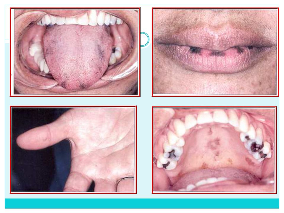
Peutz-Jeghers Syndrome
Peutz‐Jeghers syndrome is an autosomal‐dominant trait that produces the general findings of skin and/or mucosal melanotic macules with intestinal polyposis. The polypoid lesions in this condition generally behave as benign lesions although patients with carcinoma arising from adenomatous polyps have been reported. Many of these polypoid lesions are thought to be of inflammatory or hamartomatous origin and are also occasionally associated with dermatologic or oral mucosal abnormalities.

42
Clinical Presentation
In Peutz-Jeghers syndrome oral pigmentation is distinctive and is usually pathognomonic. Multiple focal melanotic brown macules are concentrated about the lips while the remaining facial skin is less strikingly involved. The macules appear as freckles or ephelides, usually measuring < 0.5 cm in diameter. Similar lesions may occur on the anterior tongue, buccal mucosa, and mucosal surface of the lips. Ephelides are also seen on the fingers and hands.

43
Microscopic Findings The lesions show basilar melanogenesis without melanocytic proliferation.

45
Differential Diagnosis
The number and locations of melanotic macules should be recorded and compared to the expected distribution. Upper and lower gastrointestinal dye radiologic series are required. Biopsy Differential Diagnosis Addison disease Albright syndrome hereditary neurofibromatosis

46
Treatment Because the malignant transformation incidence of adenomatous polyps is as high as 20% to 40%, flexible fiberoptic examinations and polyp biopsy also are valuable. Prognosis Good, but intense long‐term follow‐up is required because of a malignancy rate that is higher than previously thought and possible gastrointestinal complications.

Similar presentations