Download presentation
Presentation is loading. Please wait.

1
Embryology and Histology of the Pancreas
Prof. Abdulameer Al-Nuaimi E. mail:

2
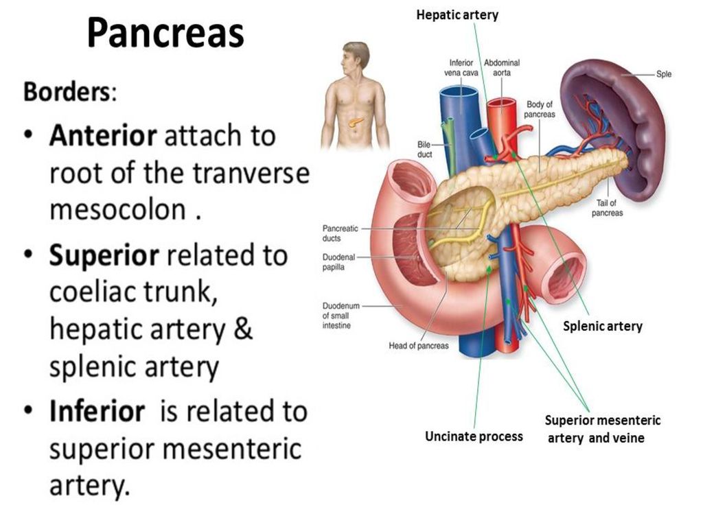
The Pancreas Pancreas is a soft lobulated organ located retroperitoneally across the posterior abdominal wall, it sits behind the stomach across the back of the abdomen. It is described as an organ having head, neck, body, and tail. The head is disc-shaped and lies within the concavity of the duodenum. Part of the head extends to the left behind the superior mesenteric vessels, it is called Uncinate process. The body extends to the left side and ends as a tail near the hilus of the spleen. The pancreas is made up of two types of glands, 1-An exocrine gland that secretes digestive enzymes and Sodium bicarbonate into the duodenum through the main and accessory pancreatic ducts. Both ducts are usually interconnected.

6
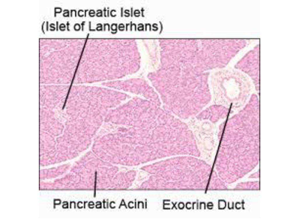
Cells of islets of Langerhans
2- An endocrine gland, which consists of the islets of Langerhans, secretes hormones into the bloodstream. Islets of Langerhans are named for the German physician Paul Langerhans, who first described them in The normal human pancreas contains about 1,000,000 islets. Cells of islets of Langerhans 1-Beta cells (β-cells), they make about 65-80% of the cells in the islets and produce Insulin. 2-alpha cells (α-cells), 15-20%, they produce an opposing hormone, Glucagon which releases glucose from the liver and fatty acids from fat tissue. 3-Delta cells (δ-cells), 3-10%, they secrete somatostatin a strong inhibitor of somatotropin, insulin, and glucagon; its role in metabolic regulation is not yet clear. Somatostatin is also produced by the hypothalamus and functions to inhibit secretion of growth hormone by the pituitary gland.
, they make about 65-80% of the cells in the islets and produce Insulin. 2-alpha cells (α-cells), 15-20%, they produce an opposing hormone, Glucagon which releases glucose from the liver and fatty acids from fat tissue. 3-Delta cells (δ-cells), 3-10%, they secrete somatostatin a strong inhibitor of somatotropin, insulin, and glucagon; its role in metabolic regulation is not yet clear. Somatostatin is also produced by the hypothalamus and functions to inhibit secretion of growth hormone by the pituitary gland.")
7
Pancreas P

8
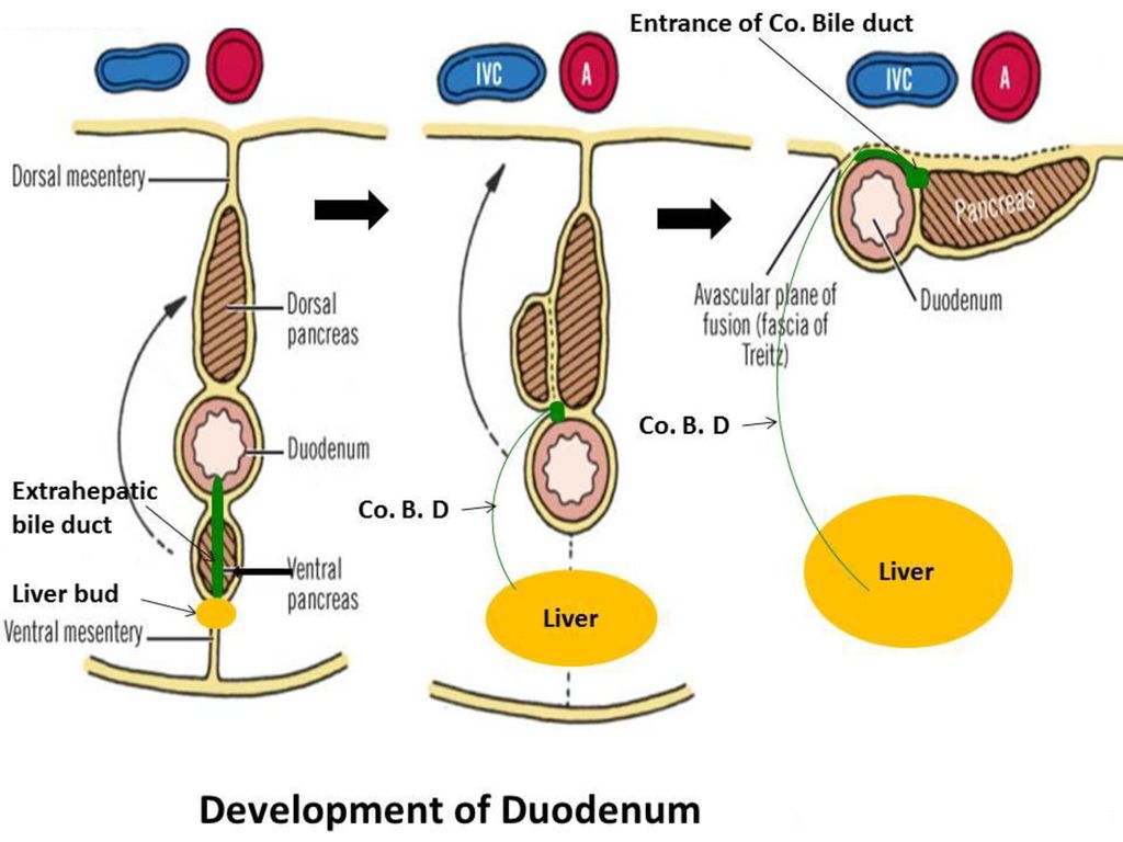
Development of Pancreas
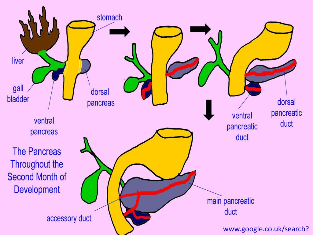
The Pancreas develops from the endodermal lining of the duodenum as a dorsal and ventral buds. The dorsal bud is in the dorsal mesentery and the ventral bud is in the ventral mesentery, close to the bile duct. When the duodenum rotates and become C-shaped, the ventral bud and the entrance of the common bile duct in the duodenum are shifted dorsally. The ventral bud comes to lie immediately below and behind the dorsal bud, finally the parenchyma and duct systems of both buds fuse together. Following swinging of the duodenum to the right, the pancreas and duodenum settles down on the posterior abdominal wall in a retroperitoneal position. The ventral pancreatic bud forms the Uncinate process and inferior part of the head of pancreas, where as the dorsal bud forms the Remaining part of pancreas.

11
Pancreatic duct The main pancreatic duct is formed by the union of the ventral pancreatic duct with the distal part of the duct of dorsal bud. The proximal part of the dorsal pancreatic duct, either obliterates or persists to form the Accessory pancreatic duct. The main pancreatic duct, together with common bile duct, enter the Ampulla of Vater which enters the posteromedial wall of the duodenum at the site of Major Papilla. Accessory pancreatic duct when persist (in 10% of cases), drains the lower part of the head and uncinate process, it opens into the duodenum at Minor Papilla, 3cm proximal to the opening of the main duct.
, drains the lower part of the head and uncinate process, it opens into the duodenum at Minor Papilla, 3cm proximal to the opening of the main duct.")
12
Pancreatic duct Access. Panc.Duct Main Panc.Duct . Minor Papil
Com. Bile Duct Access Pan. Duct Major Papilla Uncinate Process

13
Ampulla of Vater Ampulla of Vater Ampulla of Vater (Spiral sphincter(
Of Oddi Ampulla of Vater (Spiral sphincter(

14
Pancreatic islets of Langerhans develops from the parenchyma of pancreas at the third month of fetal life. Insulin secretion begins at the fifth month. Pancreatic connective tissue develops from the visceral surrounding mesoderm.

15
B- In 5% of cases, Ampulla of Vater is not present.
Variations of the opening of common bile duct and pancreatic duct into the duodenum (Contemp Surg 1987) A- In 85% of cases the common bile duct and pancreatic duct end in the Ampulla of Vater. B- In 5% of cases, Ampulla of Vater is not present. C- In 9% of cases, the common bile duct and pancreatic ducts open separately into the duodenum. 85% 5% 9%
 A- In 85% of cases the common bile duct and pancreatic duct end in the Ampulla of Vater. B- In 5% of cases, Ampulla of Vater is not present. C- In 9% of cases, the common bile duct and pancreatic ducts open separately into the duodenum. 85% 5% 9%")
16
C- The bile duct is completely covered by pancreatic tissue (30%).
variations of the third portion of common bile duct to pancreas (Smanio,1954). A, B - The bile duct is partially covered by a tongue of pancreatic tissue (44%). C- The bile duct is completely covered by pancreatic tissue (30%). D- The duct is uncovered by pancreatic tissue and located on the posterior surface of the pancreas (16.5%). E- The common bile duct is covered by two tongues of pancreatic tissue (9%). 44% Posterior surface of the Head of pancreas and common bile duct 30% 16.5% 9%
. A, B - The bile duct is partially covered by a tongue of pancreatic tissue (44%). C- The bile duct is completely covered by pancreatic tissue (30%). D- The duct is uncovered by pancreatic tissue and located on the posterior surface of the pancreas (16.5%). E- The common bile duct is covered by two tongues of pancreatic tissue (9%). 44% Posterior surface of the. Head of pancreas and. common bile duct. 30% 16.5% 9%")
17
Summary The liver bud appears as an endodermal out growth from the distal part of the Foregut (Duodenum). This gives rise to the liver and Extra hepatic biliary passages. During development, rotation and Rt swinging of duodenum, the bile duct entrance in duodenum is shifted from its initial anterior to a posteromedial position. The Pancreas develops from the endodermal lining of the duodenum as a dorsal and ventral buds. During rotation of duodenum, the ventral bud is shifted dorsally and fuses with the dorsal bud. islets of Langerhans develops from the parenchyma of pancreas at the third month of fetal life.
. This gives rise to the liver and Extra hepatic biliary passages. During development, rotation and Rt swinging of duodenum, the bile duct entrance in duodenum is shifted from its initial anterior to a posteromedial position. The Pancreas develops from the endodermal lining of the duodenum as a dorsal and ventral buds. During rotation of duodenum, the ventral bud is shifted dorsally and fuses with the dorsal bud. islets of Langerhans develops from the parenchyma of pancreas at the third month of fetal life.")
18
Thank You

Similar presentations
.>")


 Biliary system. Biliary System Liver Unique to subphylum Conservative form and function. Largest gland in body Divided into lobes.>")