Download presentation
Presentation is loading. Please wait.

1
ILO International Classification of Radiographs of Pneumoconioses David Rees June 2016

2
Purpose X-ray features standardised coding system (not necessary for dx) Clinical (only if pn or pleural) Screening and surveillance (only pn or pleural) Epi (all, 3 readers) Compensation X 20%
 Clinical (only if pn or pleural) Screening and surveillance (only pn or pleural) Epi (all, 3 readers) Compensation X 20%")
3
NIOSH form on the web

4
Learning the system B reader programme NIOSH. Examination + re- certification 4-yearly ILO training programme 4 days no examination NIOH/ILO Johannesburg November 15-18 th

5
What is it? Guidelines Guidelines for the use of the ILO International Classification Guidelines for the use of the ILO International Classificatio A box of 22 standard radiographs (reference films) A DVD with the Guidelines and the 22 standard radiographs digitalised
 A DVD with the Guidelines and the 22 standard radiographs digitalised.")
6
History Preliminary classification was first proposed at the 1930 International Conference on Silicosis held in Johannesburg South Africa First version 1950 Latest version 2011. Chapter on digital images The International Classification of High-resolution Computed Tomography (HRCT) for Occupational and Environmental Respiratory Diseases (ICOERD) Early 2000s
 for Occupational and Environmental Respiratory Diseases (ICOERD) Early 2000s.")
7
Digital analogue ILO + NIOSH preparing digital version (current is digitalised analogue films) Good agreement between soft, hard and analogue (Hard must be ≥ 2/3rds of standard-sized image 35cms x 43 cms.) Paper X Fransblau A, TeWaterNaude JM, D’Arcy H et al. The reproducibility of digital and analog chest radiology for detection and medical surveillance of silicosis and silicosis with PTB. SIM 130602 “Readings of HC images demonstrated a significantly greater prevalence of classifications of small parenchymal opacities compared to FSR and SC”

8
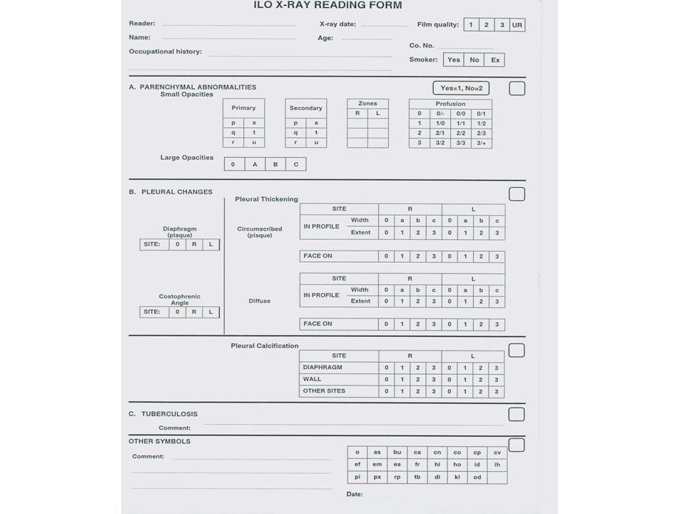
What is in the Guideline and x-ray box? Reading conditions (dim, 250 mm, 3 panels, clean) Technical quality (1-4) Parenchymal abnormalities (opacities) Distribution Profusion Size/shape Pleural abnormalities Symbols (e.g. em tb) Comments (od)
 Technical quality (1-4) Parenchymal abnormalities (opacities) Distribution Profusion Size/shape Pleural abnormalities Symbols (e.g. em tb) Comments (od).")
9
1-4 6 zones Symbols Comments

10
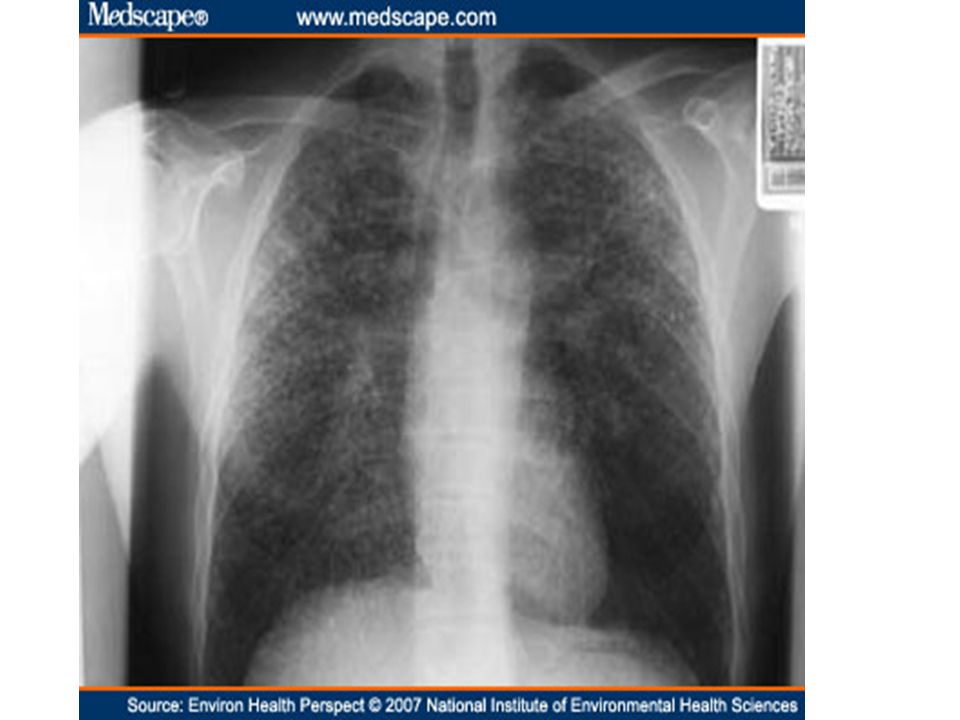
Parenchymal abnormalities: distribution of small opacities ≤ 10mm

11
6 zones

12
Parenchymal profusion small opacities ≤10mm Increasing profusion of small opacities Categories 0 1 2 3 Subcategories 0/– 0/0 0/1 1/0 1/1 1/2 2/1 2/2 2/3 3/2 3/3 3/+

15
Shape size small opacities Comment Rounded pqr DiametersUp to 1.5 mm 1.5-3 mm > 3-10 mmStandard films Irregular stuNot linear Width1.51.5 - 3> 3 – 10 mm Uncommon. Tricky

16
Primary and secondary

17
Large opacities (> 10mm) A: Sum of largest dimensions of all large opacites < 50mm B: Sum of largest dimensions of all large opacites < 50mm, but < area right upper zone C : > area RUZ
 A: Sum of largest dimensions of all large opacites < 50mm B: Sum of largest dimensions of all large opacites < 50mm, but < area right upper zone C : > area RUZ")
20
Pleural changes Plaques (parietal) Site (chest wall- in profile or face on -, diaphragm, other sites Extent - only for plaques on chest wall – relative to projection of lateral chest wall Width optional Calcified or not
 Site (chest wall- in profile or face on -, diaphragm, other sites Extent - only for plaques on chest wall – relative to projection of lateral chest wall Width optional Calcified or not")
21
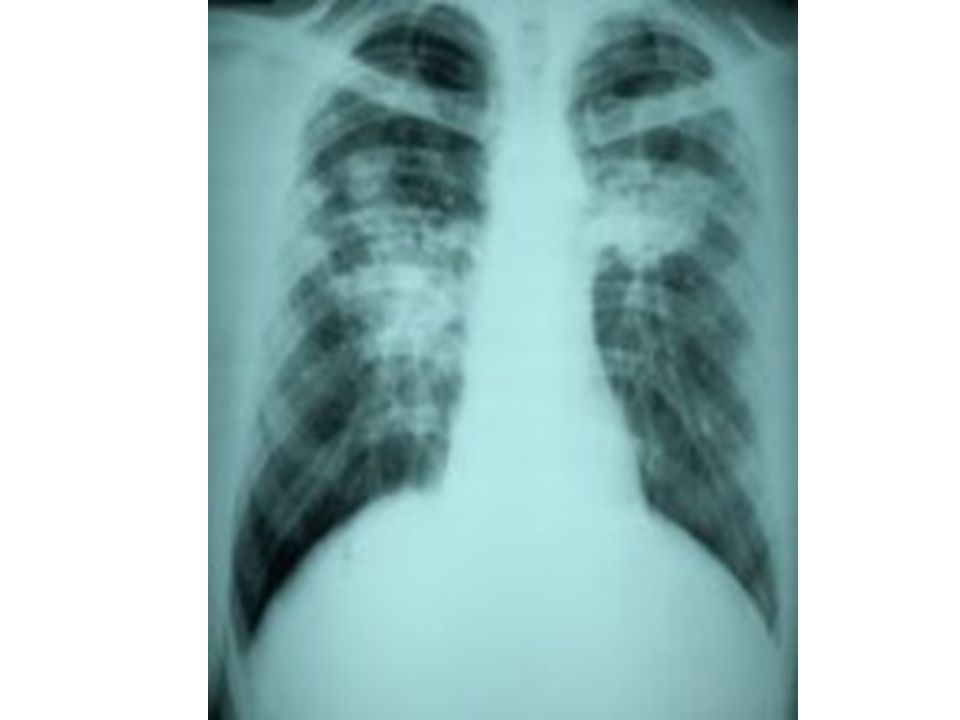
Pleural changes Diffuse pleural thickening (visceral) > 3mm In continuity with an obliterated costophrenic angle Costophrenic angle obliteration
 > 3mm In continuity with an obliterated costophrenic angle Costophrenic angle obliteration")
Similar presentations


 Phase II Year 8 Quarter 3,>")


 Panlobular Paraseptal Irregular.>")

 Occupational Health Program Andrew D. O’Brien, CSP General Manager, Safety & Health Unimin Corporation.>")

 24 – 25/7/2006.>")
