Download presentation
Presentation is loading. Please wait.

1
MAMMOGRAPHY Positioning & Anatomy
RADIOGRAPHIC IMAGING OF THE BREAST dr. Sameer Abdul Lateef

2
Mammography is the process of using low-energy-X-rays (usually around 30 kVp) to examine the human breast and is used as a diagnostic and a screening tool. The goal of mammography is the early detection of breast cancer, typically through detection of characteristic masses and/or micro calcifications. Mammography reduces deaths from breast cancer by screening programs

3
A mammogram can find breast cancer when it is very small -- 2 to 3 years before you can feel it.
No screening tool is 100% effective. Good quality mammograms can find 85-90% of cancers Some cancers are not found until they reach this size A mammogram can find cancer when it is only this size

4
Anatomy of the Breast Vary in shape & size
Cone shaped with the post surface (base) overlying the pectoralis & serratus muscles Axillaries tail extends from lat. base of the breasts to axillaries fossa Tapers ant. from the base ending in nipple, surrounded by areola
 overlying the pectoralis & serratus muscles. Axillaries tail extends from lat. base of the breasts to axillaries fossa. Tapers ant. from the base ending in nipple, surrounded by areola.")
5
Female Breast Consists of 15-20 lobes Divide into several lobules
Lobules contain acini, draining ducts and interlobular connective tissue. By teenage years each breast contains hundreds of lobules

6
POSITIONING Routine Images - CC - cranio caudad
MLO – mediolateral oblique

15
POSITIONING CC – CRANIOCAUDAD MLO – MEDIAL LATERAL OBLIQUE
“TRUE” LATERAL

16
TYPES OF BREAST TISSUE GLANDULAR DUCTS LOBES LOBULES STROMAL
MOSTLY SEEN UPPER OUTER QUADRANT STROMAL FATTY TISSUE CONNECTIVE TISSUE (COOPER’S LIGAMENTS – SUSPENSATORY LIGAMENTS

17
3 Tissue Types

18
Breast Changes with Age
Breast Classifications Breast Changes with Age

19
Fibro-glandular Breast
Dense with very little fat Females years of age Or 30 years or older without children Pregnant or lactating

20
Fibro-fatty Breast Fibro-fatty Average density
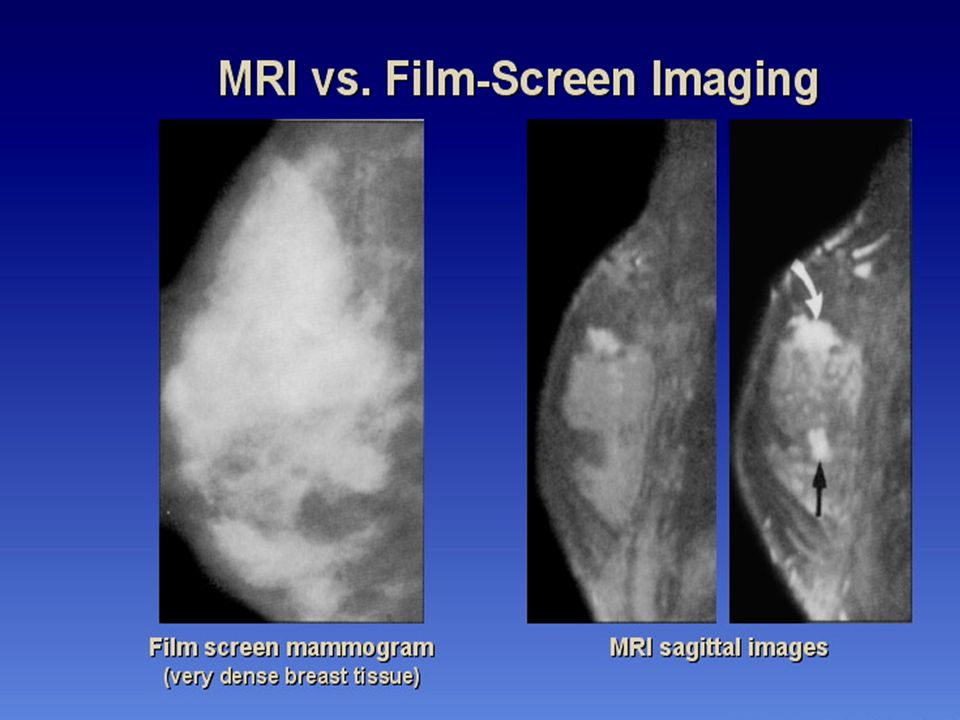
50% fat & 50% fibro-glandular Women years of age Or women with 3 or more children

21
Fatty Breast Fatty Minimal density
Women 50 and older (postmenopausal),
,")
22
How we differentiate between benign & malignant mass

23
Carcinoma of breast

24
Breast carcinoma

25
Fibroadenoma

26
Breast cyst

29
Fibrocystic disease

31
Mammograms of duct ectasia The majority of patients with duct ectasia have no diagnostic mammographic features. Occasionally the ducts are seen as tubular structures extending from the subareolar area, but this is a nonspecific sign. Still ductography can be used. The purpose of mammography in such instances is to exclude underlying malignancy.

33
Ultrasound Duct ectasia can be seen as multiple tubular structures arising from the nipple. The significance of such findings is unclear, however, as it is frequently seen in otherwise normal individuals. Ultrasound therefore has no role to play in the diagnosis of patients with nipple discharge. It should only be performed in patients who are also found to have a palpable mass

34
Male Mammography 1300 men get breast cancer per year 1/3 die
Most are 60 years or older Nearly all are primary tumors Symptoms include: Nipple retraction Crusting Discharge Ulceration

35
Gynecomastia is a benign male breast (non-cancerous) condition
Some men who have prominent breasts, or uneven breasts, often feel some embarrassment about their body image. This condition can also cause emotional conflict over sexual identity.

36
Other Imaging of the Breast

38
Xero mammography

Similar presentations