Download presentation
Presentation is loading. Please wait.

1
MCQs On Breast Imaging:
(√ ) or (X )
 or (X )")
2
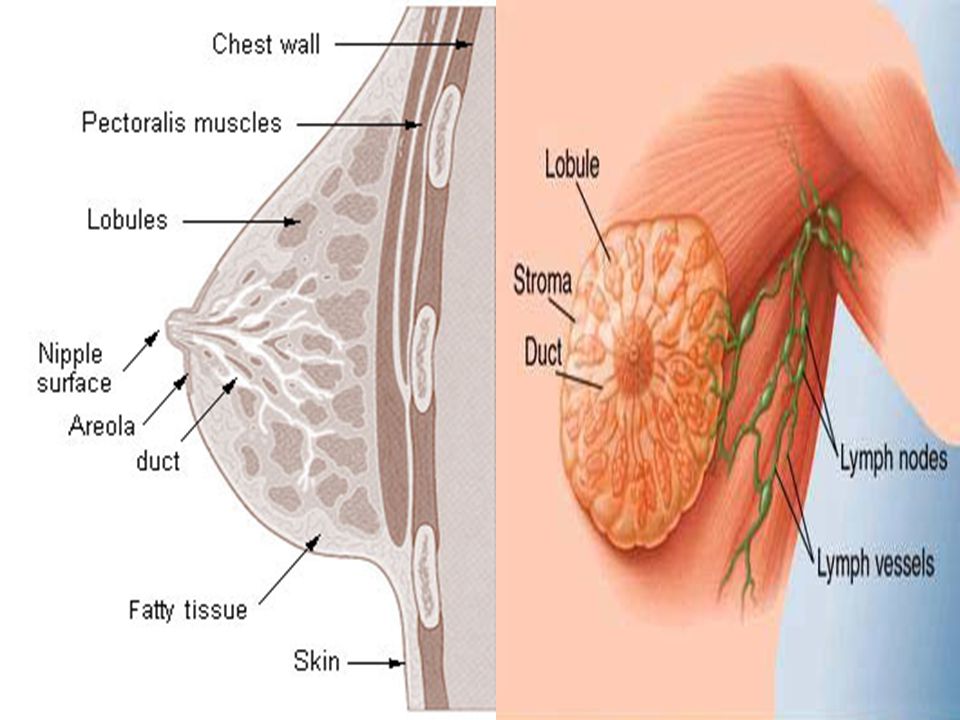
Breast Anatomy and Development:
1. The Breast is a modified apocrine sweat gland. 2. The major artery to the breast is internal mammary artery. 3. Azygos vein drains the breast 4. The terminal duct lobular unit is the basic functional unit of the breast 5. The level of lymph nodes involvement is related to pectoralis Major muscle 6. The breast extends lateraly till the anterior axillary line (√ ) (√ ) (√ ) (√ ) (X ) Pectoralis minor (X) Mid axillary line
 (√ ) (√ ) (√ ) (X ) Pectoralis minor. (X) Mid axillary line.")
4
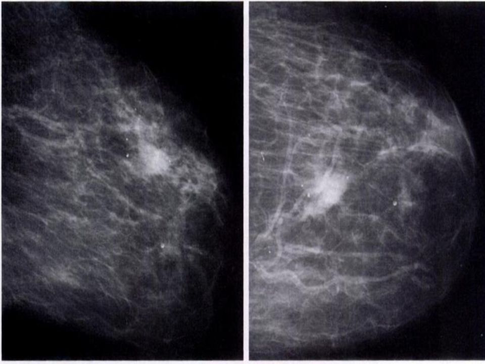
Regarding Mammography:
1. The skin is thickest over the upper outer quadrant 2. The craniocaudal view shows more breast tissue than the mediolateral view. 3. The pectoral muscle is not visualised in the craniocaudal view 4. Intramammary Lymph nodes are seen in the upper outer quadrant 5. Marked stromal proliferation is seen in the secretory phase of the menestrual cycle. 6. The breast completely lies in the superficial fascia (√ ) (X ) Craniocaudal view shows less breast tissue (√ ) (√ ) (X) Proliferative phase due to estrogen effect (√ )
 (X ) Craniocaudal view shows less breast tissue. (√ ) (√ ) (X) Proliferative phase due to estrogen effect. (√ )")
5
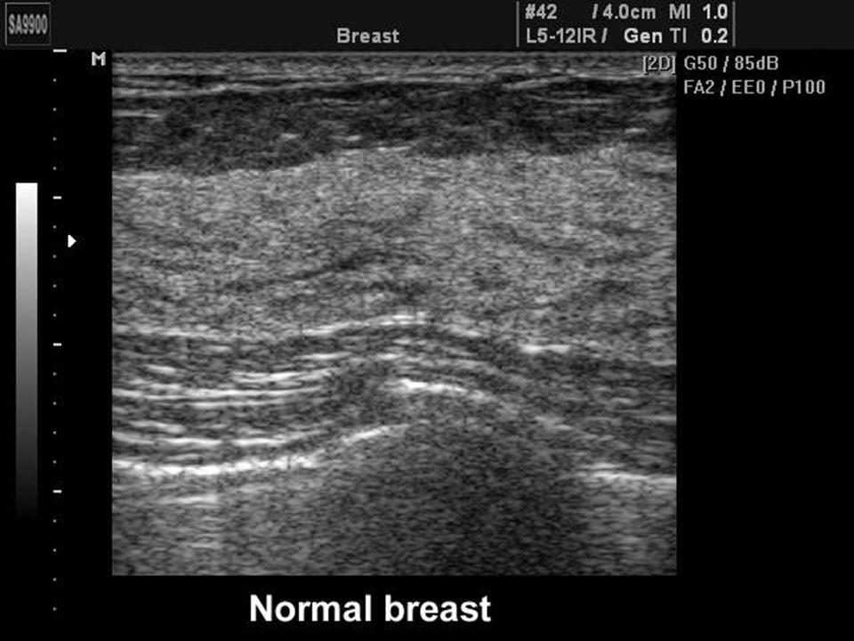
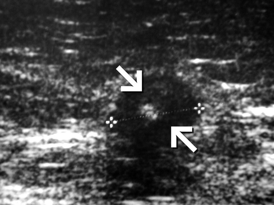
Regarding Breast Ultrasound:
1. The skin is a three layered structure 2. Coopers ligaments produce acoustic shadow 3. The Fat appear hypoechoic 4. The breast is uniformly bright due to the fibroglandular tissue 5. The ducts are clearly visualised (√ ) (√ ) (√ ) (X ) heterogenous (√ )
 (√ ) (√ ) (X ) heterogenous. (√ )")
7
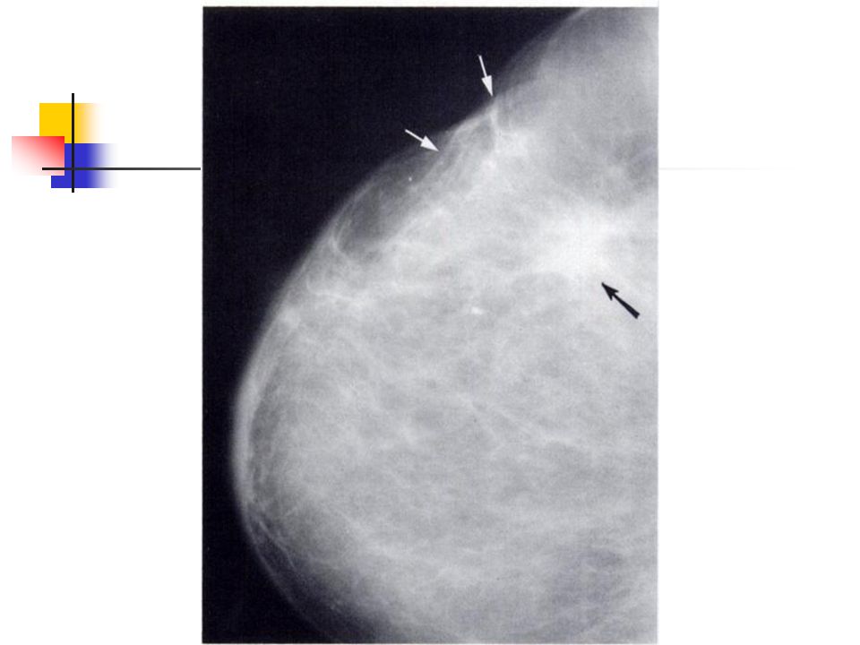
Regarding Mammography :
1. Microcalcification is calcification less than 1mm 2. Microcalcification is specific for carcinoma 3. Microcalcification is not seen in traumatic fat necrosis 4. Spiculated masses are carcinomas until proved by biopsy (X ) less than 0.5 mm (X ) not specific (X) this is a recognisable feature (√ )
 less than 0.5 mm. (X ) not specific. (X) this is a recognisable feature. (√ )")
8
The following pattern of calcifications are definitely benign:
1. Egg shell calcification 2. Floating calcification 4. Pleomorphic branching calcifications 4. Pop corn calcification 5. Tram line calcifications (√ ) Cyst (√ ) Milk Calcium Cyst (X) Feature of invasive ductal carcinoma (√ ) Fibroma (√ ) Blood vessel
 Cyst. (√ ) Milk Calcium Cyst. (X) Feature of invasive ductal carcinoma. (√ ) Fibroma. (√ ) Blood vessel.")
9
Groups of breast microcalcifications are seen in :
1. Fibroadenoma 2. Fat necrosis 3. Radiotherapy 4. Ductal carcinoma 5. Sclerosing adenitis (X ) (√ ) (X ) (√ ) (√ )
 (√ ) (X ) (√ ) (√ )")
10
Regarding Ductal Carcinoma :
1. Constitutes 40% of all breast carcinoma 2. May be associated with lobular carcinoma 3. Mammographic appearance is larger than the clinical size 4. Arises from the large ducts epithelium 5. Usually the tumour shows well defined margin , homogenous density and no acoustic shadowing (X ) (√ ) (X ) (X ) (X )
 (√ ) (X ) (X ) (X )")
11
Ductal Carcinoma It constitutes 80% of all breast cancers
Arises from epithelium of medium and small sized ducts It can be associated with invasive tubular and lobular carcinomas. The clinical size is bigger due to desmoplastic reaction Usually the tumour has ill-defined margin , heterogenous density and acoustic shadow

13
Lesions producing spiulated mass in mammography :
1. Fat necrosis 2. Sclerosing adenosis 3. Fibroadenosis 4. Abscess 5. Hamartoma (√ ) (√ ) (√ ) (√ ) (X )
 (√ ) (√ ) (√ ) (X )")
15
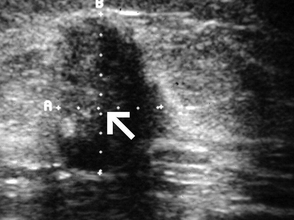
Common US features of typical breast Carcinoma:
1. Sharp margin 2. Hypoechoic 3.Long axis is perpendicular to skin 4. Hypoechoic rim of tissue 5. Echogenicity same as adjacent fibroglandular tissue but less than fat (X ) (√ ) (√ ) (X ) (X )
 (√ ) (√ ) (X ) (X )")
16
Common US features of typical breast Carcinoma:
Irregular margin Hypoechoic compared to adjacent fibroglandular tissue and fat Long axis is perpendicular to skin Hyperechoic rim of tissue (tumour, desmoplastic compressed breast tissue) Posterior acoustic shadowing.
 Posterior acoustic shadowing.")
19
High incidence of breast cancer recurrence is seen in :
1. Estrogen receptor positive 2. Comedo carcinoma 3. Age > 40 years 4. More intraductal component 5. Negative margin (X) Estrogen Receptor Negative (√ ) (X) Age < 40 years (√ ) (X) Positive Margin
 Estrogen Receptor Negative. (√ ) (X) Age < 40 years. (√ ) (X) Positive Margin.")
20
Regarding Benign Breast Disease:
1. Breast abscess is most commonly located in the upper outer Quadrant. 2. Breast abscess is most commonly due to streptococcus 3. Skin retraction is a common presentation of fat necrosis 4. Hyalinised fibroadenoma is a common feature (X) Retroareolar (X) Staphylococcus aureus (√ ) (X) Rare
 Retroareolar. (X) Staphylococcus aureus. (√ ) (X) Rare.")
21
Regarding Lymph node involvement in Breast cancer:
1. 40% of women have axillary lymph adenopathy at time of diagnosis 2. Involved lymph nodes are rounder and more reflective than normal at US 3. Internal mammary lymph nodes are more often involved than axillary lymph nodes of the inner quadrant tumors. 4. Internal mammary lymph nodes are usually resected at mastectomy 5. Supraclavicular nodal spread confers a poor prognosis (√ ) (X) Larger,more rounded and less reflective (X) Axillary lymph nodes are more comonly involved (X) (√ )
 (X) Larger,more rounded and less reflective. (X) Axillary lymph nodes are more comonly involved. (X) (√ )")
22
Regarding Male Breast cancer:
1. Most cases occur in patients with Kleinfilter 2. Frequently bilateral 3. Mammographic calcifications are fewer than in female breast cancer. 4. It is more common on left side 5. Gynaecomastia is a predisposing factor (X) (X) (X) Larger,more rounded and more scattered (√ ) (X)
 (X) (X) Larger,more rounded and more scattered. (√ ) (X)")
23
Thank You

Similar presentations