Download presentation
Presentation is loading. Please wait.

1
Study of Microbial Structure Microscopy and Specimen Preparation BIO3124 Lecture #3

2
Lenses and the bending of light light is refracted (bent) at the varying density interfaces refractive index –how greatly a substance slows the velocity of light Direction/magnitude of bending depends on the RI Incident angle Normal Refracted angle
 at the varying density interfaces refractive index –how greatly a substance slows the velocity of light Direction/magnitude of bending depends on the RI Incident angle Normal Refracted angle")
3
Light Microscopes Compound microscopes –image formed by action of 2 lenses Bright-field microscope Dark-field microscope Phase-contrast microscope Fluorescence microscope

4
The Bright-Field Microscope Dark image against a brighter bkg Several objective lenses –parfocal stays focused when objectives changed total magnification (max 1000- fold) – product of the magnifications of the ocularr lenses and the objective lenses
 – product of the magnifications of the ocularr lenses and the objective lenses")
5
Microscope Resolution Resolution: ability to distinguish two small close objects Abbe equation: d=0.5λ/nsinθ –shorter wavelength greater resolution –Numerical aperture: NA= n.sinθ –Smaller d value = more powerful optic system

6
working distance — distance between the front surface of lens and surface of cover glass or specimen when it is in sharp focus Microscope Resolution Reducing the d value (higher resolution) means increasing the θ,
 means increasing the θ,")
7
Microscope Resolution Effect of refractive index:

8
The Dark-Field Microscope Image is formed by light reflected or refracted by specimen produces a bright image against a dark bkg to observe living, unstained preparations –For eucaryotes has been used to observe internal structures –For procaryotes has been used to identify bacteria such as Treponema pallidum, the causative agent of syphilis

9
Dark field microscopy: Light path

10
Dark field microscopy Example of an insect larva examined in a dark field microscope

11
The Phase-Contrast Microscope first described in 1934 by Dutch physicist Frits Zernike enhances the contrast btw intracellular structures that have slight differences in their refractive indices excellent tool to observe living cells –bacterial components such as endospores and inclusion bodies –Eukaryotic organelles Frits Zernike (1888-1966)
")
12
The Phase-Contrast Microscope

13
Surround wave (S) Particle wave (P) Deviated wave (D) Role of phase ring
 Particle wave (P) Deviated wave (D) Role of phase ring")
14
positive (dark) and negative (bright) Ph microscopy Depends on the type of phase plate coating and material Positive (dark) phase: S and D waves are different by ½ wavelength and cancel each other (P=S+D) and the image (P) looks dark Negative (bright) phase: S and D waves are in phase and add up, so the image looks bright
 and negative (bright) Ph microscopy Depends on the type of phase plate coating and material Positive (dark) phase: S and D waves are different by ½ wavelength and cancel each other (P=S+D) and the image (P) looks dark Negative (bright) phase: S and D waves are in phase and add up, so the image looks bright")
15
Dark (positive) Phase contrast image of HeLa cells HeLa cells Reza Nokhbeh
 Phase contrast image of HeLa cells HeLa cells Reza Nokhbeh")
16
Examples of positive (dark) Ph microscopy P.aeruginosa Sporulating bacterium Contrast between spores and Vegetative forms Paramecium Intracellular organelles contrasted
 Ph microscopy P.aeruginosa Sporulating bacterium Contrast between spores and Vegetative forms Paramecium Intracellular organelles contrasted")
17
The Fluorescence Microscopy exposes specimen to ultraviolet, violet, or blue light specimens usually stained with antibodies tagged with a fluorophore shows a bright image of the object resulting from the fluorescent light emitted by the specimen Has applications in medical microbiology and molecular biology

18
The Fluorescence Microscope

19
Excitation and Emission lights

20
Poliovirus interferes with the integrity of SiRNA centres Poliovirus infected HeLa T4 cells Reza Nokhbeh Infected GW bodies disintegrate as the result of Poliovirus infection virus and GW bodies are stained with fluorochrome conjugated specific antibodies

21
Electron Microscopy Ernst Ruska and Max Hall in Germany finished the first prototype in 1931 Eli Franklin Burton (1847-1948) and his students, James Hillier, Cecil Hall and Albert Prebus, built the first functional EM in 1938 at Toronto university Louis de Broglie’s principle that electron particles also have electromagnetic (wave) property accelerated electronic beam in microscopy would enhance resolution, why? James Hillier (1915-2007)
.")
22
wavelength of electron beam is much shorter (0.005 nm or 5 A˚) than light, i.e. much higher resolution Magnification is 100,000 to 200,000 Resolution approaches 0.5 nm, ie about 1000-fold higher than light microscopes Transmission Electron Microscopy (TEM)
.")
23
Principles of light microscopy applies to TEM Thermionic Electron Gun ~300 Kev monochromatic beam

24
The Scanning Electron Microscopy (SEM) uses electrons scattered from the surface of a specimen to create image produces a 3-dimensional image of specimen’s surface features
 uses electrons scattered from the surface of a specimen to create image produces a 3-dimensional image of specimen’s surface features")
25
Interaction of M.tuberculosis with pulmonary cells Examples of TEM and SEM micrographs P. acens lytic phage TEM, 150,000x R. Nokhbeh, J. Trifkovic

26
New Techniques in Microscopy Confocal scanning laser (CLSM) microscopy and scanning probe microscopy have extremely high resolution Expanded the resolution to molecular and atomic levels i.e. 1-100 A

27
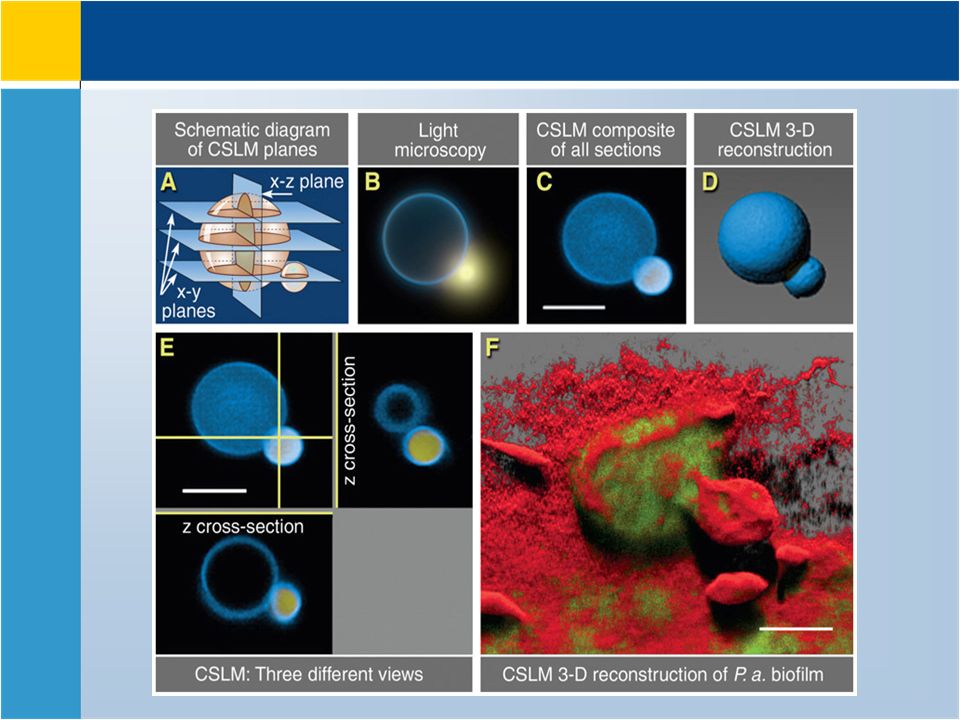
Confocal Microscopy Confocal Scanning Laser Microscope (CSLM) laser beam used to illuminate a variety of planes in the specimen computer compiles images to generate 3D image used extensively to observe biofilms Also used in studying the subcellular structures Light is only gathered from the plane of focus
 laser beam used to illuminate a variety of planes in the specimen computer compiles images to generate 3D image used extensively to observe biofilms Also used in studying the subcellular structures Light is only gathered from the plane of focus")
28
Confocal scanning laser microscope blurring does not happen since signal is gathered by scanning a thin layer of specimen, plane of focus, at each round

30
Scanning Probe Microscopy Scanning Tunneling Microscope (STM) Measures the surface features of specimen by moving a sharp probe over the surface –steady current (tunneling current) maintained between microscope probe and specimen –up and down movement of probe as it maintains current is detected and used to create image of surface of specimen – Magnification: 100 million times, capable of detecting the surface atoms
 Measures the surface features of specimen by moving a sharp probe over the surface –steady current (tunneling current) maintained between microscope probe and specimen –up and down movement of probe as it maintains current is detected and used to create image of surface of specimen – Magnification: 100 million times, capable of detecting the surface atoms")
31
Scanning Tunneling Microscope DNA double helix Atoms of MoS2, the bright spots are S atoms Silicon surface atoms enlarged 20 million times individual surface atoms and the bonds that hold them in place

32
Scanning Probe Microscopy Atomic Force Microscope (AFM) –Vertical movement of probe is followed by a laser beam –probes surfaces that are not charged
 –Vertical movement of probe is followed by a laser beam –probes surfaces that are not charged")
33
Atomic Force Microscope Membrane integral aquaporin protein captured by AFM α-synuclein protein fibers. Misfolded fibers are incolved in Parkinson disease Human mitotic chromosome spread

34
Preparation and Staining of Specimens Staining techniques are applied to increase the contrast increases visibility using bright field microscopes accentuates specific morphological features preserves specimen (due to fixation)
")
35
Fixation preserves internal and external structures and stabilizes them in position organisms usually killed and firmly attached to microscope slide heat fixation – routine use with procaryotes preserves overall morphology but not internal structures chemical fixation – used with larger, more delicate organisms protects fine cellular substructure and morphology

36
Dyes Dyes Ionizable dyes have charged groups Cationic (basic) : Cationic (basic) : Positively charged. –e.g. Methylene blue, Crystal violet, Safranine, Malachite green. Anionic (acidic): Anionic (acidic): Negatively charged –e.g. Nigrosin black, Indigo ink.
: Anionic (acidic): Negatively charged –e.g. Nigrosin black, Indigo ink..")
37
Simple and Differential staining simple staining –a single stain is used –use can determine size, shape and arrangement of bacteria Differential staining divides microorganisms into groups based on their staining properties –e.g., Gram staining –e.g., acid-fast staining

38
Staining Specific Structures endospore staining –double staining technique –bacterial endospore is one color and vegetative cell is a different color flagella staining –mordant applied to increase thickness of flagella

39
Staining Specimen staining.Positive staining: Specimen staining.

40
Staining (Contd) Negative staining:Negative staining: –Background staining, not the specimen.
 Negative staining:Negative staining: –Background staining, not the specimen.")
41
Methods Simple Staining Simple Staining : One type of stain. Cationic or Anionic stains. Able to determine the size, shape and the arrangment of bacteria.

42
Different Cell Morphologies Coccus:Coccus: –Sphere –3 planes of division –Plane of division produces different arrangements of cells. –Typical arrangements for different bacterial types. Bacillus:Bacillus: –Rods –One plane of division

43
Cocci Diplococcus Streptococcus (4-20) Tetrad Staphylococcus Division axes
 Tetrad Staphylococcus Division axes")
44
Bacilli (Bacillus) Diplobacilli Streptobacilli
 Diplobacilli Streptobacilli")
45
Other Cellular Forms Curved rods (coccobacillus) Vibrio cholerae Spirals Spirochetes
 Vibrio cholerae Spirals Spirochetes")
46
Differential Staining Techniques: Gram Staining Bacteria divided into two groups: Gram Negatives: stain red Gram Negatives: stain red –Bacilli: –Bacilli: Escherichia, Salmonella, Proteus, etc. –Cocci: –Cocci: Neisseria and Pneumococcus. Gram Positives: stain blue/purple Gram Positives: stain blue/purple –Bacilli: –Bacilli: Bacteria from the genera of Bacillus and Clostridium –Coccus: –Coccus: Streptococcus, Staphylococcus, Micrococcus

47
Gram negative Gram positive Typical examples of Gram staining reuslts

48
Mechanism of Gram staining Gram + Vs Gram -peptidoglycan Plasma membrane Lipopolysaccharide outer membrane Absent

49
Method crystal violet 1.Stain with crystal violet. Gram PositiveGram Negative 2. Add Gram’s Iodine

50
Differential stage: Ethanol wash => cell wall dehydration Dye trapped Grampositive Gram positive Dye washed away Gram negative Dehydrated wall Wall resists dehydration Alcohol Destaining

51
Safranine Counter-staining Gram Positive Gram Negative Resists counter stain Stains with counter stain

52
Mycobacteriacae Genus: –Mycobacterium Small bacilli Very difficult to grow in the lab Pathogenicity associated with leprosy and tuberculosis. lepraetuberculosis –Species: leprae and tuberculosis Resistant to conventional staining –Wall is impermeable

53
Alcohol-Acid Fast Staining Problem: Elevated levels of lipids called mycolic acids, in cell walls of mycobacteriacae, renders some bacteria impermeable to stains. Solution: Ziehl-Nelson method is used. Uses heat and phenol to drive Fuchsin into the cells. dye cannot be washed out of the cells by alcohol or acid (alcohol-acid fast)
.")
54
Method Use heat to make the cell more permeable Stain with carbol fuschin. Differential stage: Wash with acid alcohol Mycobacteria Mycobacteria retain the stain. Other bacteria lose the stain.

55
Methods of sample preparation for TEM Additional slides for your own information

56
Method and application Sample preparation: Fixation in glutaraldehyde or Osmium tetraoxide dehydration in ethanol/aceton Embedded in epoxy plastic cubes Ultramicrotomy to produce ~20 nm thick slices Stained by coating in heavey metal salt solutions of lead, uranium or osmium to form electron dense areas on cellular structures Samples are bombarded with electron beam that is transmitted through the specimen but scattered from electron dense areas Electrons passed through less electron dense areas, reach the fluorescent or digital detector and recorded

57
Heavy metal Staining for TEM Three methods: Negative staining: –good for small specimens like viruses that cannot be deposited on the copper grid support –Grid is cast with carbon followed by heavy metal salt like uranyl acetate to provide background electron dense environment –Virus solution is deposited on this supporting mesh –Electrons are scattered from the background, but pass through virus specimen –Image: dark field with bright virus Grid Carbon/Uranyl acetate coat virus T4 phage

58
Shadow casting: –Specimen is coated only on one side by vaporized heavy metal at a 45˚ angle –One side is electron dense, but not the other side –This increases the contrast –Useful for studying the viral and bacterial substructures eg. Bacterial flagellum, viral morphology Heavy metal Staining for TEM Actin filaments Cocco-bacilli

59
freeze-etching –freeze specimen in liquid nitrogen then fracture along lines of greatest weakness (e.g., membranes) – coat the fractured surfaces with layers of carbon and platinum –This forms a replica of the fractured surface –Specimen is removed chemically and studied in TEM Heavy metal Staining for TEM
 – coat the fractured surfaces with layers of carbon and platinum –This forms a replica of the fractured surface –Specimen is removed chemically and studied in TEM Heavy metal Staining for TEM")
60
TEM for freeze-etched samples Thylachoids ( freeze-etched) Bacillus bacterium (freeze-etched)
 Bacillus bacterium (freeze-etched)")
Similar presentations
 iris diaphragm.>")


