Download presentation
Presentation is loading. Please wait.

1
Meninges D.Nimer D.Rania Gabr D.Safaa D.Elsherbiny

2
Objectives Describe the arrangement of the meninges and their relationship to brain and spinal cord. Explain the occurrence of epidural, subdural and subarachnoid spaces. Locate the principal subarachnoid cisterns, and arachnoid granulations. Describe the ventricles of brain and importance of their choroids plexus. Summarize the pathway of cerebrospinal fluid (CSF) circulation Identify brain ventricles in CT scan, MRI .
 circulation. Identify brain ventricles in CT scan, MRI .")
3
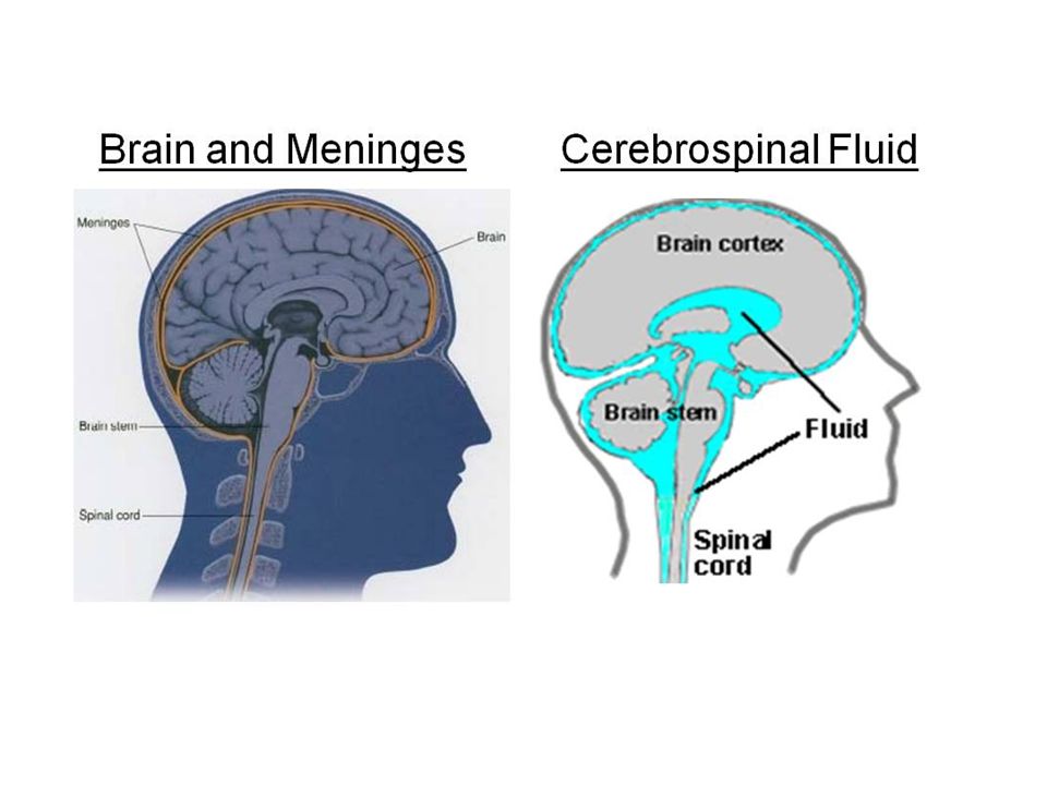
MENINGES The brain and spinal cord are surrounded by three membranes:
1. Dura mater: it is the outermost layer. 2. The arachnoid mater: the middle one. 3. The pia mater: the inner one.

6
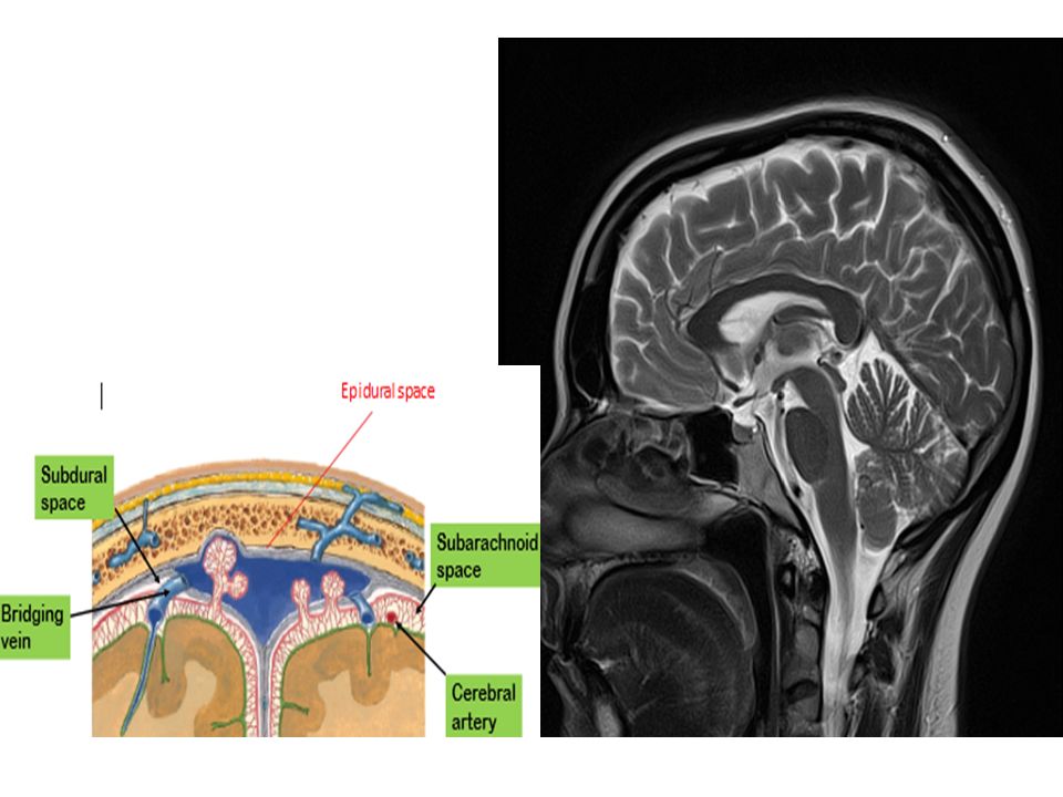
Cranial Meningeal Spaces
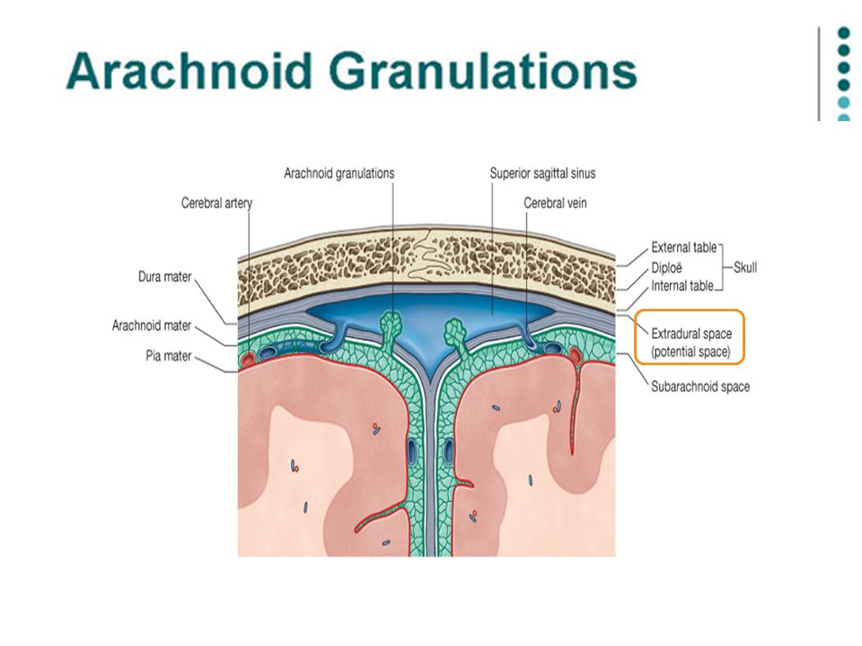
Epidural space Potential space superior to dura (between bone and dura). Normally closed since the dura is attached to the periosteum which is attached to the inner surface of the skull. In the spinal cord it contains adipose CT Subdural space Potential space between dura and arachnoid mater. Contains dural veins. Subarachnoid space Filled with CSF Contains the cerebral arteries supplying brain.
. Normally closed since the dura is. attached to the periosteum. which is attached to the. inner surface of the skull. In the spinal cord it contains adipose CT. Subdural space. Potential space between dura and arachnoid mater. Contains dural veins. Subarachnoid space. Filled with CSF. Contains the cerebral arteries supplying brain.")
7
Cranial Meningeal Spaces Cerebral arteries & CSF
Epidural space Meningeal arteries Subdural space Dural veins subarachnoid space Cerebral arteries & CSF Brain

8
Epidural hematoma

11
DURA MATER The cranial dura is a two layered tough, fibrous membrane that surrounds the brain. It is formed of two layers: The periosteal layer: is attached to the skull. The meningeal layer: it covers the brain is folded These are closely united except along certain lines, where they separate to form venous sinuses.

12
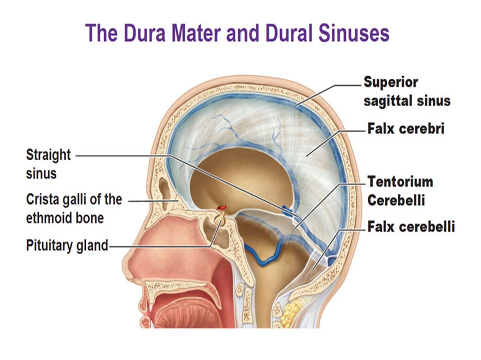
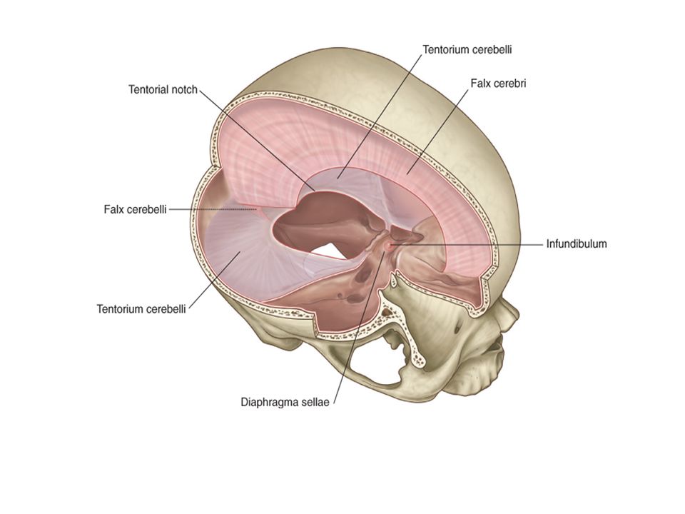
DURAL Folds The meningeal layer sends inward five septa: Falx cerebri
Falx cerebelli Tentorium cerebelli Diaphragma sellae Cavum trigeminal

14
Falx cerebri Def: Is a sickle-shaped fold of dura mater
Site: between the two cerebral hemispheres Shape: sickle shape Attachments: 1. Apex: attached to the frontal crest and the crista galli. 2. Base: with the upper surface of the tentorium cerebelli. 3. Upper border: to superior sagittal suture. 4. Lower border: free, above corpus callosum

15
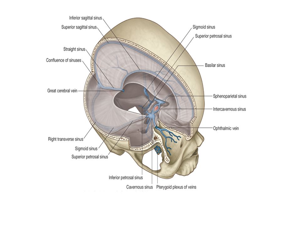
Falx cerebri Sinus contents:
The superior sagittal sinus in the upper border. The inferior sagittal sinus in the lower border (posterior 2/3). The straight sinus at the base, between the falx cerebri and the tentorium cerebelli in the median plane. 1 2 3
. The straight sinus at the base, between the falx cerebri and the tentorium cerebelli in the median plane")
16
Tentorium cerebelli Def: reduplication of inner layer of dura over the posterior cranial fossa. Shape: tent-shaped fold Site: between the cerebrum & the cerebellum. Tentorium cerebelli

17
Tentorium cerebelli Superior surface gives attachment to the falx cerebri Inferior surface gives attachment to the falx cerebelli Anterior border (free) surrounds the midbrain (tentorial notch) Posterior border (attached) attached to the upper border of the petrous part of the temporal bone and the lips of the groove for the transverse sinus.
 surrounds the midbrain (tentorial notch) Posterior border (attached) attached to the upper border of the petrous part of the temporal bone and the lips of the groove for the transverse sinus.")
18
Tentorium cerebelli Sinus contents:
The superior petrosal sinus: in the posterior border The transverse sinus: in the posterior border The straight sinus: at the junction between the flax cerebri and the tentorium cerebelli in the median plane. 3 2

19
Falx cerebelli Def: reduplication of inner layer of dura between the 2 cerebellar hemispheres. Shape: triangular in shape Site: between the 2 cerebellar hemispheres..

20
Falx cerebelli Sinus contents: The occipital sinus
Base attached to inferior surface of tentorium cerebelli Apex at posterior part of foramen magnum Anterior border (free) related to vermis Posterior border (attached) attached to internal occipital crest Sinus contents: The occipital sinus
 related to vermis. Posterior border (attached) attached to internal occipital crest. Sinus contents: The occipital sinus.")
21
Diaphragma sellae Def: reduplication of inner layer of dura that covers pituitary gland Shape: It is a small circular fold. It has a central hole which transmit the infundibulum of the pituitary gland. Site: covering sella turcica Attachment: Anterior attached to tuberculum sellae Posterior border attached to dorsum sellae Sinus content: The anterior and posterior intercavernous sinuses.

23
Arachnoid Mater& Pia Mater
The arachnoid mater is a soft, impermeable , translucent membrane loosely envelops the brain. The pia mater is the innermost, thin, delicate & highly vascular membrane that is closely adherent to the gyri and fitted into the sulci.

24
Blood supply of the dura mater Nerve supply of the dura mater
In the anterior cranial fossa: ophthalmic, anterior and posterior ethmoidal arteries. In the middle cranial fossa Middle meningeal, accessory meningeal arteries. internal carotid arteries and ascending pharyngeal arteries. In the posterior cranial fossa: Meningeal branches of the occipital, vertebral arteries. Nerve supply of the dura mater Trigeminal, facial, glossopharyngeal and vagus nerves Upper 3 cervical nerves

Similar presentations