Download presentation
Presentation is loading. Please wait.

1
Infratemporal fossa Dr A.Prasanna

2
Objectives The objective of the lecture :
Is to discuss infratemporal fossa Muscles of mastication Maxillary artery Mandibular nerve Temporomandibular joint.

3
Boundaries of infratemporal fossa
Roof: Infratemporal surface of greater wing of sphenoid bone A small part by sqamous part of temporal bone Medial wall— Lateral pterygoid plate Anterior wall—formed by posterior surface of body maxilla Lateral wall—by medial surface of ramus of mandible.

4
Contents of infratemporal fossa
Lateral and medial pterygoid muscles Maxillary artery Mandibular nerve Chorda tympani nerve Maxillary nerve

5
Muscles of mastication

6
Muscles of mastication
There are 4 muscles They are supplied by mandibular nerve They develop from 1st pharyngeal arch Muscles are : Masseter Temporalis Lateral pterygoid Medial prerygoid

7
Masseter Origin : lower border and deep surface of zygomatic arch
Insertion : lateral surface of ramus of mandible Action : elevation and protrusion of mandible

8
Zygomatic arch & ramus of mandible

9
Temporalis Origin : temporal fossa Insertion :
Coronoid process apex Anterior border of ramus of mandible Action : elevation and retraction of mandible

10
Temporal fossa & coronoid process

11
Lateral pterygoid Origin : Upper head from roof of infratemporal fossa
Lower head from lateral surface of lateral pterygoid plate Insertion : Pterygoid fovea Capsule of tempromandibular joint Articular disc Action : Depresion , protrusion and side to side movement

12
Infratemporal surface – lateral pterygoid plate & pterygoid fovea

13
Relations of Lateral pterygoid
It is triangular muscle Base in the front and apex in the back attached to pterygoid fovea From upper margin deep temporal nerves and vessels emerge Buccal nerve emerges between the two heads of muscles Maxillary artery lies superficial to the lower head of muscle Lingual nerve and inferior alveolar nerve emerge from the lower margin of muscle Middle meningeal artery and accessory middle meningeal artery passes deep to muscle

14
Lateral pterygoid relations

15
Medial pterygoid Origin : Superficial head from tuberosity of maxilla
Deep head from medial surface of lateral pterygoid plate Insertion : medial side of angle of mandible Action : Elevation , protrusion and side to side movement

16
Tuberosity of maxilla -Lateral pterygoid plate & pterygoid fovea

17
Maxillary artery

18
Maxillary artery parts

19
Maxillary artery Beginning : posterior to the neck of mandible as a large terminal branch of external carotid artery Three parts First part: runs horizontally forwards between the neck of the mandible and sphenomandibular ligament On the lower border of lateral pterygoid muscle

20
Maxillary artery Second part : runs anterosuperiorly superficial to lower head of lateral pterygoid Third part : turns medially between the heads of lateral pterygoid and ends in the pterygopalatine fossa

21
Maxillary artery The artery gives branches to :
External acoustic meatus Middle ear Muscles of the infratemporal region Skull bones Dura matter by middle meningeal artery Its branches accompanies nerves into infratemporal and pterygopalatine fossa

22
Maxillary artery The inferior alveolar artery descends with inferior alveolar nerve To enter into mandibular foramen It courses through the mandibular canal to supply teeth, gums and mandible It also supplies skin over the chin and lip through the mental artery It sends mylohyoid artery along with the mylohyoid nerve The veins drain into pterygoid venous plexus

23
Branches of the maxillary artery

24
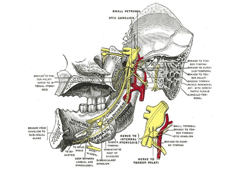
Mandibular nerve

26
Mandibular nerve It is a branch of trigeminal nerve
Arises from trigeminal ganglion Passes through foramen ovale It is joined by motor root in the foramen ovale and emerges as mixed nerve It divides immediately as anterior ( mainly motor) and posterior ( mainly sensory) divisions
 and posterior ( mainly sensory) divisions.")
27
Branches from trunk Meningeal branch enters skull with middle meningeal artery Nerve to medial pterygoid

28
Branches from anterior division
Large sensory buccal nerve Passes between the two heads of lateral pterygoid Runs on the surface of buccinator muscle It forms plexus with buccal branch of facial nerve It supplies entire thickness of cheek from skin to mucous membrane Motor branches of anterior division are : Nerve to lateral pterygoid Deep temporal nerves Nerve to masseter

29
Branches from posterior division
Auriculotemporal nerve Inferior alveolar nerve Mylohyoid nerve Lingual nerve

30
Auriculotemporal nerve
It arises by two roots It receives post ganglionic parasympathetic fibers from otic ganglion for parotid gland Preganglionic fibers reach from glossopharyngeal nerve Roots surround middle meningeal artery

31
Inferior alveolar nerve
It is a large branch of the posterior division It runs downwards with artery between the pterygoid muscles They give rise to mylohyoid branch Then enters mandibular foramen In the body of the mandible it supplies teeth and gums Sends mental branch which supplies skin of chin and lower lip Mylohyoid nerve only motor of posterior division supplies mylohyoid and anterior belly of digastric muscles The nerve is joined by submental artery in the digastric triangle Mylohyoid branch

32
Ligual nerve It is sensory to anterior two thirds of tongue , floor of mouth and gums It is medial to mandible It is in front of inferior alveolar nerve The nerve is joined by chorda tympani nerve , a branch of facial nerve Chorda tympani nerve brings preganglionic parasympathetic fibers to submandibular gland And taste sensation for anterior 2/3rds of tongue

33
Mandibular nerve

35
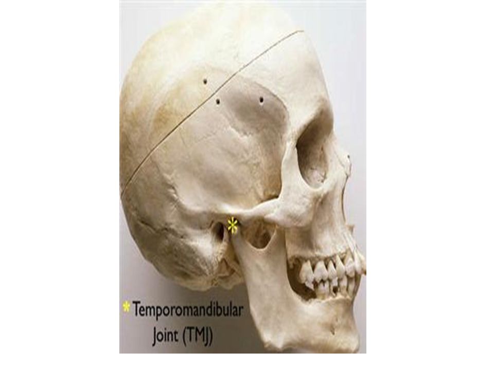
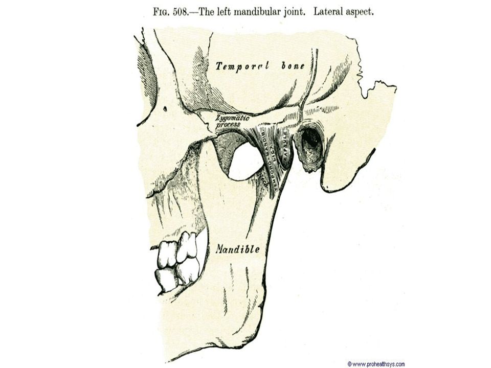
Temporomandibular joint
Type :Synovial joint Sub type : Hinge joint

38
Articular surfaces Head of the mandible
With mandibular fossa and articular tubercle of the temporal bone They are separated by articular disc Which divides joint cavity into upper and lower parts

39
Ligaments Fibrous capsule : is attached to margins of articular surfaces Laterally is thickened to form lateral ligament It is triangular with base attached to zygomatic arch With apex attached to lateral side of neck of mandible

40
Ligaments Articular disc is oval in shape
Fused with capsule around the periphery The upper surface is concavoconvex to fit articular tubercle and mandibular fossa Its concave inferior surface fits the head of he mandible

41
Ligaments The capsule and the lateral ligament are the only proper ligaments Sphenomandibular and the stylomandibular ligaments Do not add any strength to the joint Muscles of mastication provide the required strength Similar to joints maintained by muscle strength- the joint is prone for dislocation

42
Ligaments Sphenomandibular ligament extends from spine of sphenoid to lingula near the margin of the mandibular foramen It is piercred by mylohyoid nerve The ligament is the remnant of 1st pharyngeal arch

43
Relations Anterior Posterior Above Medially – Auriculotemporal nerve
Lateral pterygoid Posterior Parotid gland Above Floor of middle cranial fossa Medially – Auriculotemporal nerve

44
Blood supply Blood supply Superficial temporal artery Maxillary artery

45
Nerve supply Nerve supply
Auriculo-temporal nerve – from the posterior division of mandibular nerve Masseteric nerve – from anterior division of mandibular nerve

46
Movements of joint Protraction – pulling the jaw forwards
Retraction – it is reverse movement of protraction Depression- lowering of jaw Elevation – opposite of depresstion Side to side and grinding movement

47
Muscles acting on the joint
Protraction – lateral and medial pterygoid Retraction – posterior fibers of temporalis Depression- lateral pterygoid, digastric and infrahyoid muscles Elevation – temporalis, masseter and medial pterygoid Side to side and grinding movement – produced by muscles of opposite side acting alternately

48
Clinical anatomy Dislocation of joint

49
Learning Outcomes At the end of the lecture, students should be able to: Outline the boundaries and contents of the infratemporal fossa Describe the origin,insertion,nerve supply and action of muscles of mastication. Describe the origin,course, branches and distribution of maxillary artery Describe the origin,course, branches and distribution of mandibular nerve. Discuss the type,sub type of temperomandibular joint Articular surfaces Ligaments Relations Blood supply Nerve supply Movements Muscles acting on temperomandibular joint Outline the clinical anatomy of the temperomandibular joint

Similar presentations


>")
>")


