Download presentation
Presentation is loading. Please wait.

1
Anatomy II Head & Neck BY: DR
Anatomy II Head & Neck BY: DR.Yahya Alfarra CANADIAN BOARD IN DENTISTRY

2
Parotid region Parotid gland ::
Size : it’s the largest salivary gland & it’s compose of serous acini. Shape : it’s wedge shaped , Site & extension : it lies between the ramus of Md. & SCM It extends : Upward: to zygomatic arch Downward: to angle of Md. Anteriorly: to cover part of masseter ms. Posteriorly: to overlap SCM

3
Parotid duct(stenson’s duct)
Length : 5 cm long Beginning : at the ant. Border of the gland End: by opening into the oral cavity opposite the upper 2nd molar tooth

4
Opens in the oral cavity opposite to the second upper molar tooth.

5
Blood supply of P.G Arterial supply : small branches from ECA inside the gland Venous drainage: into retromandibular V. Lymphatic drainage : into deep & superficial parotid lymph node Nerve supply of parotid :Sensory : great auricualr N auriculotemporal N.

6
Superficial temporal artery

7
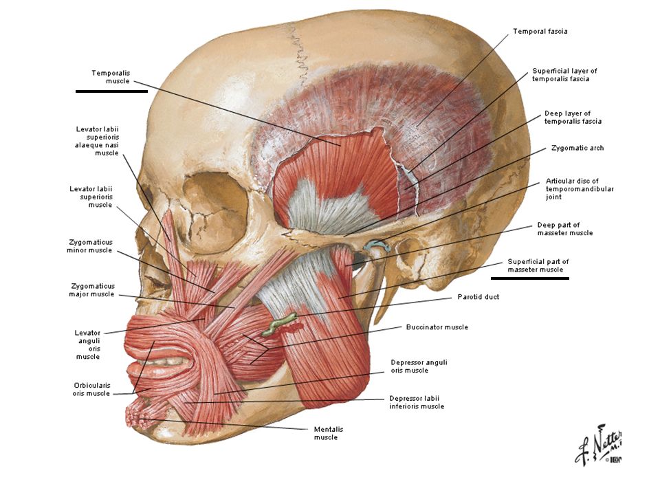
Temporal region Temporal fossa Infratemporal fossa
MuSclE OF mastications

8
Temporal fossa Boundaries : Sup. Temporal line ……… above
Zygonatic arch…………….below Frontal process of zygomatic bone……………………….ant. Contents(attachments): 2 superficial Ms of mastications Muscles : a) temporalis Ms. b) masseter Ms Nerves : a ) deep temporal N. b)auriculotemporal N. Vesseles: superficial temporal A.
: 2 superficial Ms of mastications. Muscles : a) temporalis Ms. b) masseter Ms. Nerves : a ) deep temporal N. b)auriculotemporal N. Vesseles: superficial temporal A.")
9
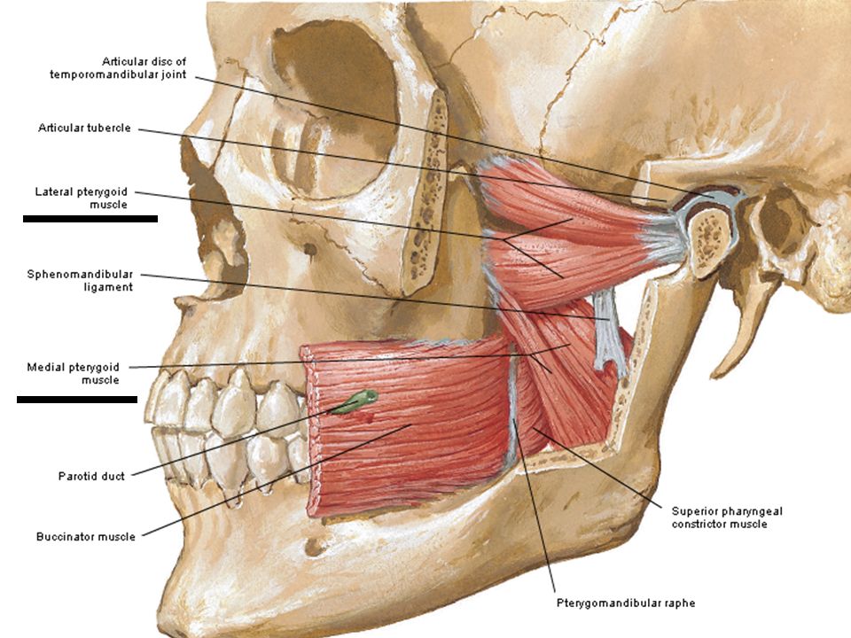
Infratemporal fossa Boundaries: Anteriorly: post. Surface of maxilla
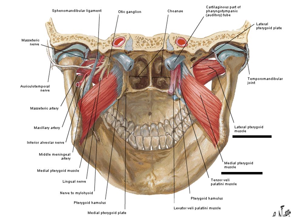
Posteriorly : styloid & mastoid process Medially: lateral pterygoid plate Laterally : coronoid process & ramus of Md. Contents: Muscles : 2 deep Ms of Mastications a) medial pterygoid Ms. b ) lateral pterygoid m. Nerves: a ) Md. N. b ) chorda tympani c ) Mx. N. Vesseles : a ) Mx. A b ) pterygoid plexus of viens
 medial pterygoid Ms. b ) lateral pterygoid m. Nerves: a ) Md. N. b ) chorda tympani c ) Mx. N. Vesseles : a ) Mx. A. b ) pterygoid plexus of viens.")
10
Muscles of mastications
Lateral Pterygoid Medial pterygoiod temporalis Masseter Arise by 2 head a)Upper head(infratemporal surfaceof greater wing of sphenoid ) b) Lower head(lat. Surface of lat. Pterygoid plate) a)superficial head(Mx.tuberosity) b) Deep head(med. Surface of lat. Pterygoid plate) Floor of temporal fossa ? origin Front of neck of Md. & articular disc of T.M.J Med. Surface of angle of Md. ?. Lateral (outer)surface of ramus of Md. Insertion
Upper head(infratemporal surfaceof greater wing of sphenoid ) b) Lower head(lat. Surface of lat. Pterygoid plate) a)superficial head(Mx.tuberosity) b) Deep head(med. Surface of lat. Pterygoid plate) Floor of temporal fossa. origin. Front of neck of Md. & articular disc of T.M.J. Med. Surface of angle of Md. . Lateral (outer)surface of ramus of Md. Insertion.")
11
Muscles of mastications
Lateral Pterygoid Medial pterygoiod temporalis Masseter Depress Md. Elevate Md. Elevate Md. To occlude teeth in mastication Action Anterior division of mandibular nerve ? Nerve supply

15
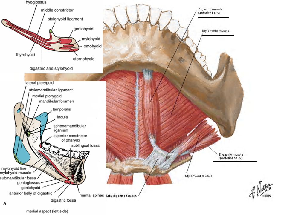
Submandibualr region Definition: it’s the regoin between Md. & hyoid bone. It contains : Muscles: digastric, mylohyoid , hyoglossus, geniohyoid , genioglossus & styloglossus Salivary gland : submandibualr & sublingual Nerves : lingual , glossopharyngeal & hyoglossal Blood vesseles: facial A. & V. , Lingual A. & V. Parasympathetic ganglion : submandibualr Lymph nodes : submandibualr group

16
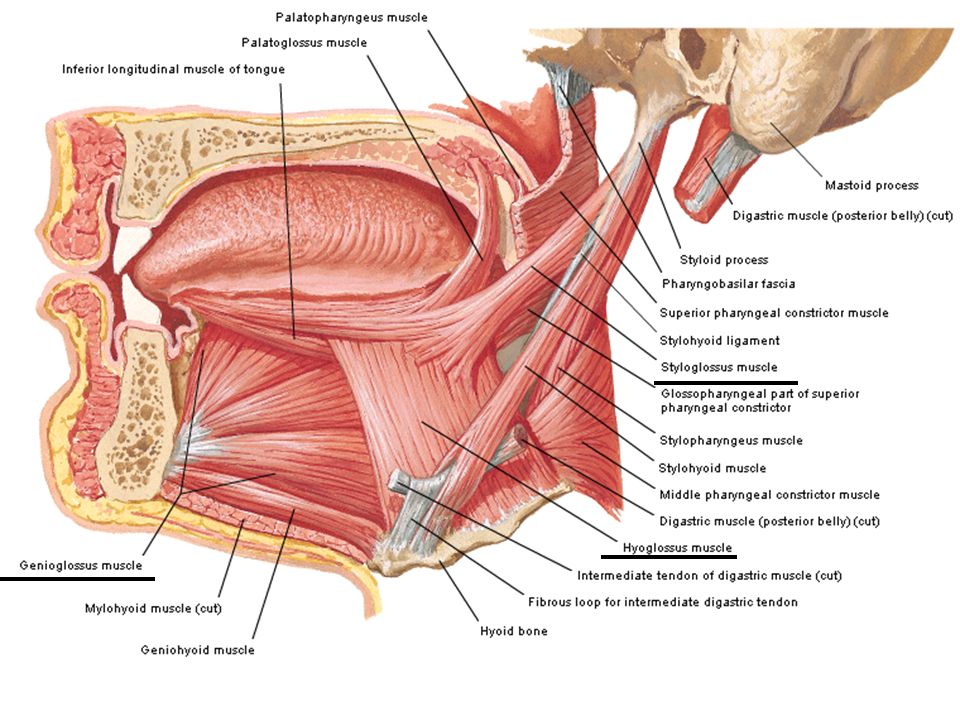
Muscles of sumandibualr region
styloglossus genioglossus hyoglossus geniohyoid mylohyoid digastric Styloid process Upper genial tubercle of Md. Body of hyoid bone it.’s a small Ms lying deep(above) to mylohyoid From genial tubercle(behind symphysis menti of Md.) Mylohyoid Md. Arise by 2 bellies: ant. Belly: lower border of Md. Close to symphysis menti Post. Belly: medial surface of mastoid process origin Whole length of side of tongue Whole length Under surface of tongue Inside tongue deep to styloglossus Upper surface of body of hyoid Into intermediate tendon that connected hyoid bone insertion
 to mylohyoid. From genial tubercle(behind symphysis menti of Md.) Mylohyoid Md. Arise by 2 bellies: ant. Belly: lower border of Md. Close to symphysis menti. Post. Belly: medial surface of mastoid process. origin. Whole length of side of tongue. Whole length Under surface of tongue. Inside tongue deep to styloglossus. Upper surface of body of hyoid. Into intermediate tendon that connected hyoid bone. insertion.")
17
Muscles of sumandibualr regoin
styloglossus genioglossus hyoglossus geniohyoid mylohyoid digastric Hypoglossal N. Hypoglossa-l N. From C1 via N. to mylohyoid a branch of Inferior alveolar N. (from Md. N.) Ant. Belly : N. to mylohyoid a branch of Inferior alveolar N. (from Md. N. ) Post. Belly: Facial Nerve Nerve supply Retract tongue upward& backward Protrude tongue into midline Depress tongue *elevate Hyoid Bone *depress Md. If Hyoid Bone is Fixed(open Mouth) *support Floor of mouth Ant. Belly : opening Md. Retract hyoid bone upward& backward Action
 Ant. Belly : N. to mylohyoid a branch of Inferior alveolar N. (from Md. N. ) Post. Belly: Facial Nerve. Nerve supply. Retract tongue upward& backward. Protrude tongue into midline. Depress. tongue. *elevate. Hyoid Bone. *depress. Md. If Hyoid. Bone is. Fixed(open. Mouth) *support. Floor of. mouth. Ant. Belly : opening Md. Retract hyoid bone upward& backward. Action.")
20
Salivary gland

21
Submandibular gland Site & extension: It lies below Md.
It extends to : Mylohyoid line : a bove Digastric muscle : below Mental foramen : anterior Angle of Md. : posterior Size: about ½ the size of P.G. Shape: wedge shape Structure: it consists of 2 parts,a large superficial part & a small deep part

22
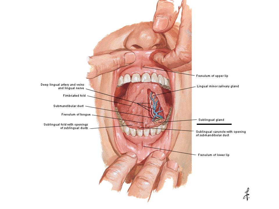
Submandibular duct It’s 5 cm long It’s duct open into
the floor of mouth close to frenulum of tongue.

23
Parasympathetic: from facial N.
Blood supply of gland: The arterial supply: the gland is supplied by branches of facial & lingual A. The viens drain into facial & lingual V. Nerve supply : sympathetic : from plexus arround lingual A. Parasympathetic: from facial N.

24
Sublingual gland Site: beneath tongue
Size: it’s the smallest of the 3 S.G. Shape: almond-shaped Relation : Ant. : the gland of the opposite side Post. : the deep part of submandibualr gland Med. : genioglossus Ms. , lingual N. & submand. Duct Laterally: sublingual fossa of Md. Sup. : mucous membrane of mouth(which elevated to form sublingual fold) Inf. : mylohyoid Ms.
 Inf. : mylohyoid Ms.")
26
Blood supply : the gland is supplied by branch of facial & lingual A.
Sublingual duct : It opens directly in the floor of mouth on sublingual fold Blood supply : the gland is supplied by branch of facial & lingual A. The viens drain into facial & lingual V. Nerve supply : sympathetic :from plexus arround lingual A. Parasympathetic: from facial N.

27
Nerves OF submandibualr regoin
lingual , glossopharyngeal & hyoglossal N.B. : lingual N. : branch of post. Division of Md. N. in the infratemporal fossa Blood vesseles submandibualr regoin : facial A. & V. , Lingual A. & V. N.B. Facial A. : Branch of ECA Facial V. : It units ant. Division of retromandibualr V. TO drain into IJV Lingual A. : Branch of ECA Lingual V. : drain into IJV

28
Good luck

Similar presentations


>")


