Download presentation
Presentation is loading. Please wait.

1
Lower limb fractures and dislocation
DR. MOHAMAD KHAIRUDDIN BIN ABDUL WAHAB M.B.B.S (Univ. Malaya), MS Ortho (UKM) ORTHOPAEDIC SURGEON FACULTY OF MEDICINE CUCMS
, MS Ortho (UKM) ORTHOPAEDIC SURGEON. FACULTY OF MEDICINE. CUCMS.")
2
Learning outcome: The student should be able to:
Discuss on the mechanism, clinical presentation, classification, radiological findings, and its complications of fractures and joint dislocation Derive treatment option of the common lower limb fractures and joint dislocation

3
Contents: FRACTURE NECK OF FEMUR INTERTROCHANTERIC FRACTURE
HIP JOINT DISLOCATION FEMUR SHAFT FRACTURE DISTAL FEMUR FRACTURE KNEE JOINT DISLOCATION PATELLA FRACTURE TIBIAL PLATEAU FRACTURE

4
CONT’: TIBIA SHAFT FRACTURE MALLEOLI FRACTURE TALUS FRACTURE
CALCANEUM FRACTURE

5
Fracture neck of femur Common in elderly following fall (osteoporosis)
Young adult is due to high energy impact such as road traffic accident May accompanied hip joint dislocation (high impact injury) Demonstrated radiological (AP view of hip joint) as: Loss of Shenton’s line Disruption of proximal femur trabecula
 Demonstrated radiological (AP view of hip joint) as: Loss of Shenton’s line. Disruption of proximal femur trabecula.")
6
Classification: Garden’s classification (4 stages) for femur neck fracture Help to determine the management and predict the prognosis on complication (avascular necrosis of the femoral head)
")
7
Garden’s classification
Stage I Incomplete # (impacted) Stage II Complete and undisplaced Stage III Complete and moderately displaced Stage IV Severely displaced
 Stage II. Complete and undisplaced. Stage III. Complete and moderately displaced. Stage IV. Severely displaced.")
8
Anatomical classification:
Also can describe the pattern of neck fracture Subcapital region Transcervical region Basal region Prognosis for AVN worsen in subcapital and transverse fracture

9
Radiological features of neck of femur fracture
Shenton’s line

10
Complication: Avascular necrosis of the femur head
Non-union of the fracture General complications following prolong bedridden for conservative treatment (bedsore, DVT, pneumonia, stiffness)
")
11
Treatment: Depend on the age of the patient, patient’s health and fracture stages & duration Non-operative reserve for: Poor health (unfit for surgery) patient Require on Traction for 3 – 6 weeks then start ambulate
 patient. Require on Traction for 3 – 6 weeks then start ambulate.")
12
Cont’: Operative treatment is the main goal:
Younger age group with acute # and elderly with impacted # (preserved the head) usage of fracture fixation devices eg. Screw fixation, Dynamic Hip Screw Elderly patient with displaced # or chronic # subjected to hip replacement (hemiarthroplasty or total arthroplasty of the hip joint)
 usage of fracture fixation devices eg. Screw fixation, Dynamic Hip Screw. Elderly patient with displaced # or chronic # subjected to hip replacement (hemiarthroplasty or total arthroplasty of the hip joint)")
13
Intertrochanteric fracture
Commonly occur in elderly patient (osteoporosis) following trivial fall Extension to subtrochanteric region May presented as comminuted fracture pattern
 following trivial fall. Extension to subtrochanteric region. May presented as comminuted fracture pattern.")
14
Radiograph shows intertrochanteric
fracture of the femur

15
Complications: Mal-union of the fracture
Failure in fixation for the fracture due to osteoporotic bone General complications following prolong bedridden

16
Treatment Operative is the main goal except unfit patient for anaesthesia or extreme osteoporotic bone Choices of implant for fracture fixation: Dynamic Hip Screw Proximal femoral nail (PFN)
")
17
Fixation of fracture intertrochanteric fracture

18
Hip joint dislocation Direction: posterior is more common than anterior Mechanism: ‘dash-board’ injury Limb attitude: Posterior dislocation (flexed, adducted, internally rotated, short limb) Anterior dislocation (flexed, externally rotated, abducted) Association with acetebular fractures of femoral head fractures
 Anterior dislocation (flexed, externally rotated, abducted) Association with acetebular fractures of femoral head fractures.")
19
Left side Radiograph shows left hip dislocation

20
Complications: Sciatic nerve injury leading muscle paralysis and loss of sensory below the knee Prolong dislocation can also result in avascular necrosis of the femoral head

21
Treatment Emergency CMR under sedation Failure in CMR open reduction
Failure in CMR to obtain acceptable reduction is due to: Inverted limbus of the acetebular rim Intra-articular fracture fragment

22
Femoral shaft fractures
Area that is well padded with muscles leading to fracture displacement and difficulty in CMR and maintain the reduction Associated with soft tissue injury due to high-energy injury risk of getting compartment syndrome Long bones – segmental # Occasionally associated with # neck of femur

23
Radiographs show femur shaft fractures
Distal 1/3 supracondyalar Proximal 1/3

24
Complication Vascular injury (femoral artery) Fat embolism
Delayed and non-union of the fracture Mal-union of the fracture Joint stiffness (knee)
")
25
Treatment Less preference for non-operative treatment (as the bone is weight bearing region) in adult Operative fracture fixation used : Intramedullary-Locking-Nail Plating (DCP)
")
26
Intramedullary locking
nail

27
Distal femur #: Supracondylar & intercondylar
Supracondylar # can be isolated or combination with intercondylar # Result from high energy force Risk of vascular injury (femoral artery) Intercondylar extension may involved articular region of the knee
 Intercondylar extension may involved articular region of the knee.")
28
Complications Joint stiffness and arthrosis if involve the articular region Risk of femoral artery injury

29
Treatment Open Reduction Internal Fixation is a goal standard treatment Fixation devices: Angled blade plate CDS (condylar dynamic screw) Supracondylar inter-locking nail Buttress plating (locking plate)
 Supracondylar inter-locking nail. Buttress plating (locking plate)")
30
Angled blade plate for fixation
of supracondylar fracture of the femur

31
Knee joint dislocation
Result from violence injury force Involve more than two of knee ligaments injury Can presented as ‘self-reduction’ joint dislocation Associated with popliteal vessel injury and common peroneal nerve injury Urgent attention for vascular assessment

32
Radiographs show anterior
dislocation of the knee

33
Risk of vascular injury
Transected or thrombosis (following intimal injury) Vascular assessment or surveillance Angiogram as indicated
 Vascular assessment or surveillance. Angiogram as indicated.")
34
Directions of dislocation
Reference to the position of tibia Anteromedial dislocation (risk of associated intimal injury of popliteal artery) Posterolateral dislocation (highly associated with transected popliteal artery)
 Posterolateral dislocation (highly associated with transected popliteal artery)")
35
artery

36
Complications Neurovascular injury
Knee ligaments injury (result in joint instability) Stiffness of the joint Arthrosis formation following cartilage damage
 Stiffness of the joint. Arthrosis formation following cartilage damage.")
37
Treatment Immediate reduction and immobilization
Artery exploration and repair in the evidence of arterial injury Immobilization in cast (FLPOP) or external fixation Ligaments repair or reconstruction for multiple ligaments injury resulting in instability
 or external fixation. Ligaments repair or reconstruction for multiple ligaments injury resulting in instability.")
38
Tibial plateau fractures
Mechanism: varus or valgus force combined with axial loading Also known as ‘bumper fracture’ Tibial condyle can be crushed or split Presentation: haemathrosis, instability, associated neurovascular injury

40
Types of TP # Simple split lateral condyle
Depressed, comminuted lateral condyle Crushed comminuted lateral condyle Split medial condyle Bicondylar fractures Bicondylar and subcondylar

41
Complications Compartment syndrome Joint stiffness Deformity arthrosis

42
Treatment Undisplaced or minimally displaced
Traction until swelling subsided, apply cast immobilization Displaced and depressed Open reduction and internal fixation (buttress plate, inter-fragmentary screw) May need bone grafting in depressed fractures
 May need bone grafting in depressed fractures.")
43
Patella fractures Direct injury (dash board, direct fall onto the knee) produced ‘stellate’ fracture Indirect injury (forced flexion knee) produce avulsion type or simple transverse pattern Loss of extensor mechanism Haemathrosis
 produce avulsion type or simple transverse pattern. Loss of extensor mechanism. Haemathrosis.")
44
Complications Joint stiffness Patellofemoral arthrosis
reduced knee extensor mechanism

45
Treatment Undisplaced fracture
Cylinder cast immobilization for 6 weeks Displaced fracture ORIF (tension band wiring) Severely comminuted Cerclage wiring or patellectomy
 Severely comminuted. Cerclage wiring or patellectomy.")
46
Tibial shaft fractures
Proximal, middle, distal region Compartment syndrome (proximal 1/3) Affecting union (distal 1/3) Spiral, oblique (indirect force) Transverse, comminuted (direct force) With or without fibular shaft #
 Affecting union (distal 1/3) Spiral, oblique (indirect force) Transverse, comminuted (direct force) With or without fibular shaft #")
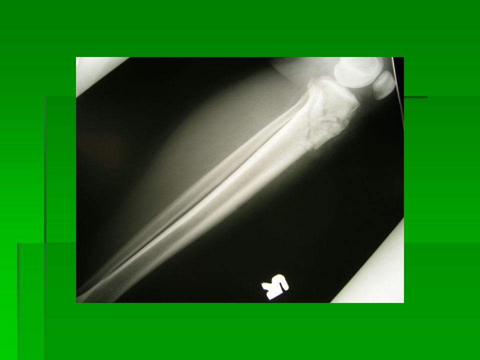
47
Radiographs show tibial shaft fracture

48
Complications Compartment syndrome
Malunion (leading to shortening and arthrosis) Nonunion
 Nonunion.")
49
Treatment Acceptable displacement with less comminuted (stable)
Apply Full Length POP immobilization for 6 weeks Comminuted, segmental (unstable reduction alignment) Internal fixation (ILN, Plating)
 Internal fixation (ILN, Plating)")
50
Intramedullary Locking nail for Tibia shaft fracture

51
Malleoli fractures Forces to the ankle region
External rotation, abduction, adduction, Ankle joint dislocation or subluxation Ankle ligaments injury including syndesmosis

52
Classification Danis & Weber (Muller et al 1991):
Type A: # below the tibiofibular syndesmosis abduction or adduction force Medial malleolus may #ed or rupture of deltoid ligament

53
Cont’: Type B: # level with syndesmosis Oblique fibular #
External rotation force Disrupted medial structures Syndesmosis intact

54
Cont’: Type C: # above the syndesmosis
Abduction alone or combination of abduction and external rotation force Disruption of syndesmosis and interosseous membrane (widened mortise) Unstable tibiofibular region
 Unstable tibiofibular region.")
55
Fracture of lateral malleolus

56
Complications Dislocated or subluxated ankle joint Stiffness
Arthrosis of ankle joint Ankle instability Nonunion fracture (displaced medial malleolus) Malunion of the fracture
 Malunion of the fracture.")
57
Treatment Undisplaced # Cast immobization (boot POP)
Displaced # with or without subluxation joint or loss of normal ankle mortise ORIF (fibular plating, screw fixation of medial malleoli, syndesmotic screw)
")
58
Plating of the lateral malleolus fracture
with 1/3 tubular plate

59
Talus fractures Rare injury
Violence injury (following inversion force or axial loading) +/- dislocation of the ankle joint or subtalar joint Regions affected: head, neck, body, and lateral process Risk of developing avascular necrosis of talus dome
 +/- dislocation of the ankle joint or subtalar joint. Regions affected: head, neck, body, and lateral process. Risk of developing avascular necrosis of talus dome.")
60
Talus fractures Dome of talus fracture showed Through CT-scan
Neck of talus fracture

61
Complications Skin damage or necrosis due to pressure from the underling bone Nonunion of the fracture AVN following fracture at the neck region Arthrosis (ankle and subtalar)
")
62
Treatment Undisplaced #: cast immobilization (boot POP)
Displaced # +/- dislocation: ORIF screw fixation If AVN developed later may consider arthrodesis of the ankle joint

63
Screw fixation of the talus fracture at the neck region

64
Calcaneum fractures Result from axial loading
Traction through Achilles tendon lead to avulsion fracture Can be extra-articular or intra-articular fracture (referring to subtalar joint) Result in loss of foot arch (Bohler’s angle: 25 –40 degrees) lead to flat foot
 Result in loss of foot arch (Bohler’s angle: 25 –40 degrees) lead to flat foot.")
65
Extra-articular fracture of calcaneum

66
Complications Skin necrosis (intense swelling)
Malunion of the fracture Peroneal tendon impingement Flat and broad foot (shoe fitting) Subtalar arthrosis
 Subtalar arthrosis.")
67
Treatment Extra-articular fractures or undisplaced intra-articular fractures may require Robert-Jones bandaging for 1 week then followed by boot POP cast for 5 weeks No weight bearing is allowed Displaced intra-articular # or avulsion of Achilles insertion: ORIF screw or recon plate

68
Exercise for student: After reviewing the lecture notes, you are
require to do some exercises. The answers to the exercise need to be submitted via (address:

69
Questions: Briefly discuss on the classification used to describe neck of femur fracture. With regards to dislocated knee, describe the direction of dislocation in relation to vascular injury pattern. Briefly discuss on the complications following calcaneum fracture.

70
Reference for further reading:
Orthopaedic Surgery Essential: Trauma; Charles Court-Brown, Lippincott Williams & Wilkins; 2005 Turek’s Orthopaedics: Principles & their application; Stuart L. Wienstein, Joseph A. Backwalter: 5th Edition Lippincott Williams & Wilkins 2005 Practical Fracture Treatment; Ronald McRae, Max Esser; 4th Edition, Churchill Livingstone 2002

71
Enjoy reading….. For further questions or enquiry , please contact through: Handphone:

Similar presentations


, F.R.C.S.(C )>")


![Thigh and knee. CLASSIFICATION FRACTURES OF THE FEMUR [1 ]Fracture of the neck of the femur, and [2]Fracture of the trochanteric region [3] Fracture of.](/14/4311083/big_thumb.jpg "Thigh and knee. CLASSIFICATION FRACTURES OF THE FEMUR [1 ]Fracture of the neck of the femur, and [2]Fracture of the trochanteric region [3] Fracture of.>")


 guidelines with attention.>")