Download presentation
Presentation is loading. Please wait.

1
DNA RESEARCH AND APPLICATIONS

2
DNA STRUCTURE

3
A2.6.1 Crick and Watson’s discovery of the structure of DNA using model making. ■Watson and Crick used evidence to develop possible structures for DNA and then tested their theories by building models. ■1 st model- triple helix, bases on the outside, magnesium holding the strands together. This was falsified for 2 reasons. –Ratio of adenine to thymine was not 1:1 (as discovered by Chargaff) –It required too much magnesium as identified by Franklin
 –It required too much magnesium as identified by Franklin.")
4
2 nd model- ■Base Pairs –Watson and Crick had to take into account Chargaff’s findings that the amount of adenine bases equal the amount of thymine bases, and the amount of guanine equals the amount of cytosine. –They cut cardboard models of the nitrogen bases and showed that base pairs could be formed, with hydrogen bonds linking them. ■Antiparallel strands –Based on setbacks from first model and Xray diffraction patterns, they knew DNA must be a double helix –They realized that the 2 strands had to run in opposite directions in order to fit together (antiparallel) –They built a model to scale and quickly convinced all who saw it. –The model also suggested a mechanism for copying DNA and led to the realization that the genetic code must consist of triplets of bases.
 –They built a model to scale and quickly convinced all who saw it. –The model also suggested a mechanism for copying DNA and led to the realization that the genetic code must consist of triplets of bases..")
5
A7.1.1 Rosalind Franklin’s and Maurice Wilkins’ investigation of DNA structure by X-ray diffraction. ■X-ray diffraction –When X-rays are directed at a material, of the X-rays are scattered by the material. This scattering is called diffraction. In order to work well, the material should be crystallized in order to diffract the x-rays in a regular pattern. –Rosalind Franklin X-ray diffractionRosalind Franklin X-ray diffraction

6
Rosalind Franklin ■Franklin improved the resolution of the camera in order to take more detailed measurements of X-ray diffraction patterns. In addition, she provided high quality samples of DNA that could be used to obtain good images. (DNA can’t be crystallized, so it is harder to get a good image.) ■The images taken allowed Franklin to deduce that DNA is a double helix and that the phosphate groups are on the outside of the molecule. ■Measurements taken by Franklin and Wilkins allowed them to determine the basic dimensions of the DNA molecule. Maurice Wilkins ■Produced clear X-ray diffraction patterns of DNA. ■Gave Watson one of Franklin’s DNA images, which was used by him and Crick to determine the structure of DNA.
 ■The images taken allowed Franklin to deduce that DNA is a double helix and that the phosphate groups are on the outside of the molecule. ■Measurements taken by Franklin and Wilkins allowed them to determine the basic dimensions of the DNA molecule. Maurice Wilkins ■Produced clear X-ray diffraction patterns of DNA. ■Gave Watson one of Franklin’s DNA images, which was used by him and Crick to determine the structure of DNA..")
7
S2.7.1 Analysis of Meselson and Stahl’s results to obtain a support for the theory of semi-conservative replication of DNA. ■Before Meselson and Stahl’s work, there were different theories as to how DNA replicated, but they provided evidence that DNA replication is semi-conservative. ■The link below provides a detailed animation. A step by step description of the process is also described in the following slides. ■http://highered.mheducation.com/sites/9834092339/student_view0/chapter14/ meselson_and_stahl_experiment.htmlhttp://highered.mheducation.com/sites/9834092339/student_view0/chapter14/ meselson_and_stahl_experiment.html ■

8
■Bacterial cells were grown in a medium containing a heavy isotope of nitrogen ( 15 N), so the DNA in the bacteria contain “heavy” nitrogen. ■After many generations of the bacteria were grown, the cells were moved to a medium containing the more common isotope of nitrogen ( 14 N). Samples of the bacteria were then collected at different time intervals. ■DNA was extracted from the collected cells and dissolved in a solution of cesium chloride and then spun in a centrifuge. ■A concentration gradient is established and the DNA molecules travel to a place in the solutions where their density is equal to the cesium. DNA containing 14 N and DNA containing 15 N travel to different places because 15 N is more dense than 14 N.
. Samples of the bacteria were then collected at different time intervals. ■DNA was extracted from the collected cells and dissolved in a solution of cesium chloride and then spun in a centrifuge. ■A concentration gradient is established and the DNA molecules travel to a place in the solutions where their density is equal to the cesium. DNA containing 14 N and DNA containing 15 N travel to different places because 15 N is more dense than 14 N..")
9
■After one generation in 14 N medium, the bacteria made a single type of DNA whose density was in between the density of 14 N and 15 N. This indicated that only one strand of each DNA molecule contained 15 N. ■After 2 generations, two different types of DNA were made, one with a density in between 14 N and 15 N and one with a density matching that of 14 N. ■Conclusion: Meselson and Stahl determined that the replication of DNA involves using each strand from the original DNA molecule as a template to form a new strand. This results in 2 new molecules, each containing an original strand and a newly synthesized strand.

10
S7.1.2 Analysis of results of the Hershey and Chase experiment providing evidence that DNA is genetic material. ■Hershey and Chase proved that DNA is genetic material. ■They used a phage (bacterial virus) which consisted of a DNA molecule surrounded by a protein coat. ■Experiment- –phage produced in a medium containing radioactive sulfur (35S), which resulted in a phage population containing radioactively-labeled proteins. –The phage were then allowed to infect the bacteria. Another phage population was produced in a medium containing radioactive phosphorus (32P), which resulted in radioactively-labeled DNA. –The phage were allowed to infect a different group of bacterial cells. –Both groups of bacterial cells were subjected to vigorous shaking (blender) and concentrated (centrifuged) into a pellet. Hershey and Chase measured the radioactivity of the cells and determined that DNA had been injected into the cells, providing evidence that DNA is genetic material. ■http://highered.mheducation.com/sites/9834092339/student_view0/chapter14/hershe y_and_chase_experiment.htmlhttp://highered.mheducation.com/sites/9834092339/student_view0/chapter14/hershe y_and_chase_experiment.html
 which consisted of a DNA molecule surrounded by a protein coat. ■Experiment- –phage produced in a medium containing radioactive sulfur (35S), which resulted in a phage population containing radioactively-labeled proteins. –The phage were then allowed to infect the bacteria. Another phage population was produced in a medium containing radioactive phosphorus (32P), which resulted in radioactively-labeled DNA. –The phage were allowed to infect a different group of bacterial cells. –Both groups of bacterial cells were subjected to vigorous shaking (blender) and concentrated (centrifuged) into a pellet. Hershey and Chase measured the radioactivity of the cells and determined that DNA had been injected into the cells, providing evidence that DNA is genetic material. ■ y_and_chase_experiment.htmlhttp://highered.mheducation.com/sites/ /student_view0/chapter14/hershe y_and_chase_experiment.html.")
11
A2.7.1 Use of Taq DNA polymerase to produce multiple copies of DNA rapidly by the polymerase chain reaction (PCR) ■Polymerase Chain Reaction –Typically used to copy a segment of DNA (not the whole genome) –used to amplify small samples of DNA for DNA profiling, recombination, species identification, or other research ■Virtual LabVirtual Lab ■PCR animation walkthroughPCR animation walkthrough ■https://www.dnalc.org/resources/animations/pcr.htmlhttps://www.dnalc.org/resources/animations/pcr.html
 ■Polymerase Chain Reaction –Typically used to copy a segment of DNA (not the whole genome) –used to amplify small samples of DNA for DNA profiling, recombination, species identification, or other research ■Virtual LabVirtual Lab ■PCR animation walkthroughPCR animation walkthrough ■")
12
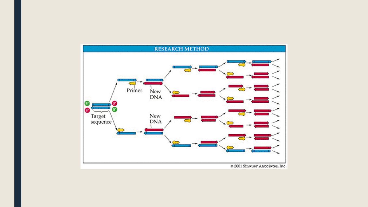
How PCR works ■PCR requires –Target DNA (containing the segment you wish to copy) –free nucleotides –DNA primers- short strands of DNA nucleotides, complimentary to the ends of the target DNA sequence –enzyme Taq polymerase- DNA polymerase taken from the bacteria Thermus aquaticus, which is found in hot springs and can survive the high temps in PCR
 –free nucleotides –DNA primers- short strands of DNA nucleotides, complimentary to the ends of the target DNA sequence –enzyme Taq polymerase- DNA polymerase taken from the bacteria Thermus aquaticus, which is found in hot springs and can survive the high temps in PCR")
13
Steps of PCR ■1. Denaturation: the mixture is heated to 95 degrees Celsius, separating the DNA strands. ■2. Annealing: The temperature is reduced and the primers can form hydrogen bonds (anneal) with their complimentary sequences in the target DNA. ■3. Elongation: Temp is raised to 72 degrees C. Taq polymerase begins adding nucleotides to the 3’ end of each primer attached to the DNA strand. After one complete cycle, there are 2 double stranded copies of the target DNA. ■Repeat: The mixture is heated up again, and the cycle repeats. Thirty cycles can amplify the segment of DNA by a factor of one billion, while using only a short amount of time.
 with their complimentary sequences in the target DNA. ■3. Elongation: Temp is raised to 72 degrees C. Taq polymerase begins adding nucleotides to the 3’ end of each primer attached to the DNA strand. After one complete cycle, there are 2 double stranded copies of the target DNA. ■Repeat: The mixture is heated up again, and the cycle repeats. Thirty cycles can amplify the segment of DNA by a factor of one billion, while using only a short amount of time..")
15
DNA structure links and animations ■http://learn.genetics.utah.edu/content/molecules/builddna/http://learn.genetics.utah.edu/content/molecules/builddna/ ■http://www.johnkyrk.com/DNAanatomy.htmlhttp://www.johnkyrk.com/DNAanatomy.html

16
DNA replication links and animations ■http://highered.mheducation.com/sites/0072943696/student_view0/chapter3/an imation__dna_replication__quiz_1_.htmlhttp://highered.mheducation.com/sites/0072943696/student_view0/chapter3/an imation__dna_replication__quiz_1_.html ■http://sites.fas.harvard.edu/~biotext/animations/replication1.swfhttp://sites.fas.harvard.edu/~biotext/animations/replication1.swf ■http://www.stolaf.edu/people/giannini/flashanimat/molgenetics/dna-rna2.swfhttp://www.stolaf.edu/people/giannini/flashanimat/molgenetics/dna-rna2.swf ■http://link.hhmi.org/services/player/bcpid1876525637001?bckey=AQ~~,AAAArZFA _pE~,D1__qX58jerI2jGitJBc_zBPvdgXTuAq&bctid=2116972003001http://link.hhmi.org/services/player/bcpid1876525637001?bckey=AQ~~,AAAArZFA _pE~,D1__qX58jerI2jGitJBc_zBPvdgXTuAq&bctid=2116972003001

Similar presentations


 When do reviews start? 2) What days of the week are the reviews? 3) Write 5 things you learned from this week’s reading? 4) When is the genetics.>")


. PCR produces billions of copies of a specific piece of DNA from trace amounts of starting material. (i.e. blood, skin.>")

 Ribonucleic Acid (RNA)>")


 and Replication of DNA (chapter 12, pages 318 – 334)>")