Download presentation
Presentation is loading. Please wait.

2
Lower limb

4
QUADRICEPS FEMORIS RECTUS FEMORIS : Quadriceps tendon in to petella
Origin : Straight Head: A.I.I.S Reflected Head: Ilium (Above Acetabulum) Insertion : Quadriceps tendon in to petella Via lig.patellae in to tibial tubercle N. supply: Femoral nerve Action : Extension of Leg at Knee joint Flexion of thigh at hip joint
 Insertion : Quadriceps tendon in to petella. Via lig.patellae in to tibial tubercle. N. supply: Femoral nerve. Action : Extension of Leg at Knee joint. Flexion of thigh at hip joint.")
5
VASTUS LATERALIS petella via Lig.patellae in to tubercle of tibia
Origin : Upper end & shaft of femur Insertion : Quadriceps tendon in to petella via Lig.patellae in to tubercle of tibia N. supply: Femoral nerve Action : Extension of Leg at Knee joint

6
VASTUS MEDIALIS petella Via Lig.patellae in to tubercle of tibia
Origin : Upper end & shaft of femur Insertion : Quadriceps tendon in to petella Via Lig.patellae in to tubercle of tibia N. supply: Femoral nerve Action : Extension of Leg at Knee & Stabilizes Patella

7
VASTUS INTERMEDIUS of shaft of femur petella Via Lig.patellae
Origin : Anterior & lateral surface of shaft of femur Insertion : Quadriceps tendon in to petella Via Lig.patellae in to tubercle of tibia N. supply: Femoral nerve Action : Extension of Leg at Knee joint

8
VASTUS INTERMEDIUS of shaft of femur petella Via Lig.patellae
Origin : Anterior & lateral surface of shaft of femur Insertion : Quadriceps tendon in to petella Via Lig.patellae in to tubercle of tibia N. supply: Femoral nerve Action : Extension of Leg at Knee joint

9
SARTORIUS shaft of tibia N. supply: Femoral nerve
Origin : A.S.I.S. Insertion : Upper medial surface of shaft of tibia N. supply: Femoral nerve Action :1.Flexion, abduction and Lateral rotation of thigh at hip joint. 2. Flexion leg at knee joint

10
ILIACUS lesser trochanter of femur N. supply: Femoral nerve
Origin : Iliac fossa of hip bone Insertion : Along with psoas into lesser trochanter of femur N. supply: Femoral nerve Action :Flexes thigh on trunk Eg.: Sitting up from lying down

11
PSOAS I.V.D.’S of T12 & L 1-L5 vertebrae trochanter of femur
Origin : Trasnverse process, bodies & I.V.D.’S of T12 & L 1-L5 vertebrae Insertion : Along with Iliacus into lesser trochanter of femur N. supply: Lumbar plexus Action : Flexes thigh on trunk Eg.: Sitting up from lying down T12 12345

12
PECTINEUS Upper end of linea aspera of shaft of femur
Origin : Superior ramus of Pubis Insertion : Upper end of linea aspera of shaft of femur N. supply: Femoral nerve Action :Flexion, adduction ofthigh at hip joint

13
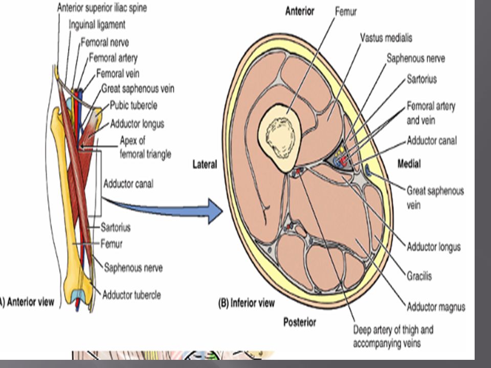
Femoral Triangle The femoral triangle, a subfascial space, is a triangular landmark useful in dissection and in understanding relationships in the groin. In living people it appears as a triangular depression inferior to the inguinal ligament when the thigh is flexed, abducted, and laterally rotated.

15
Femoral Triangle Inguinal ligament Iliopsoas Sartorius Adductor longus
Pectineus Sartorius Adductor longus Adductor longus

16
The femoral triangle is bounded
Superiorly by the inguinal ligament (the thickened inferior margin of the external oblique aponeurosis) that forms the base of the femoral triangle. Medially by the adductor longus. Laterally by the sartorius. Apex is where the lateral border of the sartorius crosses the medial border of the adductor longus. Roof of the femoral triangle is formed by the fascia lata, subcutaneous tissue, and skin. Floor of the femoral triangle is formed by the iliopsoas laterally and the pectineus medially.
 that forms the base of the femoral triangle. Medially by the adductor longus. Laterally by the sartorius. Apex is where the lateral border of the sartorius crosses the medial border of the adductor longus. Roof of the femoral triangle is formed by the fascia lata, subcutaneous tissue, and skin. Floor of the femoral triangle is formed by the iliopsoas laterally and the pectineus medially.")
19
The contents of the femoral triangle, from lateral to medial
Femoral nerve and its (terminal) branches. Femoral sheath and its contents: Femoral artery and several of its branches. Femoral vein and its proximal tributaries (e.g., the great saphenous and deep femoral veins). Deep inguinal lymph nodes and associated lymphatic vessels. NAVL(Lateral to medial)
 branches. Femoral sheath and its contents: Femoral artery and several of its branches. Femoral vein and its proximal tributaries (e.g., the great saphenous and deep femoral veins). Deep inguinal lymph nodes and associated lymphatic vessels. NAVL(Lateral to medial)")
21
Femoral Sheath The femoral sheath is a funnel-shaped fascial tube of varying length (usually 3 to“4 cm) that passes deep to the inguinal ligament. The sheath is formed by an inferior prolongation of transversalis and iliopsoas fascia from the abdomen/greater pelvis. The femoral sheath is subdivided internally into three compartments. 1. Lateral compartment for the femoral artery. 2. Intermediate compartment for the femoral vein. 3. Medial compartment, which constitutes the femoral canal

23
Femoral canal The femoral canal is the smallest of the three compartments. It is short (approximately 1.25 cm). The base of the femoral canal, formed by the small (approximately 1 cm wide) proximal opening at its abdominal end, is the oval femoral ring.
 proximal opening at its abdominal end, is the oval femoral ring.")
24
Femoral Hernia Femoral Hernias are much more common in women, but can occur in men. A weakness in the lower groin allows an intestinal sac to drop into the femoral canal.

26
Femoral Nerve Cutaneous nerve: saphenous nerve and medial and intermediate cutaneous nerve of thigh. Motor nerves: Quadriceps femoris muscles (rectus femoris, vastus lateralis, vastus intermedius, and vastus medialis muscles) and the sartorius muscle
 and the sartorius muscle.")
27
Femoral Nerve

28
Femoral Artery Branches
1.Superficial circumflex iliac artery 2.Superficial epigastric artery 3.Superficial external pudendal artery 4.Deep external pudendal artery 5.Deep artery of thigh OR Profunda femoris artery 6.Medial circumflex femoral 7.Lateral circumflex femoral

30
Femoral Vein The femoral vein is the continuation of the popliteal vein proximal to the adductor hiatus. Tributaries The tributaries of the femoral vein are the great saphenous vein and veins that correspond to the branches of the femoral artery. The superficial circumflex iliac vein, the superficial epigastric vein, and the external pudendal veins drain into the great saphenous vein.

31
Femoral vein and artery cathetarization

32
Adductor Canal, subsartorial canal; Hunter canal
Approximately 15 cm. It extends from the apex of the femoral triangle, where the sartorius crosses over the adductor longus, to the adductor hiatus in the tendon of the adductor magnus. Contents:Femoral artery and vein, the saphenous nerve, and the nerve to vastus medialis.

34
Medial compartment of thigh
Muscles: Gracilis, adductor longus, adductor brevis, adductor magnus, and obturator externus. Blood supply: Profunda femoris artery and obturator artery. Nerve supply: Obturator nerve

36
GRACILIS ramus of Ischium part of shaft of tibia
Origin : Inferior ramus of Pubis & ramus of Ischium Insertion :Medial aspect of upper part of shaft of tibia N. supply: Obturator nerve Action : Adduction of thigh at hip joint Flexion of Leg at Knee joint

37
ADDUCTOR LONGUS to pubic tubercle shaft of femur
Origin : Body of Pubis, medial to pubic tubercle Insertion : Posterior surface of shaft of femur N. supply: Obturator nerve Action : Adduction of thigh at hip joint Assists Lateral rotation

38
ADDUCTOR BREVIS shaft of femur N. supply: Obturator nerve hip joint
Origin : Inferior ramus of pibis Insertion : Posterior surface of shaft of femur N. supply: Obturator nerve Action : Adduction of thigh at hip joint Assists Lateral rotation

39
ADDUCTOR MAGNUS Ramus of ischium Ischial tuberosity shaft of femur
Origin : Inferior ramus of pubis Ramus of ischium Ischial tuberosity (HAMSTRING PORTION) Insertion :Posterior surface of shaft of femur Adductor tubercle of femur N. supply: Obturator + Sciatic nerves Action : Adduction of thigh at hip Assists Lateral rotation & Extension of thigh at hip joint
 Insertion :Posterior surface of. shaft of femur. Adductor tubercle of femur. N. supply: Obturator + Sciatic. nerves. Action : Adduction of thigh at hip. Assists Lateral rotation & Extension of thigh at hip joint.")
40
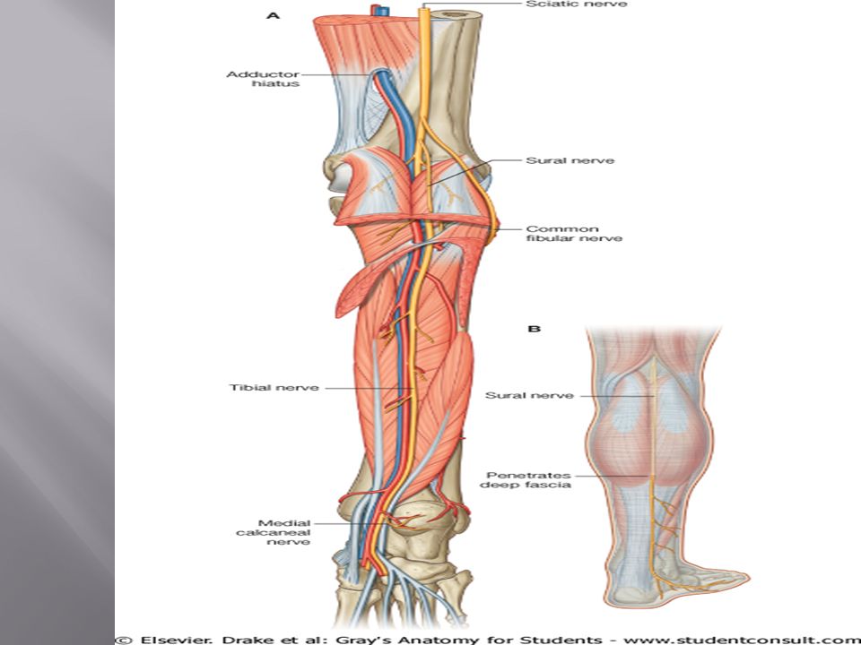
Adductor Hiatus The adductor hiatus is an opening or gap between the aponeurotic distal attachment of the adductor part of the adductor magnus and the tendinous distal attachment of the hamstring part. The adductor hiatus transmits the femoral artery and vein from the adductor canal in the thigh to the popliteal fossa posterior to the knee.

41
OBTURATOR EXTERNUS Obturator membrane Pubic & Ischial rami
Origin : Outer surface of Obturator membrane Pubic & Ischial rami Insertion :Medial surface of greater trochanter N. supply: Obturator nerve Action : Lateral rotation of thigh at hip joint

42
Obturator nerve As the obturator nerve enters the thigh, it divides into two branches, an anterior branch and a posterior branch, which are separated by the adductor brevis muscle: Anterior branch: adductor longus, gracilis, and adductor brevis muscles, and often contributes to the supply of the pectineus muscle, and cutaneous branches innervate the skin on the medial side of the thigh. Posterior branch: obturator externus and adductor brevis muscles and the part of adductor magnus that attaches to the linea aspera

43
Obturator nerve

44
Obturator nerve

45
Deep artery of thigh (Profunda femoris artery)
Branches :lateral and medial circumflex femoral branches and four perforating branches. Lateral circumflex femoral artery: 3 branches 1. Ascending branch 2. Descending branch 3. Transverse branch Medial femoral circumflex artery: It takes part in the formation of the cruciate anastomosis.

47
Obturator Artery The obturator artery is a branch of the internal iliac artery. It gives off muscular branches and an articular branch to the hip joint.

48
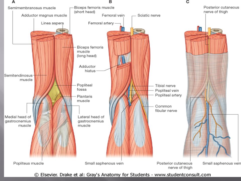
THE POSTERIOR THIGH MUSCLES

49
There are four muscles in the posterior aspect of the thigh
These are; The harmstrings: Semitendinosus Semimembranosus Long head of biceps femoris 2. Short head of biceps femoris All except the short head of biceps femoris cross both the hip and knee joints

51
THE HAMSTRINGS They have common features These are;
Supplied by the tibial division of the sciatic nerve Act on two joints- knee and hip, producing extension at the hip and flexion at the knee Prox. Attach is to the ischial tuberosity, deep to G. maximus

52
Muscles of the posterior compartment of thigh. Posterior view.

54
The two actions of the hamstrings cannot be performed maximally at the same time
When the thighs and the legs are fixed, the hamstrings help extend the trunk at the knee joint They are the hip extensors involved in walking on flat ground, when the G. max demonstrate minimal activity. Here contraction is eccentric

55
Length varies but it’s a matter of conditioning
TEST; Flex the leg against resistance The muscles especially the tendons on each side of the popliteal fossa should be prominent as you bend the knee

57
SEMITENDINOSUS Its a semitendinous muscle as the name implies
Has a fusiform belly About 2/3rd of its distance down the thigh, it becomes tendinous Distal attachment is in the medial surface of superior part of tibia It forms part of the pes anserinus

58
SEMIMEMBRANOSUS Has a flattened membraneous proximal attachment to the ischial tuberosity Tendon forms around the middle of the thigh Tendon descend to the posterior part of the tibial condyle and divides into 3 parts; 1.Blends with the popliteal fossa 2. Attach directly to the posterior aspect of the tibial condyle 3. A reflected part that reinforces the intercondylar part of the joint capsule of the KJ as the oblique popliteal ligament

59
BICEPS FEMORIS Has two prox. attachment LONG AND SHORT HEAD
In the inferior part of the thigh, long head becomes tendinous and joined by short head The round common tendon attaches to the head of fibular and The long head crosses and provide protection for the sciatic nerve The lateral branch of the SN continue the relationship- runs with the biceps femoris

60
The short head is attached proximally to the lateral lip of the inferior 1/3rd of the linea aspera
Also to the supracondylar ridge of the femur

61
N/S The hamstring muscle is supplied by the tibial branch of the SN
The biceps femoris is innervated by the fibular branch Thus since each nerve have different nerve supply a wound in the posterior thigh with nerve injury may paralyzed one head and not the other

62
B/S From the internal iliac artery through perforating branches
These branches enter the posterior compartment by perforating aponeurotic portion of the adductor magnus attachment and medial intermuscular septum,

63
CLINICAL A person with paralyzed harmstrings tend to fall forward cos the G. max cannot maintain the necessary muscle tone to stand straight Harmstring strains are common in individuals who run and kick hard. They are two times as common as quadriceps strains HURDLER’S INJURY; Avulsion of the ischial tuberosity. May result from forceful flexion of the hip with knee extended [e.g. in kicking football]

64
Popliteal Fossa

65
Popliteal fossa The popliteal fossa is a diamond-shaped space behind the knee joint formed between muscles in the posterior compartments of the thigh and leg.

66
POPLITEAL FOSSA Boundaries Laterally :Biceps femoris above,
and Plantaris and lateral head of Gastrocnemius below. Medially : Semitendinous & Semimembranosus above, and by the medial head of Floor is formed by the popliteal surface of the femur, upper end of the tibia, oblique popliteal lig. and the fascia covering the Popliteus; Roof is formed by the fascia lata.

67
Contents: Tibial nerve and common peroneal nerve
Popliteal vein & Termination of the small saphenous vein Popliteal artery Popliteal lymph nodes Post. Cutaneous branches.

71
popliteal artery Branches 1.superior lateral 2.superior medial
3.Middle 4.inferior lateral 5.inferior medial genicular

72
Popliteal Vein Tributaries
1.Veins that correspond to branches given off by the popliteal artery 2. Small saphenous vein, which perforates the deep fascia and passes between the two heads of the gastrocnemius muscle to end in the popliteal vein

73
Nerves in the Popliteal Fossa
The sciatic nerve usually ends at the superior angle of the popliteal fossa by dividing into the tibial and common fibular nerves. Cutaneous: Sural nerve and sural communicating branch

75
LOWER LIMB LEG

76
LEG The leg is that part of the lower limb between the knee joint and ankle joint . The leg is divided into anterior (extensor), posterior (flexor), and lateral (fibular) compartments .
, posterior (flexor), and lateral (fibular) compartments .")
77
Leg movements by compartment

78
Anterior Compartment Muscles : 1.Tibialis anterior
2.Extensor hallucis longus 3.Extensor digitorum longus 4.Fibularis tertius ACTION:Dorsiflex the foot at the ankle joint, extend the toes, and invert the foot Blood supply: Anterior tibial artery Nerve supply: Deep peroneal nerve

79
Anterior Leg (deep fibular n.)
Fibularis (peroneus) longus Extensor digitorum longus Extensor hallicus longus Tibialis anterior
 longus. Extensor digitorum longus. Extensor hallicus longus. Tibialis anterior.")
80
Tibialis anterior Origin: Lateral surface of tibia and adjacent interosseous membrane Insertion: Medial cuneiform and base of first metatarsal bone Nerve Supply:Deep peroneal nerve Action:Dorsiflexion of foot at ankle joint; inversion of foot

81
Extensor digitorum longus
Origin: Anterior surface of shaft of fibula Insertion: Extensor expansion of lateral four toes Nerve Supply: Deep peroneal nerve Action: Extension of lateral four toes and dorsiflexion of foot

82
Extensor hallucis longus
Origin: Anterior surface of shaft of fibula Insertion: Base of distal phalanx of great toe Nerve Supply: Deep peroneal nerve Action: Extension of great toe and dorsiflexion of foot

83
Fibularis tertius Origin: Anterior surface of shaft of fibula
Insertion: Base of fifth metatarsal bone Nerve Supply: Deep peroneal nerve Action: Dorsiflexion and eversion of foot

84
Arteries Anterior tibial artery:
In the distal leg, it lies between the tendons of the tibialis anterior and extensor hallucis longus muscles.

85
Nerves Deep fibular nerve:
This nerve originates in the lateral compartment of leg as one of the two divisions of the common fibular nerve.

87
Posterior compartment of leg
Muscles in the posterior (flexor) compartment of leg are organized into two groups, superficial and deep. Blood supply: Posterior tibial artery. Nerve supply: Tibial nerve
 compartment of leg are organized into two groups, superficial and deep. Blood supply: Posterior tibial artery. Nerve supply: Tibial nerve.")
88
Muscles of the Posterior Compartment of the Leg
Superficial group of muscles 1.Gastrocnemius 2.Soleus 3.Plantaris Deep group of muscles 1.Popliteus 2.Tibialis posterior 3.Flexor digitorum longus 4.Flexor hallucis longus Muscles mainly plantarflex and invert the foot and flex the toes.

89
Gastrocnemius Origin: Lateral head from lateral condyle of femur
Medial head from above medial condyle Insertion:Via tendo calcaneus into posterior surface of calcaneum Innervation:Tibial nerve Function:Plantarflexes foot and flexes knee

90
Plantaris Origin: Lateral supracondylar ridge of femur
Insertion: Posterior surface of calcaneum Innervation: Tibial nerve Function: Plantarflexes foot and flexes knee

91
Soleus Origin:Shafts of tibia and fibula
Insertion:Posterior surface of calcaneum Innervation:Tibial nerve Function:Plantarflexes the foot

93
Popliteus Origin:Lateral surface of lateral condyle of femur
Insertion:Posterior surface of shaft of tibia above soleal line Innervation:Tibial nerve Function: Flexes leg at knee joint; unlocks knee joint by lateral rotation of femur on tibia and slackens ligaments of joint

94
Flexor digitorum longus
Origin: Posterior surface of shaft of tibia Insertion: Bases of distal phalanges of lateral four toes Innervation: Tibial nerve Function: Flexes distal phalanges of lateral four toes; plantar flexes foot at ankle joint

95
Flexor hallucis longus
Origin: Posterior surface of shaft of fibula Insertion: Base of distal phalanx of big toe Innervation: Tibial nerve Function:Flexes distal phalanx of big toe; plantar flexes foot at ankle joint

96
Tibialis posterior Origin:Posterior surface of shafts of tibia and fibula and interosseous membrane Insertion:Tuberosity of navicular bone Innervation: Tibial nerve Function: Inversion and plantarflexion of foot

98
Posterior Compartment
Nerve supply: Tibial nerve: Supplies all muscles in posterior compartment. Divides into medial and lateral plantar nerves inferior and posterior to medial malleolus. Gives off medial sural cutaneous nerve. Joins with communicating branch of common peroneal (fibular) nerve to form: Sural nerve: Cutaneous.
 nerve to form: Sural nerve: Cutaneous.")
100
Posterior Compartment
Blood supply: Posterior tibial artery: Largest branch of popliteal artery. Divides into medial and lateral plantar arteries deep to origin of abductor hallucis muscle. Peroneal artery: Most important branch of posterior tibial artery. Supplies lateral compartment and popliteus muscles. Supplies other muscles in posterior compartment.

101
Arteries in the posterior compartment of leg.

102
Lateral compartment of leg
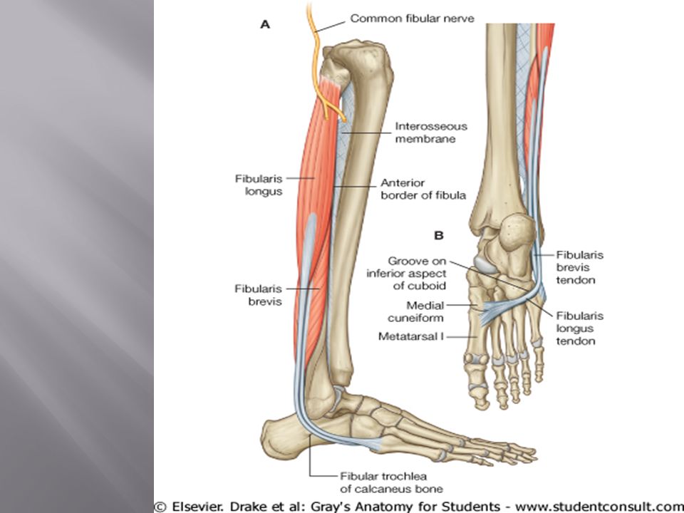
Muscles: Peroneus longus and peroneus brevis Blood supply: Branches from the peroneal artery Nerve supply: Superficial peroneal nerve

103
Peroneus longus Origin:Lateral surface of shaft of fibula
Insertion:Base of first metatarsal and the medial cuneiform Innervation:Superficial peroneal nerve Function:Plantar flexes foot at ankle joint; everts foot

105
Peroneus brevis Origin:Lateral surface of shaft of fibula
Insertion:Base of fifth metatarsal bone Innervation:Superficial peroneal nerve Function:Plantar flexes foot at ankle joint; everts foot

106
Lateral Compartment Nerve supply:
Superficial peroneal (fibular) nerve: Deep to peroneus longus. Inserts on lateral tuberosity. Blood supply: No major arteries in lateral compartment. Muscular branches arise from the peroneal artery: Branch of posterior tibial.
 nerve: Deep to peroneus longus. Inserts on lateral tuberosity. Blood supply: No major arteries in lateral compartment. Muscular branches arise from the peroneal artery: Branch of posterior tibial.")
Similar presentations


.>")


 Joint>")
>")