Download presentation
Presentation is loading. Please wait.

1
Heart Pt. II

2
Heart Actions The heart chambers function in coordinated fashion
Atria contract (atrial systole) while ventricles relax (ventricular diastole); Followed by contraction of the ventricles (ventricular systole) while atria relax (atrial diastole)
 while ventricles relax (ventricular diastole); Followed by contraction of the ventricles (ventricular systole) while atria relax (atrial diastole)")
3
Cardiac Cycle During a cardiac cycle, the pressure in the heart chambers rises and falls In atrial systole and ventricular diastole: Blood flows passively into the ventricles The remaining 30% of blood is pushed into the ventricles The A-V valves open and the semilunar valves close The ventricles relax This causes an increase in ventricular pressure In ventricular systole and atrial diastole: The A-V valves close The chordae tendinae prevent the cusps of the valves from bulging too far into the atria The atria relax The blood flows into atria The ventricular pressure increases and opens the semilunar valves The blood flows into pulmonary trunk and aorta

4
Heart Sounds A heart beat through a stethoscope sounds like “lub-dub” or “lub-dup” The “lub” The first heart sound It occurs during ventricular systole The A-V valves are closing The “dup” The second heart sound It occurs during ventricular diastole The pulmonary and aortic (semilunar) valves are closing Murmur – abnormal heart sound from the cusps not completely closing
 valves are closing. Murmur – abnormal heart sound from the cusps not completely closing.")
5
Myocardial muscle fibers Cardiac Muscle Fibers Cardiac muscle fibers form a functional syncytium This is a mass of cells that function as a unit, connected by intercalated discs Two such areas exist in the heart: In the atrial walls called the atrial syncytium In the ventricular walls called the ventricular syncytium

6
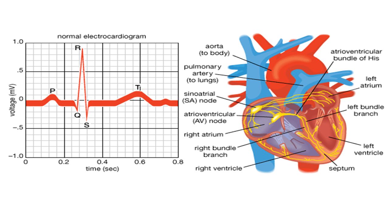
Cardiac Conduction System
Atrial syncytium Junctional fibers Bundle branches Purkinje fibers SA node AV node AV bundle Ventricular syncytium Cardiac Conduction System Heart is autorhythmic 1% of heart tissue specialized to not contract Transmits signals to cells that contract Self-excitable Generate and transmit impulses Clumps or strands of specialized cardiac muscle tissue which initiate and distribute impulses throughout the myocardium The cardiac conduction system coordinates the events of the cardiac cycle A node is just a compact mass of cells.

7
Ventricular syncytium
Purkinje fibers Interatrial septum Left bundle branch Interventricular septum Right bundle Junctional fibers AV node SA node AV bundle Atrial syncytium Junctional fibers Bundle branches Purkinje fibers SA node AV node AV bundle Ventricular syncytium

8
Regulation of the Cardiac Cycle
SA Node – controls heart rate Also, sympathetic & parasympathetic fibers, and regulatory reflex centers Additional factors that may influence heart rate: Physical exercise Body temperature Concentration of various ions (including K+, Ca2+) Parasympathetic impulses decrease heart action Sympathetic impulses increase heart action Cardiac center regulates autonomic impulses to the heart
 Parasympathetic impulses decrease heart action. Sympathetic impulses increase heart action. Cardiac center regulates autonomic impulses to the heart.")
9
Internal Anatomy of the Heart: Terms to Know
Aortic semilunar valve Atrioventricular node Atrioventricular septa Bicuspid valve Chordae tendinae Coronary sinus Coronary arteries Foramen ovale Fossa ovalis Interatrial septum Interventricular septum Moderator band Papillary muscles Pectinate muscles Pulmonary semilunar valve Sinoatrial node Trabeculae carnae Tricuspid valve

10
EKG/ECG (Electrocardiogram)
Recording of the electrical charges in the myocardium Electrodes placed on skin Assesses heart’s ability to conduct impulses

11
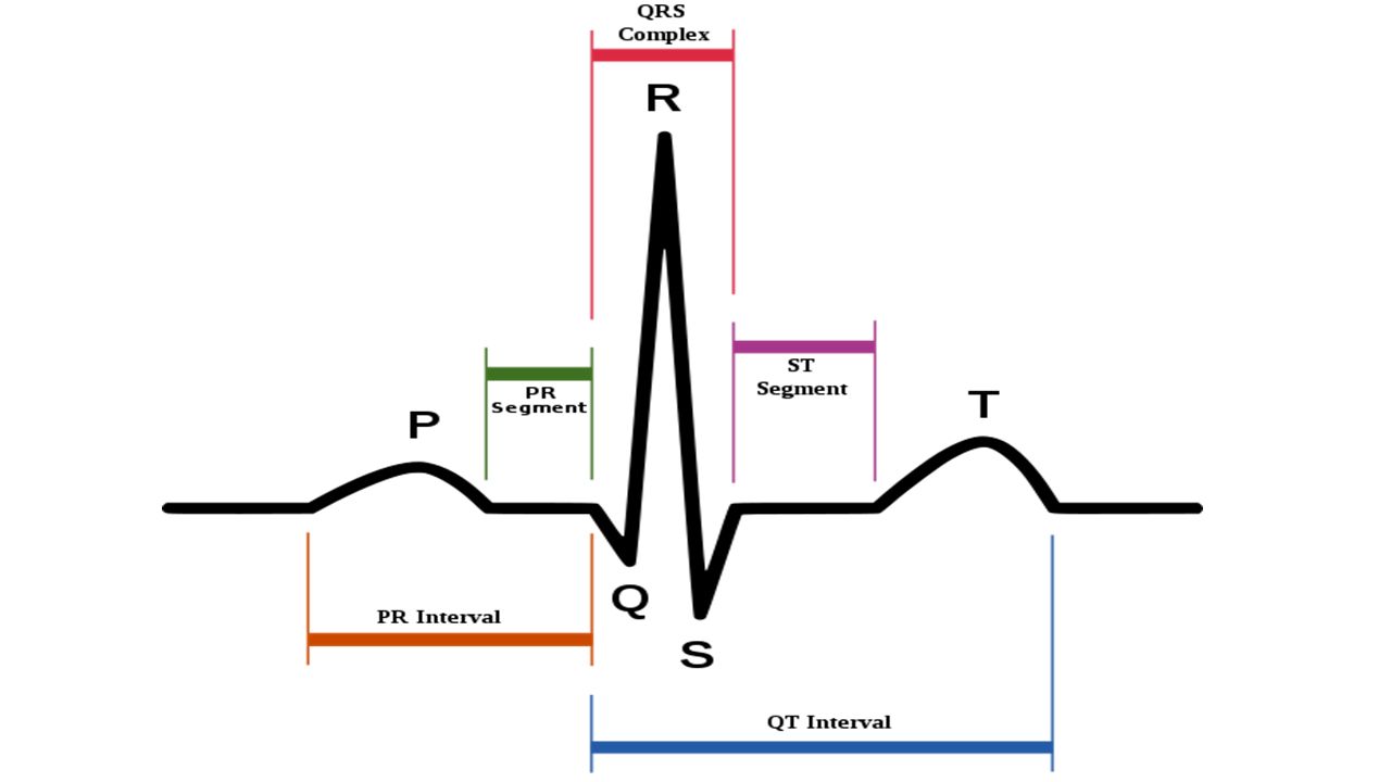
EKG Deflections or Waves: P Wave – atrial depolarization
Reduction in voltage across cell membrane Indicates stimulation/receiving impulse SA Node impulse – spreads through atria, atria contract (atrial systole) QRS Complex (3 waves) – ventricular depolarization Electrical impulses spreading through ventricles More muscle – more charges Ventricular systole (contraction) T wave – ventricular repolarization Restores ventricles to resting state Ventricles relax (ventricular diastole)
 QRS Complex (3 waves) – ventricular depolarization. Electrical impulses spreading through ventricles. More muscle – more charges. Ventricular systole (contraction) T wave – ventricular repolarization. Restores ventricles to resting state. Ventricles relax (ventricular diastole)")
14
+1 –1 120 100 80 60 40 20 160 P Q S T R 0.3 0.6 0.9 seconds Heart sounds Electrocardiogram (ECG) Pressure changes Aortic pressure Atrial pressure Volume (mL) Millivolts Pressure (mm Hg) One cardiac cycle Atrial systole Ventricular diastole Aortic semilunar valve opens AV valve closes AV valve opens Ventricular pressure Aortic semilunar valve closes Ventricular volume Lubb: AV valves close Dupp: Semilunar
 Pressure changes. Aortic pressure. Atrial pressure. Volume (mL) Millivolts. Pressure (mm Hg) One cardiac cycle. Atrial. systole. Ventricular. diastole. Aortic. semilunar. valve. opens. AV valve. closes. AV valve opens. Ventricular pressure. Aortic semilunar. valve closes. Ventricular volume. Lubb: AV. valves close. Dupp: Semilunar.")
Similar presentations


 –Contracts.>")


 Transport O 2, nutrients, hormones, cell wastes, etc…>")


