Download presentation
Presentation is loading. Please wait.

1
BONE TUMORS Dr.ZEENAT NASEERUDDIN m.d pathology

2
LECTURE 48 BONE FORMING TUMORS
OBJECTIVES a.Enlist and classify common types of bone tumors. b.Discuss their epidemiology. c.Describe their morphology and location. d.Describe their clinical features and course.

3
Bone tumors Bone tumors are classified into:
Primary bone tumors Secondary bone tumors ( Metastasis) Most are classified according to the normal cell of origin and apparent pattern of differentiation
 Most are classified according to the normal cell of origin and apparent pattern of differentiation.")
4
Bone tumors Bone-forming tumors Cartilage-forming tumors
Miscellaneous tumors Hematopoietic tumors Fibrous tumors

5
Primary Bone Tumors Bone-Forming tumors Osteoma
Osteoid osteoma and Osteoblastoma Osteosarcoma Cartilage-Forming tumors Chondroma (Enchondroma) Osteochondroma Chondrosarcoma Miscellaneous tumors Ewing’s sarcoma Giant cell tumor of bone
 Osteochondroma. Chondrosarcoma. Miscellaneous tumors. Ewing’s sarcoma. Giant cell tumor of bone.")
6
Bone tumors: Etiology ??? Although the cause of most bone tumors is unknown. Genetic alterations e.g. bone sarcomas in the Li-Fraumeni and hereditary retinoblastoma which are linked to mutations in p53 and Rb genes. Bone infarcts Chronic osteomyelitis Paget’s disease Radiation and Metal prostheses are also associated with increased incidence of bone neoplasia.

7
Diagnosis of Bone Tumors:
1. Age of patient 2. Location of tumor 3. Radiological appearance 4. Histological features

8
Bone-Forming Tumors

9
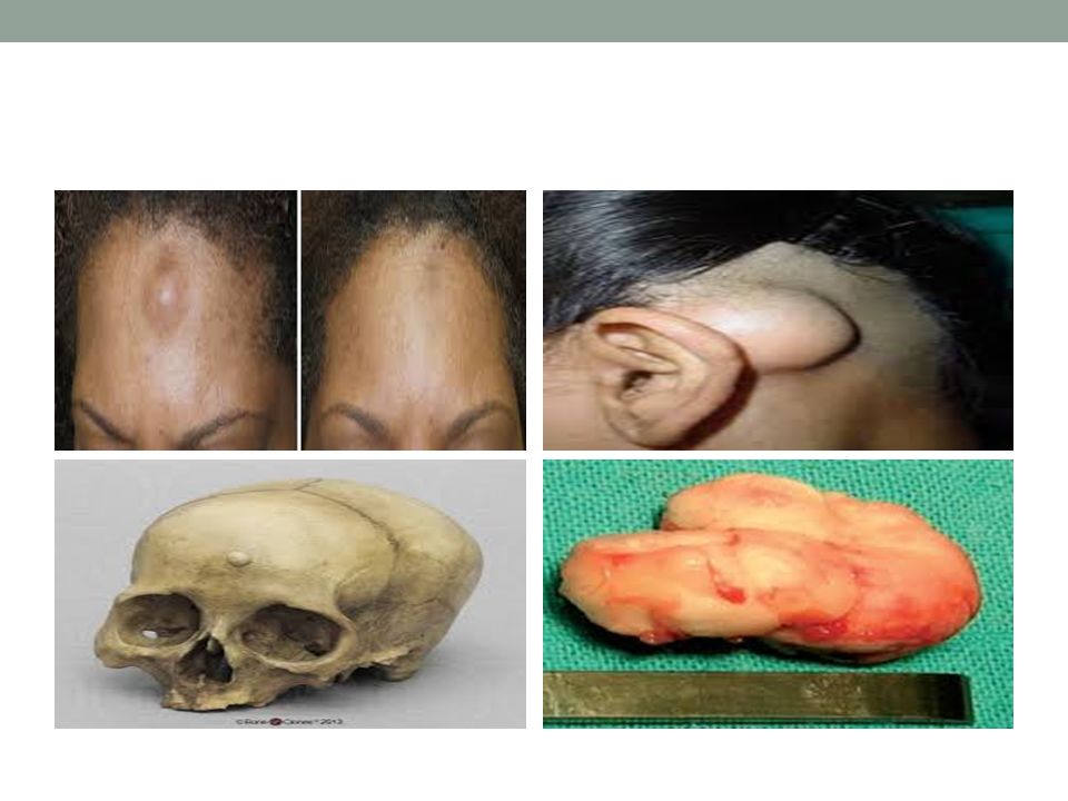
Osteoma Osteoma are benign lesions of bone that in many cases represent developmental aberrations or reactive growths rather than true neoplasms. Site; Age; Gross: Histology:

10
Osteoma Small bosselated benign tumor.
Usually solitary and detected in the middle age. Multiple in the setting of Gardner syndrome. Do not transform into osteosarcoma.

13
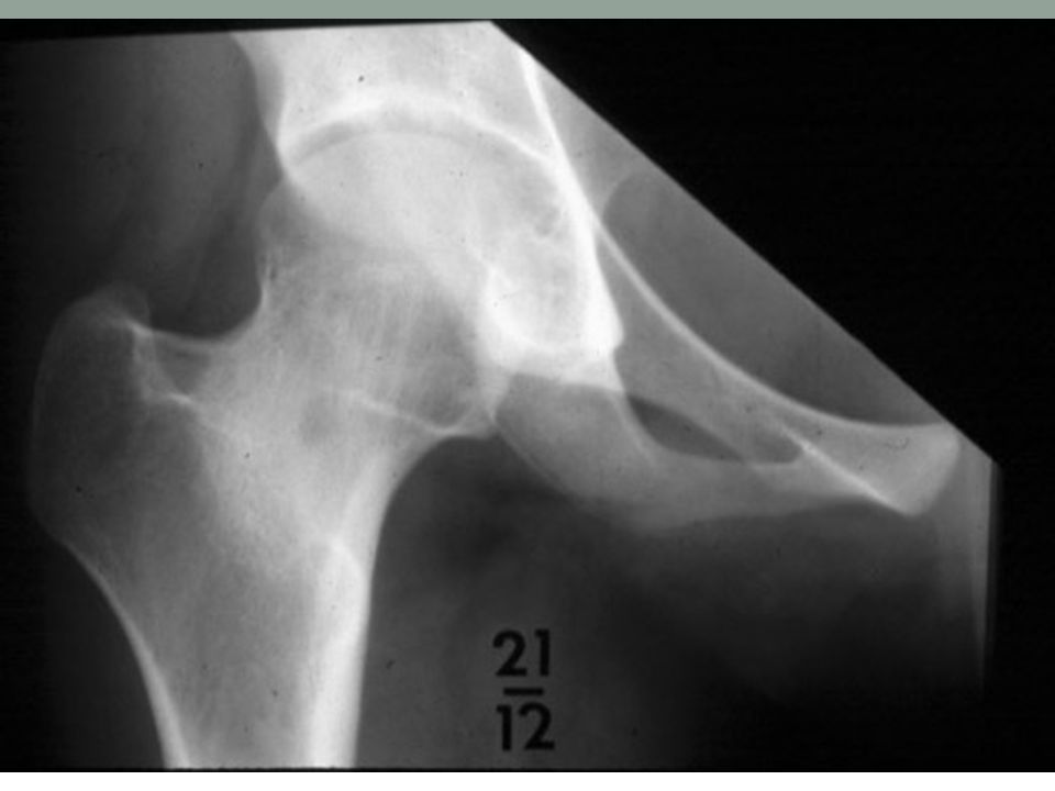
Osteoid Osteoma Signs/Symptoms: Age: Sex: Anatomic Distribution:
Pain, characteristically more intense at night, relieved by NSAID (PG-E) and eliminated by excision Vertebral lesions may cause scoliosis Age: 10-30 years Sex: M > F (2:1) Anatomic Distribution: Nearly every location, most frequent in femur, tibia, humerus, bones of hands and feet, vertebrae and fibula Over 50% of cases in femur or tibia Metaphysis of long bones By definition less than 2 cm in diameter.
 and eliminated by excision. Vertebral lesions may cause scoliosis. Age: years. Sex: M > F (2:1) Anatomic Distribution: Nearly every location, most frequent in femur, tibia, humerus, bones of hands and feet, vertebrae and fibula. Over 50% of cases in femur or tibia. Metaphysis of long bones. By definition less than 2 cm in diameter.")
14
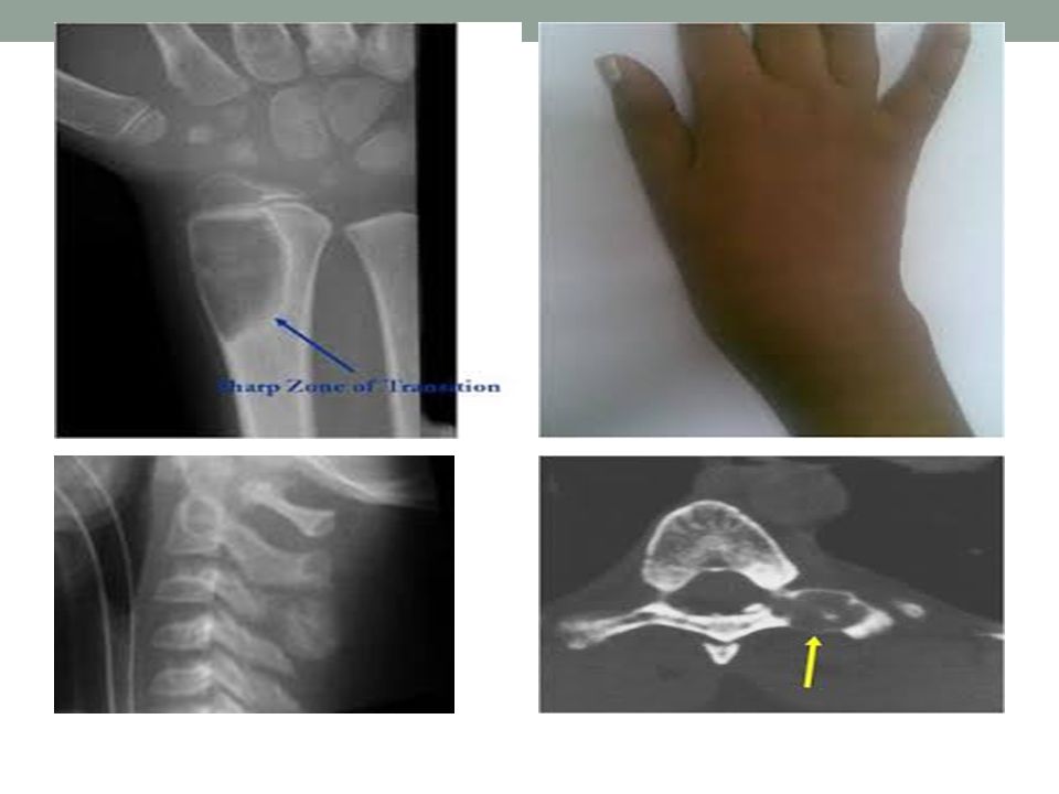
Central radiolucent nidus with or without a radiodense center; surrounded by thickened sclerotic bone

16
Central hemorrhagic nidus surrounded by dense rim of sclerotic bone

17
Nidus contains interlacing network of osteoid and bony trabeculae with variable amount of mineralization, lying in vascular fibrous tissue

19
Osteoid Osteoma Ancillary Testing: Prognosis/Treatment: N/A
Surgical excision is treatment of choice Recurrence unlikely with complete excision

20
Osteoblastoma (Giant Osteoid Osteoma)
Signs/Symptoms: Pain Gait disturbances Age: 80% of patients < 30 years Sex: M >> F (3:1) Anatomic Distribution: Predilection for vertebral column Metaphysis of long bones
 Anatomic Distribution: Predilection for vertebral column. Metaphysis of long bones.")
22
Osteoblastoma (Giant Osteoid Osteoma)
Radiographic Findings: Similar to osteoid osteoma, though much larger (up to 11.0 cm) Gross and Microscopic Findings: Similar to osteoid osteoma, though much larger nidus Ancillary Testing: N/A Prognosis/Treatment: Curettage followed by bone grafting If incompletely removed, tumor may recur Malignant change to osteosarcoma has been rarely reported
 Gross and Microscopic Findings: Similar to osteoid osteoma, though much larger nidus. Ancillary Testing: N/A. Prognosis/Treatment: Curettage followed by bone grafting. If incompletely removed, tumor may recur. Malignant change to osteosarcoma has been rarely reported.")
23
Osteosarcoma Osteosarcoma is a bone-producing malignant mesenchymal tumor .

24
Osteosarcoma Incidence: Age: Sex: Site :

25
Osteogenic Sarcoma (Osteosarcoma)
Most frequent primary malignant bone tumor Malignant cells must produce osteoid Most tumors arise de novo (primary), though others arise in the setting of: Paget’s disease Previous RT Previous chemo (especially alkylating agents) Fibrous dysplasia Osteochondromatosis Chondromatosis Chronic osteomyelitis
, though others arise in the setting of: Paget’s disease. Previous RT. Previous chemo (especially alkylating agents) Fibrous dysplasia. Osteochondromatosis. Chondromatosis. Chronic osteomyelitis.")
26
Osteogenic Sarcoma (Osteosarcoma)
Signs/Symptoms: Pain and swelling Pathologic fracture is uncommon Age: Bi-modal age group Peak in 2nd decade with gradual decrease thereafter. Sex: M > F Anatomic Distribution: 50% arise around the knee Metaphysis of long bones

27
Osteosarcoma Distribution

28
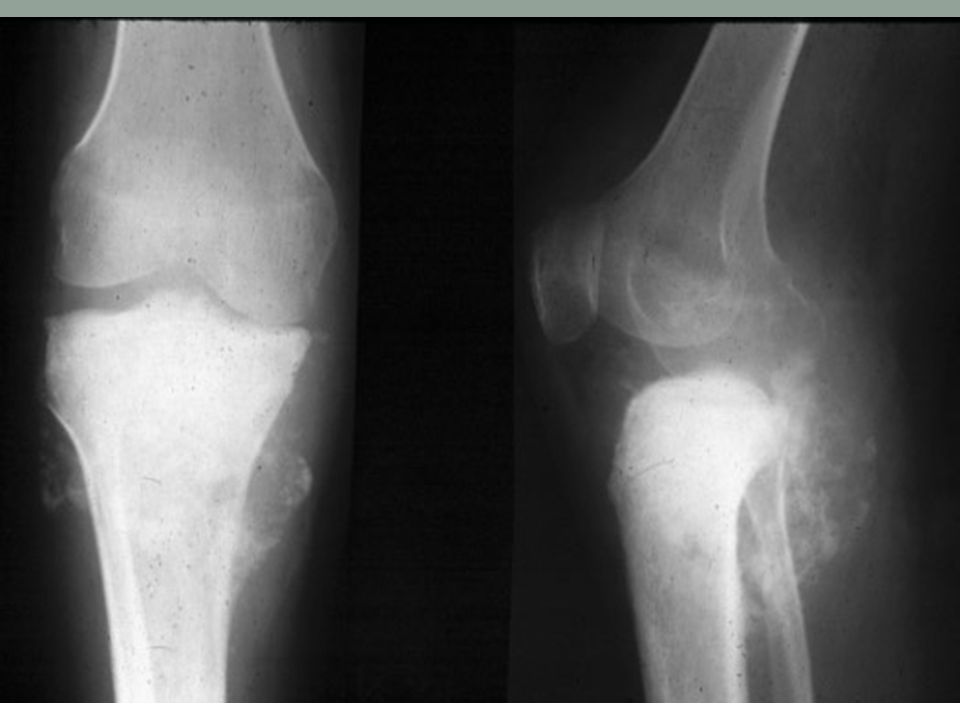
Osteosarcoma Radiograph

31
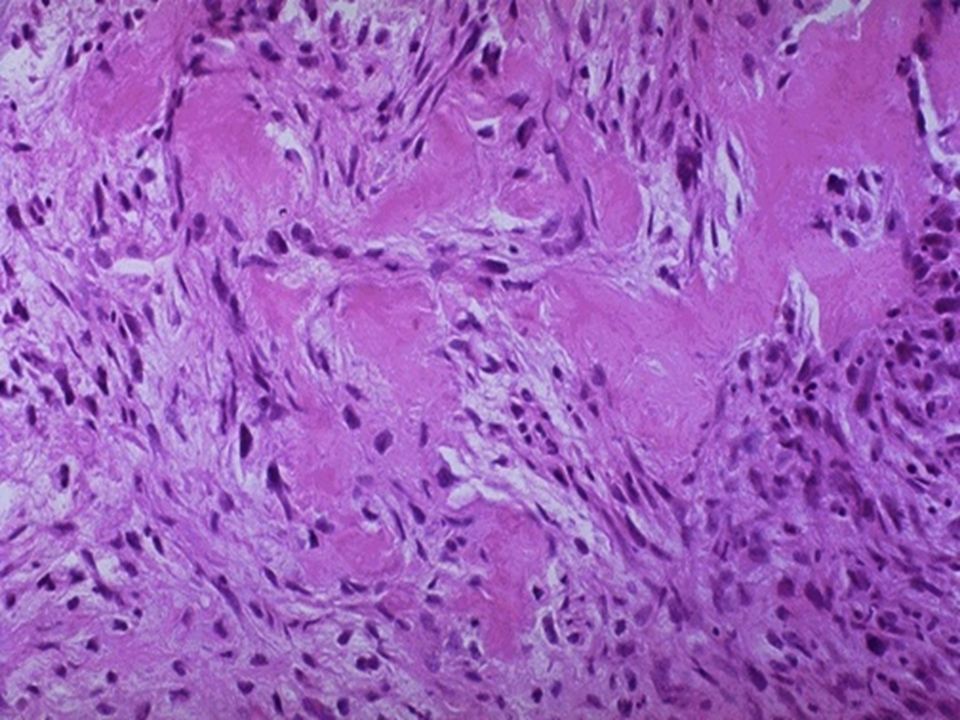
Osteogenic Sarcoma Morphology:
Most commonly involves metaphysis of long bones. Gross features: Big bulky tumors, grey white often containing areas of hemorrhage and cystic degeneration. Micro: Pleomorphic tumor cells with large hyper chromatic nuclei ,mitotic figures. Formation of pink homogenous bone is the most characteristic feature of osteogenic sarcoma

32
Osteosarcoma Gross features

35
Bone-Forming tumors; Tumor Type Locations Age Morphology BENIGN
Osteoma Facial bones, skull 40-50 Exophytic growths attached to bone surface; histologically resemble normal bon Osteoid osteoma Metaphysis of femur and tibia 10-20 Cortical tumors, characterized by pain; histologically interlacing trabeculae of woven bone Osteoblastoma Vertebral column vertebral processes; histologically similar to osteoid osteoma MALIGNANT Primary osteosarcoma Metaphysis of distal femur, proximal tibia, and humerus Grow outward, lifting periosteum, and inward to the medullary cavity; microscopically malignant cells form osteoid. Secondary osteosarcoma Femur, humerus, pelvis >40 Complications of polyostotic Paget disease; histologically similar to primary osteosarcoma

36
An 11-year-old male was seen in consultation for an increasingly painful distal femoral lesion associated with a soft tissue mass. Plain radiograph shows an ill-defined destructive tumor in the distal femur. Fluffy radio-dense infiltrates represent malignant tumor osteoid. Biopsy material shows two major components of this neoplasm: highly pleomorphic cells and haphazard deposits of osteoid. Note that the malignant cells fill the spaces between osteoid deposits. Lace-like osteoid deposition is very characteristic of this neoplasm.

37
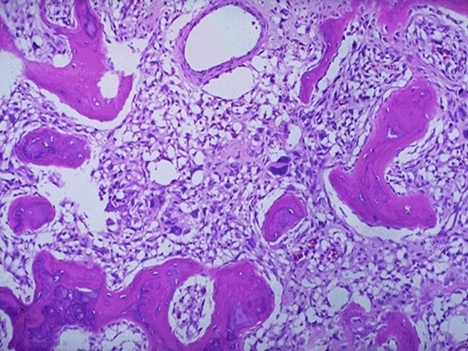
A 16-year-old boy was seen in consultation for increasing pain in the mid upper arm. Characteristically, the pain intensified at night and subsided with aspirin. Plain film shows a small, intracortical, radiolucent focus (nidus), surrounded by dense reactive periosteal bone. The lesion is located in the mid portion of the humeral shaft. If the nidus is removed intact, it appears as a circumscribed portion of red, trabecular bone, usually less than 1cm in size. Low-power view shows the lesional tissue ("nidus"), well demarcated from the surrounding sclerotic bone. The lesion is composed of thin, often interconnected spicules of osteoid and woven bone rimmed by osteoblasts. Osteoclast-like giant cells can be seen. Intervening fibrous stroma shows prominent vascularity.
, surrounded by dense reactive periosteal bone. The lesion is located in the mid portion of the humeral shaft. If the nidus is removed intact, it appears as a circumscribed portion of red, trabecular bone, usually less than 1cm in size. Low-power view shows the lesional tissue ( nidus ), well demarcated from the surrounding sclerotic bone. The lesion is composed of thin, often interconnected spicules of osteoid and woven bone rimmed by osteoblasts. Osteoclast-like giant cells can be seen. Intervening fibrous stroma shows prominent vascularity.")
38
METASTATIC BONE TUMORS
Metastatic tumors are the most common malignant tumor of bone. Pathways of spread: Origin: The radiologic appearance of metastases

41
THANK YOU

Similar presentations


>")


 - Genetic factors may play a role (p53 and RB mutations) - Bone infarcts, trauma, osteomyelitis,>")


 normal cell of origin Most are classified.>")

: Principal Modality (2): Musculoskeletal.>")

. General considerations Primary bone tumors are much less than secondary tumors. All age groups affected,>")


