Download presentation
Presentation is loading. Please wait.

1
Congenital Heart Diseases Dr. Usha Singh Department of Pediatrics

2
Aims & Objectives At the end of this session the student should be able to: understand the hemodynamics of common congenital heart diseases (CHD) recognize a child with congenital heart disease classify the heart disease and give a provisional diagnosis provide basic essential management to a case of congenital heart disease.
 recognize a child with congenital heart disease classify the heart disease and give a provisional diagnosis provide basic essential management to a case of congenital heart disease.")
3
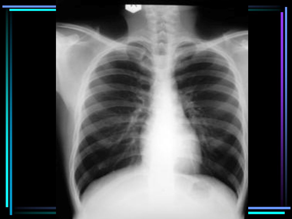
The Normal Heart

5
When to suspect the infant has a CHD History: –Maternal: diabetes, age, drugs, SLE –Infant: cyanosis, respiratory distress, prematurity, failure to thrive, forehead sweating, suck-rest-suck cycle Examination: –Dysmorphic facies –Anthropometry –Heart failure –Vitals –Murmurs

6
Classification Congenital Heart Defect Finally look for the chamber enlargement in ECG Cyanosis -ntCyanosis +nt Pulmonary blood flow increaseddecreasedincreaseddecreased

7
Acyanotic Congenital Heart Disease Volume overload: L→ R shunts A-V valve regurgitation Cardiomyopathy Pressure overload: Ventricular outflow obstruction Coarctation of aorta

8
Cyanotic congenital heart disease Increased pulmonary blood flow: –TGV –Truncus arteriosus –Single atrium/ventricle Decreased pulmonary blood flow: –Tetrology of Fallot –Tricuspid atresia –Single ventricle with PS

9
Atrial Septal Defect Hemodynamics Clinical Picture –Right ventricular hypertrophy –Loud 1 st heart sound –2 nd heart sound widely split and fixed (upper left sternal edge) Complications Treatment
 Complications Treatment")
10
.

12
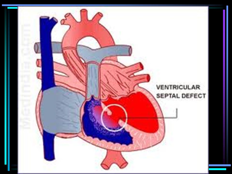
Ventricular Septal Defect Hemodynamics Clinical picture –Left/biventricular hypertrophy –Pansystolic murmur-lower left sternal edge, thrill Complications Treatment

15
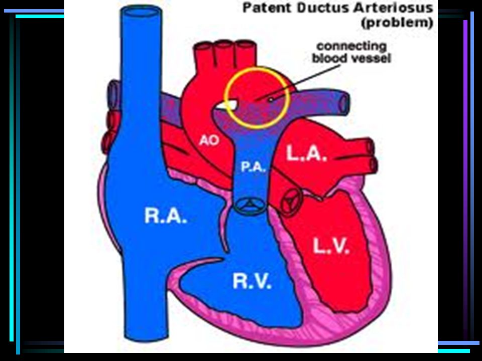
Patent Ductus Arteriosus Hemodynamics Clinical picture –High volume pulse –Continuous murmur/machinery murmur in left sternal edge, may radiate to clavicle Complications Treatment

18
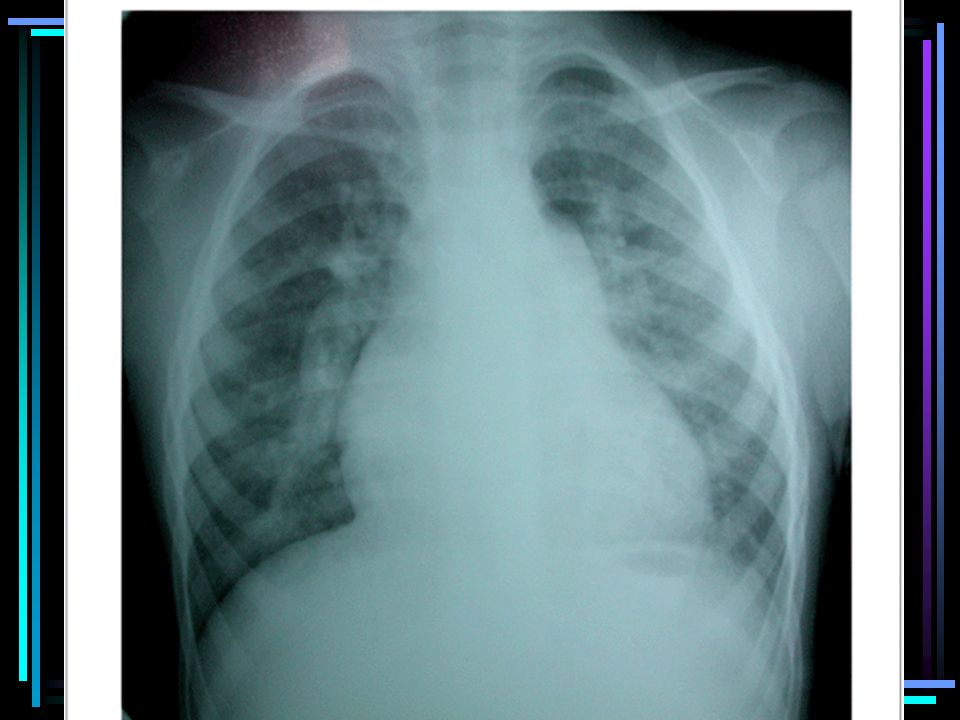
Tetrology of Fallot 4 components Clinical Picture: –Cyanosis –Squatting –Tet spells –Systolic murmur in L 3 rd -4 th space X-ray Complications

20
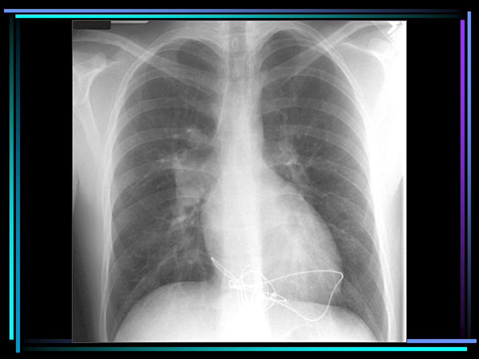
Transposition of Great Vessels Narrow pedicle giving the “egg- on-string” appearance due to the antero – posterior relationship of the great vessels

22
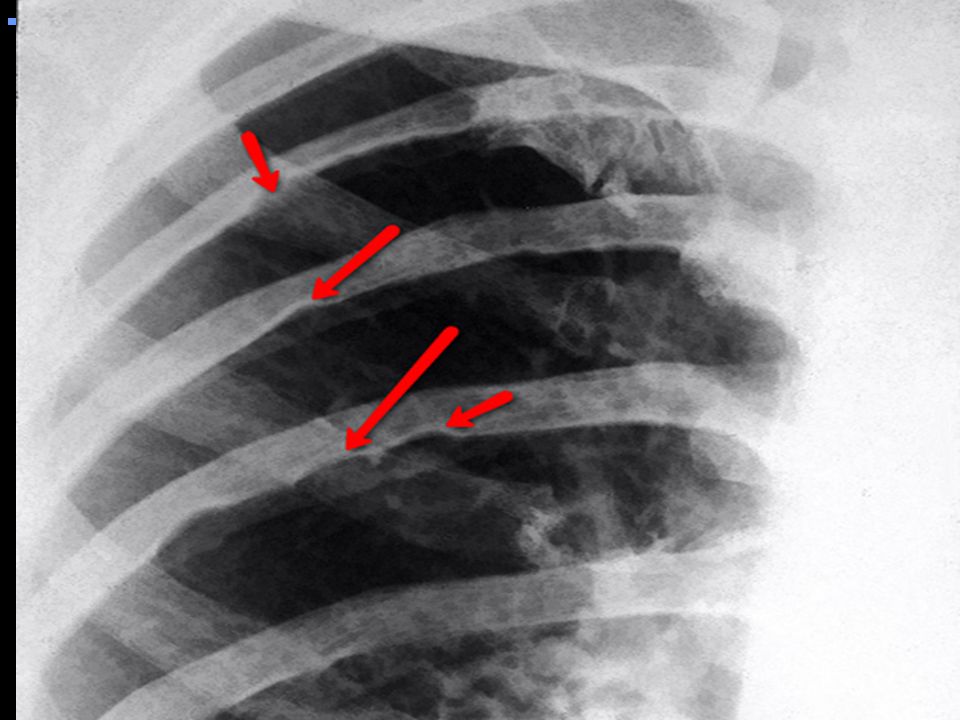
Coarctation of Aorta Hemodynamics Clinical picture –Asymptomatic –Hypertension –Weak lower limb pulses –Systolic murmur along L sternal border –Differential cyanosis Treatment

Similar presentations


 tricuspid valve 2. Hypoplastic right ventricle 3. Ventricular septal defect 4. Atrial.>")


