Download presentation
Presentation is loading. Please wait.

1
Chapter 19 Human Parasitosis Peking Union Medical College Hospital Lu Zhaohui Central South University XiangYa School of Medicine Wen Jifang

2
Definition human parasitosis is a diseases caused by the pathogen of parasites Source of infection, route of transmission and susceptible populationRegionalSeasonal Activity of the natural foci

3
Amoebiasis Amoebiasis is a human entamoeba histolyticaIntestinal amoebiasis Amoebiasis is a human parasitosis mostly caused by entamoeba histolyticaIntestinal amoebiasis Amoebic liver abscess Amoebic pulmonary abscess Amoebic cerebral abscess

4
Intestinal amoebiasis Pathogenesis mechanical injury and phagocytosis Contact lysis Cyto-toxicBacterial Susceptibility Immune escape Immune suppression and Immune escape

5
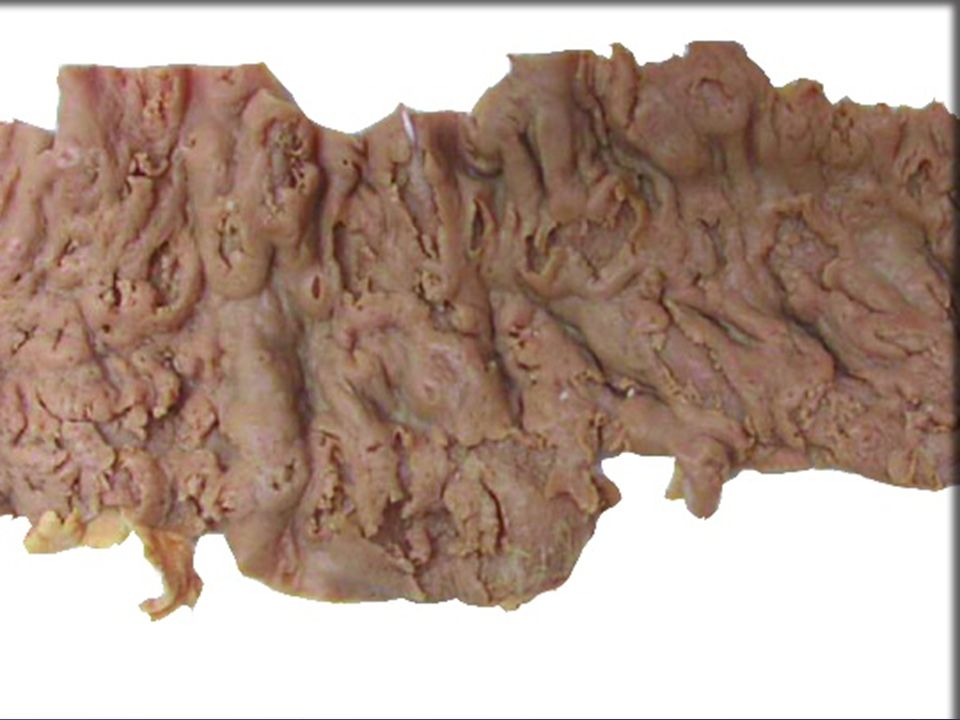
Morphology Location: firstly involves secondly in some cases involve the whole colon and Location: firstly involves cecum, colon ascendens, secondly sigmoid colon and rectum, in some cases involve the whole colon and inferior segment of small intestine flask shaped ulcer Acute phase Chronic phase

6
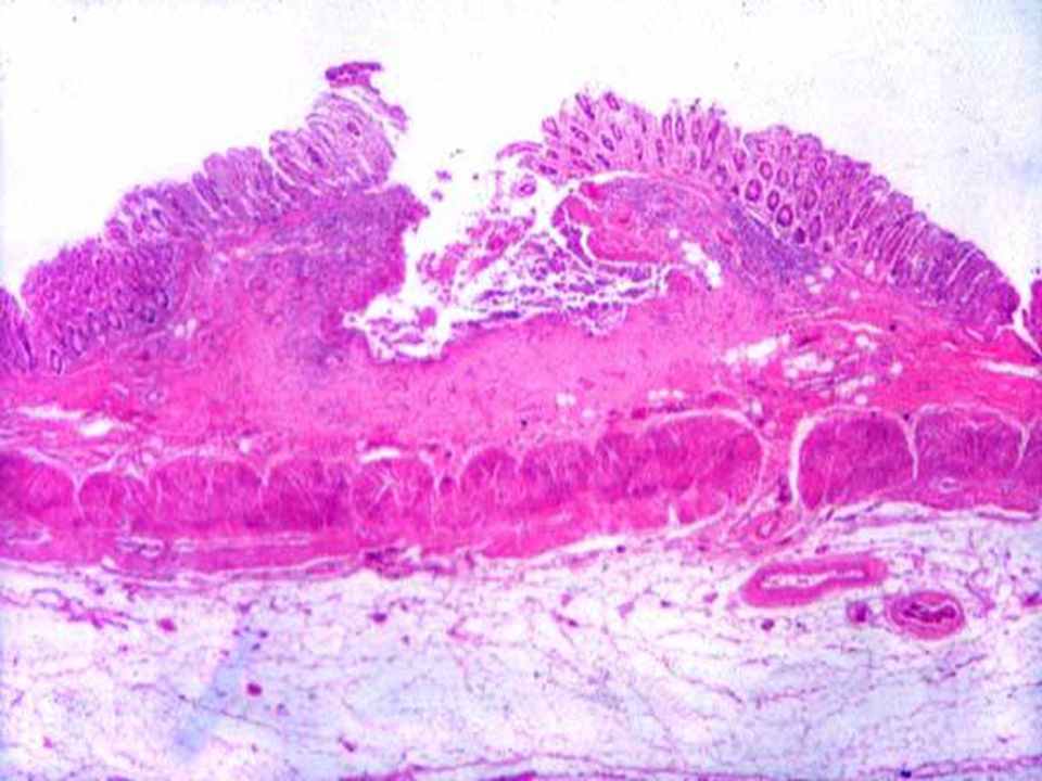
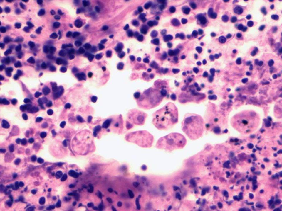
Acute Phase Gross Yellowish spotty necrosis/ulcer Swelling, edema and bleeding zone Map necrosis, filled with yellowish-brown mucous purulence substanceHistology, only minor inflammation nearby liquefaction necrosis, only minor inflammation nearby Large amoebic Large amoebic trophozoite can be seen in the juncture of necrosis and normal tissue vacant space around trophozoite

10
Chronic Phase Mixture of fresh and old lesion Necrosis, ulcer with proliferation epithelial proliferation and polyp Fibrous proliferation, scar, thicken and harden of the enteric wall, stricture of enteric cavity

11
Amoebic liver abscess →→→liver Amoeba trophozoite penetrate the vein of enteric wall→vena mesenterica→Vena portalis hepatis→liverGross Single, the size of abscess varied Single, the size of abscess varied The liquification, The fruit jam substance consists of liquification, oboslete bleeding Abscess wall consists of the connect tissue of vena portalis hepatis, like old cotton Abscess wall consists of the connect tissue of vena portalis hepatis, like old cotton Minor inflammation around the abscess Minor inflammation around the abscessHistology Pus cells, necrosis Pus cells, necrosis Amoeba trophozoite around the wall of abscess

13
Amoebic pulmonary abscess Rare, developed from Amoebic liver abscess, which penetrate the Rare, developed from Amoebic liver abscess, which penetrate the disphragmatic muscle Most commonly single, often located Most commonly single, often located lower lobe of the right lung, may connect with liver abscess The liquification necrosis may be discarded, and the The liquification necrosis may be discarded, and the pulmonary cavity formed Dark brown Dark brown purulent expectoration, Amoeba trophozoite may be found

14
Achistosomiasis Etiology and route of infection Pathogenesis and morphology cercarial dermatitis cercarial dermatitis Damage by the schistosomulum Damage by the schistosomulum Damage by prosopon Damage by prosopon Damage by egg Damage by egg Damage by the antigens and immune compounds in circulation Damage by the antigens and immune compounds in circulation

19
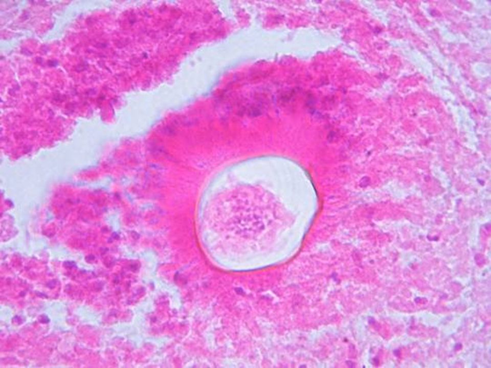
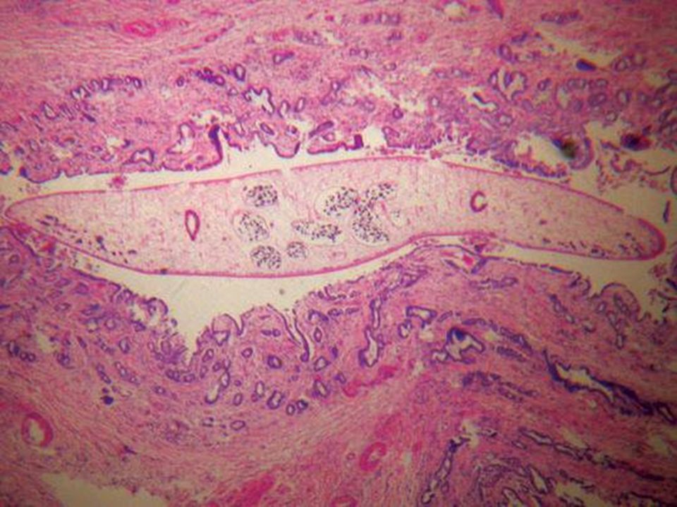
Morphology of organs Colon Acute phase Congestion, edema, gray-yellowish or yellow-whitish nodule, 0.5 ~ 4mm in diameter Congestion, edema, gray-yellowish or yellow-whitish nodule, 0.5 ~ 4mm in diameter The nodule may break →ulcer The nodule may break →ulcer The eggs in the ulcer may be discarded and become pollution The eggs in the ulcer may be discarded and become pollution Chronic phase Eggs, fibrous proliferation, scaring Eggs, fibrous proliferation, scaring Ulcer, necrosis, narrowing or obstruction of the enteric cavity Ulcer, necrosis, narrowing or obstruction of the enteric cavity

21
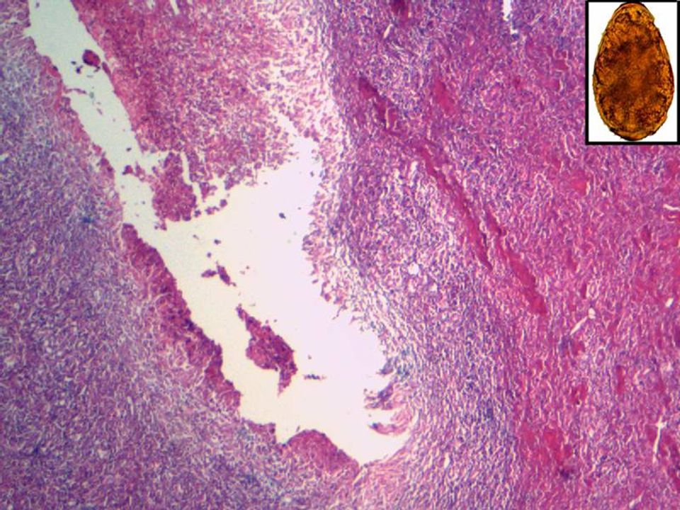
Morphology of organs Liver Acute phase Swelling of liver, not smooth in the surface, numerous Swelling of liver, not smooth in the surface, numerous millet or mung bean sized yellow or whitish nodules in the surface or the cutting surface Histologically, the acute egg can be seen Histologically, the acute egg tubercle mainly located around portal area, the atrophy, degeneration and focal necrosis of hepatocytes can be seen Chronic phase Eggs, fibrous proliferation, calcification Eggs, fibrous proliferation, calcification Fibrous proliferation around the portal area, thicken of the wall of veins, thrombosis Fibrous proliferation around the portal area, thicken of the wall of veins, thrombosis Stem live Stem live liver cirrhosis Portal hypertension and relative symptoms

23
Morphology of organs Ectopic Lung Lung Brain Brain Others Others Schistosoma dwarfism Acute schistosomiasis

24
Clonorchiasis Etiology and Etiology and route of infection Pathogenesis and morphology

25
Morphology of organs LiverBladderPancreas

27
Paragonimiasis Etiology and Etiology and route of infection Pathogenesis and morphology hydrohymenitis hydrohymenitis Fistula Fistula Abscess and cyst Abscess and cyst fibrous scar fibrous scar acute pulmonary paragonimiasis acute pulmonary paragonimiasis

29
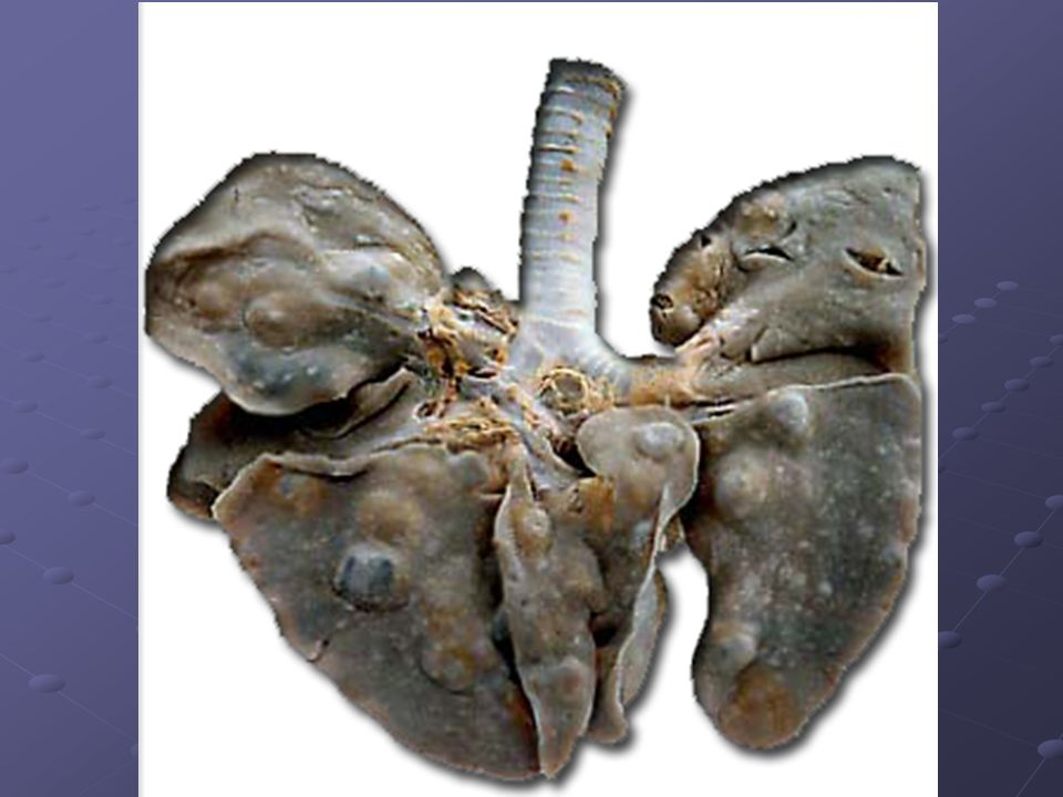
Morphology of organs Lung cystic nodule BrainOthers

31
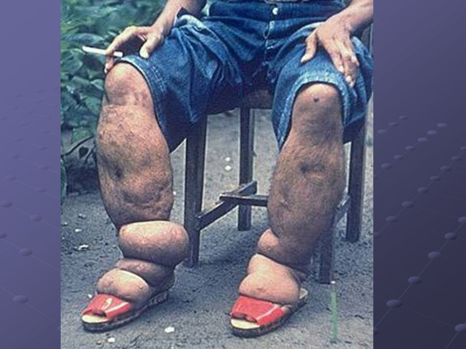
Filariasis Etiology and Etiology and route of infection Pathogenesis and morphology tuberuloid granuloma acute filariasis and chronic filariasis centrifugal lymphangioitis centrifugal lymphangioitis filarial erysipelatous dermatitis filarial erysipelatous dermatitis Lymphotics Lymphotics Lymph noditis Lymph noditis lymph edema lymph edema elephantiasis elephantiasis iridocyclitis, optic neuritis, optic nerve hemorrhage

33
Echinococcosis Etiology and Pathogenesis Morphology

34
Morphology of organs Hydatidocystis of liver Hydatidocystis of lung

35
Alveococcosis Etiology and Pathogenesis Morphology

Similar presentations


>")
 Chapter: Hemodynamic disorders, Thrombosis and Shock - Edema - Hemorrhage - Hyperemia.>")
 Environmental.>")


 Doç.Dr.Hrisi BAHAR.>")


:>")
