Download presentation
Presentation is loading. Please wait.

1
Class: Trematodes (flukes)
")
2
Phylum Platyhelminthes – Class Trematoda General characters:
Dorsoventrally flattened typically leaf shaped have an oral and a ventral sucker used for attachment and movement. Bifurcate intestine complex life cycle with multiple hosts. mollusc is usually 1st intermediate host definitive host is a vertebrate most are hermaphroditic except blood flukes.

5
Trematoda According to their habitat Blood flukes, Schistosoma species
Liver flukes, Fasciola species Intestinal flukes, Fasciolopsis buski Lung flukes, Paragonimus species

6
Blood flukes Schistosoma species

7
Blood flukes Schistosoma species:
They differ from other trematodes in that they have separate sexes. Only trematodes that live in the blood stream of warm-blooded hosts Definitive host: Human Intermediate host: Snail Reservoirs: monkeys, rodents, cats, dogs, cattle, horses, swine, wild mammals The human disease known as shistosomiasis , Bilharzia, Bilhariaziasis In 1852 the German parasitologist, Theodor Bilharz, made the first discovery of schistosomes (S. haematobium) while working in Egypt Eggs are responsible for the pathology associated with infections
 while working in Egypt. Eggs are responsible for the pathology associated with infections.")
8
Human Blood flukes: Three major species
Schistosoma haematobium Schistosoma japonicum Schistosoma mansoni

10
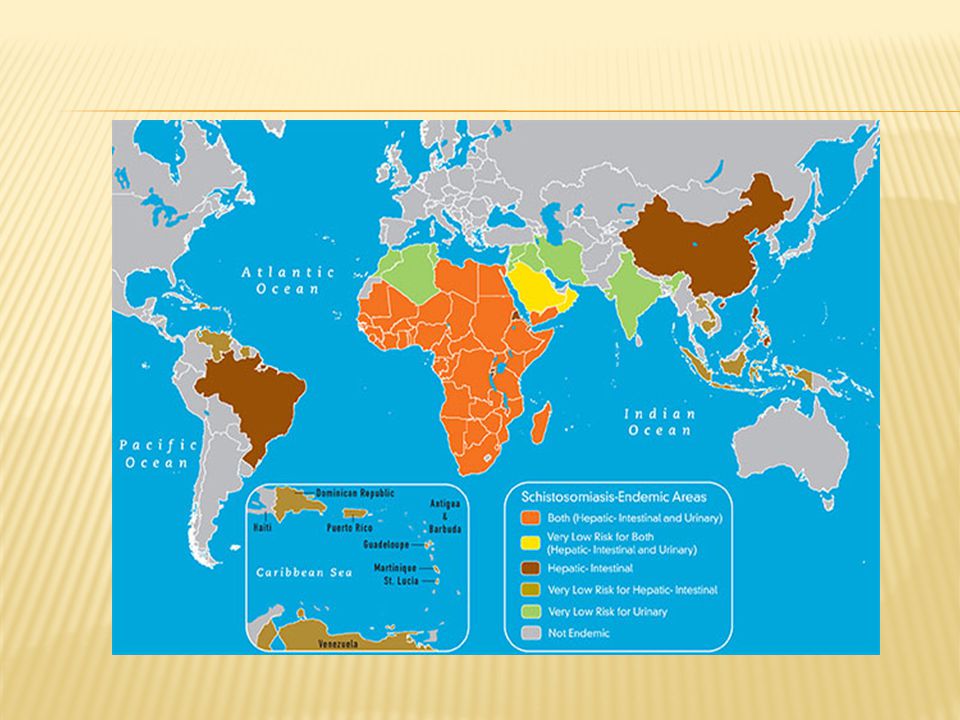
Blood flukes Transmission

11
Morphology: 1- All species roughly similar.
2- Sexes are separate (dioecious). 3- size cm in length. 3- Males short and wide, females long, thin. 4- Female adults reside in gynecophoral canal formed by male body.
. 3- size cm in length. 3- Males short and wide, females long, thin. 4- Female adults reside in gynecophoral canal formed by male body.")
12
Morphology

13
Eggs are responsible for the pathology associated with infections
eggs/day Diagnostic stage

14
Morphology eggs S. haematobium S. mansoni S.japonicum Login: Register
Athens/Institution Login Not Registered? User Name: Password: Remember me on this computer Forgotten password? Home Browse My Settings Alerts Help Quick Search Title, abstract, keywords Author e.g. j s smith Journal/book title Volume Issue Page Trends in Immunology Volume 24, Issue 4, April 2003, Pages Abstract Full Text + Links PDF (59 K) Related Articles in ScienceDirect Hepatic Changes in Congenital Schistosoma japonicum Inf... Journal of Comparative Pathology Hepatic Changes in Congenital Schistosoma japonicum Infections in Pigs Journal of Comparative Pathology, Volume 136, Issue 4, May 2007, Pages T. Iburg, M.V. Johansen, P.S. Leifsson, A.L. Willingham and R. Lindberg Abstract Summary The inflammatory response in liver tissue from piglets congenitally infected with Schistosoma japonicum was examined at two different timepoints after infection. The piglets, which were the offspring of three sows infected with 9000 S. japonicum cercariae in the 10th week of gestation, were allocated into two groups (n=9 and 17) killed 5 or 11 weeks after birth, respectively. All piglets developed a low level infection,with no significant difference between the groups. Inflammatory lesions in the liver consisted mainly of granulomas in portal areas, often obliterating the portal veins, and frequently with central eggs or egg remnants. The granulomatous reaction consisted of epithelioid cells and occasional giant cells surrounded by layers of lymphocytes, eosinophils, plasma cells, and various amounts of collagen and fibroblasts. Mild to moderate infiltration of portal and septal connective tissue with eosinophils and lymphocytes was common, but the connective tissue was generally not increased. At the two timepoints, slight differences were observed in the numbers of eosinophils and lymphocytes in the granulomas and in the size of the granulomatous reaction. The same pattern of immunohistochemical labelling was seen in both groups. CD79α+ B cells were scarce except in granuloma-associated lymphoid follicles;the majority of lymphocytes in granulomas and at other sites were CD3ε+ T cells. The granulomatous reaction in the livers of piglets to schistosoma eggs from prenatal S. japonicum infection was similar to that seen in postnatal infection. Signs of immunomodulation of granulomas between the two timepoints of infection were not demonstrable. Abstract | Full Text + Links | PDF (860 K) The schistosome egg granuloma: immunopathology in the c... Transactions of the Royal Society of Tropical Medicine ... The schistosome egg granuloma: immunopathology in the cause of host protection or parasite survival? Transactions of the Royal Society of Tropical Medicine and Hygiene, Volume 80, Issue 4, 1986, Pages M. J. Doenhoff, O. Hassounah, H. Murare, J. Bain and S. Lucas Abstract The granulomatous inflammatory response induced by schistosome eggs entrapped in the microvasculature of host tissues is considered responsible for much of the symptomatology of schistosomiasis. However, the evolutionary role of the egg granuloma in the host-parasite relationship is not yet well defined. Some evidence indicates that the lesion may protect the host, either by shielding tissues against toxic egg products, or by interfering with the migration patterns of secondary infections, and thereby non-specifically contributing to the host's acquired “immunity”. We here review earlier work concerned with the role of the egg granuloma in the host-parasite relationship in schistosomiasis, and we present new experimental evidence to suggest that the function of this cell-mediated immune response might, in addition to its putative host protective function, facilitate the extravasation of parasite eggs in the mesenteries, and thereby contribute directly to the continuation of the schistosome life-cycle. Abstract | Abstract + References | PDF (2801 K) The host's genetic background determines the extent of ... Acta Tropica The host's genetic background determines the extent of angiogenesis induced by schistosome egg antigens Acta Tropica, Volume 99, Issues 2-3, October 2006, Pages Koen K. Van de Vijver, Cecile G. Colpaert, Werner Jacobs, Kristel Kuypers, Cornelis H. Hokke, André M. Deelder and Eric A. Van Marck Abstract Schistosomiasis is characterised by periovular granuloma formation within the portal tract and presinusoidal venules. As inflammation wanes, continued attempts to wall off and repair hepatic injury, lead to the development of extensive fibrosis. The codependence of chronic inflammation and angiogenesis is a well-known phenomenon. Neovascularisation is a complex process of endothelial cell proliferation and remodelling of the extracellular matrix. Previous studies demonstrated the ability of schistosome soluble egg antigens (SEAs) to stimulate endothelial cell activation in vitro. In the present study, we investigated the angiogenic potential of SEA in Swiss and BALb/c mice, after infection with Schistosoma mansoni or S. haematobium and by implanting SEA-coated beads into the murine liver. Anti-CD34 and anti-Ki-67 immunohistochemical stainings demonstrated newly formed blood vessels within and at the periphery of the granulomas. However, in one third of the granulomas the pre-existing portal stroma was not destroyed by the granulomatous inflammation, angiogenesis was minimal or absent and further growth of the granuloma was prevented. In C57BL/6J and C3H/HeN inbred mice, this polarisation was even more pronounced. In 91% of the granulomas in C57BL6/J mice the portal stroma was preserved. These mice had significantly smaller granulomas, less fibrosis and less mortality as compared to the high pathology C3H/HeN mice, where 87% of the granulomas were of the angiogenic type with destruction of the pre-existing stroma, leading to more severe chronic pathology. Thus, host's genetic mechanisms regulating the degree of angiogenesis and fibrosis, determine the severity of schistosome-induced pathology. Abstract | Full Text + Links | PDF (890 K) Immunoregulation within the granulomas of murine schist... Microbes and Infection Immunoregulation within the granulomas of murine schistosomiasis mansoni Microbes and Infection, Volume 1, Issue 7, June 1999, Pages Joel V. Weinstock, David Elliott, Ahmed Metwali, Arthur Blum, Jie Li, Khurram Qadir and Matyas Sandor Abstract | Full Text + Links | PDF (230 K) Subcutaneous implantation of the spleen as a new techni... Transactions of the Royal Society of Tropical Medicine ... Subcutaneous implantation of the spleen as a new technique for experimental induction of hepatic Schistosoma mansoni egg granulomas Transactions of the Royal Society of Tropical Medicine and Hygiene, Volume 80, Issue 4, 1986, Pages SanaàS. Botros, Nawal El-badrawi and E. H. El-raziky Abstract Schistosoma mansoni egg granulomas were induced in the livers of mice by injecting eggs into spleens which had been surgically exposed with their vascular beds intact. Subsequent return of the spleens to a subcutaneous site allows repeated injections of eggs into the liver if necessary. Abstract | Abstract + References | PDF (177 K) View More Related Articles View Record in Scopus Cited By in Scopus (1) doi: /S (03) Copyright © 2003 Elsevier Science Ltd. All rights reserved. Letters Granulomas are not just gizmos for immunologists S.japonicum
 Related Articles in ScienceDirect Hepatic Changes in Congenital Schistosoma japonicum Inf... Journal of Comparative Pathology. Hepatic Changes in Congenital Schistosoma japonicum Infections in Pigs Journal of Comparative Pathology, Volume 136, Issue 4, May 2007, Pages T. Iburg, M.V. Johansen, P.S. Leifsson, A.L. Willingham and R. Lindberg Abstract Summary. The inflammatory response in liver tissue from piglets congenitally infected with Schistosoma japonicum was examined at two different timepoints after infection. The piglets, which were the offspring of three sows infected with 9000 S. japonicum cercariae in the 10th week of gestation, were allocated into two groups (n=9 and 17) killed 5 or 11 weeks after birth, respectively. All piglets developed a low level infection,with no significant difference between the groups. Inflammatory lesions in the liver consisted mainly of granulomas in portal areas, often obliterating the portal veins, and frequently with central eggs or egg remnants. The granulomatous reaction consisted of epithelioid cells and occasional giant cells surrounded by layers of lymphocytes, eosinophils, plasma cells, and various amounts of collagen and fibroblasts. Mild to moderate infiltration of portal and septal connective tissue with eosinophils and lymphocytes was common, but the connective tissue was generally not increased. At the two timepoints, slight differences were observed in the numbers of eosinophils and lymphocytes in the granulomas and in the size of the granulomatous reaction. The same pattern of immunohistochemical labelling was seen in both groups. CD79α+ B cells were scarce except in granuloma-associated lymphoid follicles;the majority of lymphocytes in granulomas and at other sites were CD3ε+ T cells. The granulomatous reaction in the livers of piglets to schistosoma eggs from prenatal S. japonicum infection was similar to that seen in postnatal infection. Signs of immunomodulation of granulomas between the two timepoints of infection were not demonstrable. Abstract | Full Text + Links | PDF (860 K) The schistosome egg granuloma: immunopathology in the c... Transactions of the Royal Society of Tropical Medicine ... The schistosome egg granuloma: immunopathology in the cause of host protection or parasite survival Transactions of the Royal Society of Tropical Medicine and Hygiene, Volume 80, Issue 4, 1986, Pages M. J. Doenhoff, O. Hassounah, H. Murare, J. Bain and S. Lucas Abstract The granulomatous inflammatory response induced by schistosome eggs entrapped in the microvasculature of host tissues is considered responsible for much of the symptomatology of schistosomiasis. However, the evolutionary role of the egg granuloma in the host-parasite relationship is not yet well defined. Some evidence indicates that the lesion may protect the host, either by shielding tissues against toxic egg products, or by interfering with the migration patterns of secondary infections, and thereby non-specifically contributing to the host s acquired immunity . We here review earlier work concerned with the role of the egg granuloma in the host-parasite relationship in schistosomiasis, and we present new experimental evidence to suggest that the function of this cell-mediated immune response might, in addition to its putative host protective function, facilitate the extravasation of parasite eggs in the mesenteries, and thereby contribute directly to the continuation of the schistosome life-cycle. Abstract | Abstract + References | PDF (2801 K) The host s genetic background determines the extent of ... Acta Tropica. The host s genetic background determines the extent of angiogenesis induced by schistosome egg antigens Acta Tropica, Volume 99, Issues 2-3, October 2006, Pages Koen K. Van de Vijver, Cecile G. Colpaert, Werner Jacobs, Kristel Kuypers, Cornelis H. Hokke, André M. Deelder and Eric A. Van Marck Abstract Schistosomiasis is characterised by periovular granuloma formation within the portal tract and presinusoidal venules. As inflammation wanes, continued attempts to wall off and repair hepatic injury, lead to the development of extensive fibrosis. The codependence of chronic inflammation and angiogenesis is a well-known phenomenon. Neovascularisation is a complex process of endothelial cell proliferation and remodelling of the extracellular matrix. Previous studies demonstrated the ability of schistosome soluble egg antigens (SEAs) to stimulate endothelial cell activation in vitro. In the present study, we investigated the angiogenic potential of SEA in Swiss and BALb/c mice, after infection with Schistosoma mansoni or S. haematobium and by implanting SEA-coated beads into the murine liver. Anti-CD34 and anti-Ki-67 immunohistochemical stainings demonstrated newly formed blood vessels within and at the periphery of the granulomas. However, in one third of the granulomas the pre-existing portal stroma was not destroyed by the granulomatous inflammation, angiogenesis was minimal or absent and further growth of the granuloma was prevented. In C57BL/6J and C3H/HeN inbred mice, this polarisation was even more pronounced. In 91% of the granulomas in C57BL6/J mice the portal stroma was preserved. These mice had significantly smaller granulomas, less fibrosis and less mortality as compared to the high pathology C3H/HeN mice, where 87% of the granulomas were of the angiogenic type with destruction of the pre-existing stroma, leading to more severe chronic pathology. Thus, host s genetic mechanisms regulating the degree of angiogenesis and fibrosis, determine the severity of schistosome-induced pathology. Abstract | Full Text + Links | PDF (890 K) Immunoregulation within the granulomas of murine schist... Microbes and Infection. Immunoregulation within the granulomas of murine schistosomiasis mansoni Microbes and Infection, Volume 1, Issue 7, June 1999, Pages Joel V. Weinstock, David Elliott, Ahmed Metwali, Arthur Blum, Jie Li, Khurram Qadir and Matyas Sandor Abstract | Full Text + Links | PDF (230 K) Subcutaneous implantation of the spleen as a new techni... Transactions of the Royal Society of Tropical Medicine ... Subcutaneous implantation of the spleen as a new technique for experimental induction of hepatic Schistosoma mansoni egg granulomas Transactions of the Royal Society of Tropical Medicine and Hygiene, Volume 80, Issue 4, 1986, Pages SanaàS. Botros, Nawal El-badrawi and E. H. El-raziky Abstract Schistosoma mansoni egg granulomas were induced in the livers of mice by injecting eggs into spleens which had been surgically exposed with their vascular beds intact. Subsequent return of the spleens to a subcutaneous site allows repeated injections of eggs into the liver if necessary. Abstract | Abstract + References | PDF (177 K) View More Related Articles View Record in Scopus Cited By in Scopus (1) doi: /S (03) Copyright © 2003 Elsevier Science Ltd. All rights reserved. Letters. Granulomas are not just gizmos for immunologists. S.japonicum.")
15
Schistosoma haematobium
Habitat: venules of the urinary bladder Disease: Urinary schistosomiasis Egg: Ellipsoidal with terminal spine. Diagnostic stage Intermediate hosts: are fresh water snails Bulinus

16
Schistosoma mansoni Habitat: in venules of the large intestine
Disease: intestinal schistosomiasis Egg: Ellipsoidal with lateral spine Diagnostic stage Intermediate hosts: are fresh water snail Biomphalaria

18
Schistosoma japonicum
Habitat: venules of the small intestine Disease: oriental schistosomiasis Egg:round with rudimentary spine laterally Diagnostic stage Intermediate host: are fresh water snail Oncomelania

19
Life stages

20
Cercaria in the water Infective stage

23
Symptoms: The first symptom is a localized dermatitis, often observed following cercarial penetration of the skin It is characterized by itching and local edema, which usually disappear after 4 days Following skin penetration. The symptoms of human schistosomiasis appear in 3 phases: Migration phase, Acute phase Chronic phase

24
1- The migration phase (from penetration to egg production)
There are often no symptoms It can be characterized by toxic reactions and pulmonary congestion accompanied by fever. This phase may last 4-10 weeks, during which the worms migrate from the lungs to the liver where they reach sexual maturity and mate. 2. The acute phase (begins at egg production) Symptoms such as blood stools (S. mansoni and S. japonicum) and hematuria (S. hematobium) are caused by passage of eggs through the intestinal and urinary bladder walls.
 Symptoms such as blood stools (S. mansoni and S. japonicum) and hematuria (S. hematobium) are caused by passage of eggs through the intestinal and urinary bladder walls.")
25
Chronic Disease Hepatosplenic Schistosomiasis :
S. mansoni and S. japonicum Several years after initial infection Granulomas form within the liver and biliary tree Hepatosplenomegaly portal hypertension – liver failure Intestinal Schistosomiasis Adults migrate to intestinal wall Lay eggs which migrate into intestinal lumen and out into stool Severe anemia from chronic GI blood loss

26
Chronic Disease S. haematobium Several years after infection
Adults migrate to small venules around the bladder and ureter Eggs are deposited into surrounding tissue and penetrate out into bladder Causes calcifications where eggs are trapped Characterized by hematuria Leads to cancer of bladder

27
Granuloma or pseudotubercle:
forms around each egg or cluster of eggs; the result of leukocyte infiltration and secretion of fibroblast growth factors Small abscesses, accompanied by occlusion of small blood vessels, lead to necrosis and ulceration. Schistosoma egg in the liver : granuloma formation Intestinal schistosomiasis: eggs in the wall of the gut

28
Clinical Diagnosis Haematuria

29
Laboratory Diagnosis:
1- Microscopic identification of eggs in stool or urine is the most practical for the diagnosis: S. haematobium eggs in urine are ellipsoidal with a terminal spine. S. mansoni eggs in feces are also ellipsoidal but with a lateral spine. S. japonicum eggs are more round with rudimentary spine laterally. 2- Tissue biopsy (rectal biopsy for all species and biopsy of the bladder for S. haematobium) may demonstrate eggs when stool or urine examinations are negative. 3- Serological tests are of value in the diagnosis of schistosomiasis when eggs cannot be found.
 may demonstrate eggs when stool or urine examinations are negative. 3- Serological tests are of value in the diagnosis of schistosomiasis when eggs cannot be found.")
30
Swimmers Itch: An interesting aspect of schistosome biology concerns cercarial dermatitis or swimmer’s itch The condition is caused when cercariae of blood flukes that normally parasitize aquatic birds and mammals penetrate the human skin, sensitizing the areas of attack and causing pustules and an itchy rash Since humans are not suitable definitive hosts for these flukes, the cercariae do not normally enter the blood stream and mature Instead, after penetrating the skin, they are destroyed by the victim’s immune response Allergenic material released from dead and dying cercariae produce a localized inflammatory reaction

31
Cercarial Dermatitis (Swimmer’s Itch)
Due to cercaria of avian schistosomes Avian (bird) schistosomiasis
 schistosomiasis.")
Similar presentations


 Doç.Dr.Hrisi BAHAR.>")


