Download presentation
Presentation is loading. Please wait.

1
Appendix A The Microscope Lab Exercise 3 Mitosis p. 367-373 all p. 24 Activities 6-7

2
Objectives Identify all of the parts of a microscope and the function of each part Demonstrate the proper focusing of a microscope up to high power Determine total magnification for each objective lens Practice skills in microscope use on several prepared slides Practice preparing a wet mount using cheek cells Identify major organelles in the cell using illustrations in the book and wet mount slides Define interphase, mitosis, and cytokinesis, and identify and describe the stages of mitosis using prepared whitefish blastulae slides Perform activity demonstrating stages of mitosis using pop beads

3
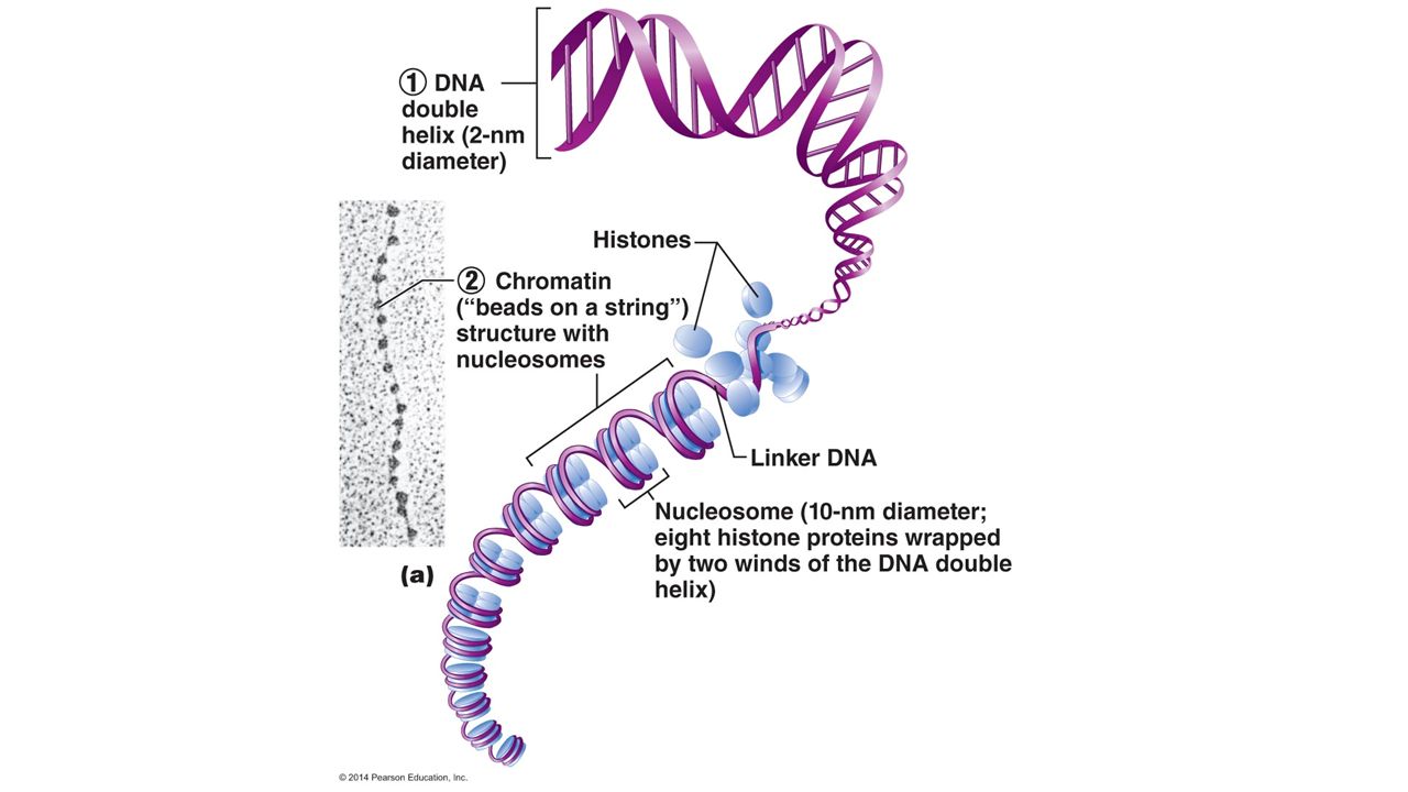
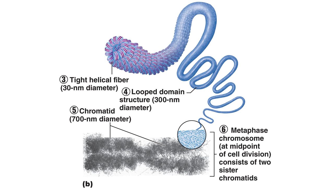
Terms to know: Microscope: Mitosis : Interphase Mitosis Cytokinesis Phases: prophase, metaphase, anaphase, telophase Chromosomes Chromatids Centromere Spindle Base Substage light Stage Condenser Iris diaphragm lever Coarse adjustment knob Fine adjustment knob Head Arm Ocular lens Nosepiece Objective lenses Scanning power Low-power High-power Oil immersion

4
Care and Structure of the Compound Microscope Carry microscope upright with one hand on microscope base and one hand on microscope arm, avoid jarring Use lens paper and lens cleaner only Always begin on the lowest power objective Never use coarse focus knob on high or oil immersion power Always use a coverslip with wet mount slips Before returning the microscope to storage, remove slide, return to low power, and replace cover Inform your instructor if mechanical problems occur

5
Activity: Identify parts of a Microscope Iris diaphragm control knob

6
Determining Total Magnification Total magnification of a specimen is equal to the power of the ocular lens multiples by the power of the objective lens Resolution is the ability to discriminate two close objects as separate Resolving power (RP) increases with increasing light, so you need to increase light intensity at higher magnifications Ocular lens=_____XScanning powerLow powerHigh powerOil Immersion Magnification of Objective lens______X_____X______X Total Magnification______X Field size (diameter)______μm Working distance______mm
 increases with increasing light, so you need to increase light intensity at higher magnifications Ocular lens=_____XScanning powerLow powerHigh powerOil Immersion Magnification of Objective lens______X_____X______X Total Magnification______X Field size (diameter)______μm Working distance______mm")
7
Activity Viewing Objects through the Microscope On scanning (40X) power, use a mm ruler to measure the “working distance” between the specimen (letter “e”) and the objective lens. Record the distance on page 369, sketch the “e” on scanning power. Move the “e” and observe the motion of the image Focus with the fine focus knob on low and high power

8
Activity Determining the Size of the Microscope Field of View Measure the field of view on scanning power using a mm ruler 1000μm in 1 mm To find the diameter of the low and high power fields of view, use the formula: diameter (scanning) X mag. (scanning) = diameter (low) X mag. (low) 40 x 2.5 mm = 100 X _____
 = diameter (low) X mag. (low) 40 x 2.5 mm = 100 X _____.")
9
Perceiving Depth Determine with thread is on bottom and which tread is on top of the stack of three by focusing up through the stack. Repeat on low and high power

10
Preparing and Observing a Wet Mount Prepare a wet mount slide of you cheek cells using a toothpick, saline, methylene blue, a clean slide and cover slip. Measure your cell under 400X

11
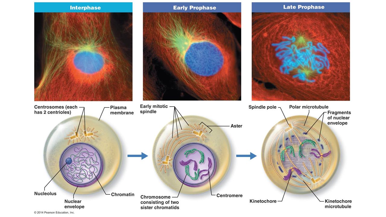
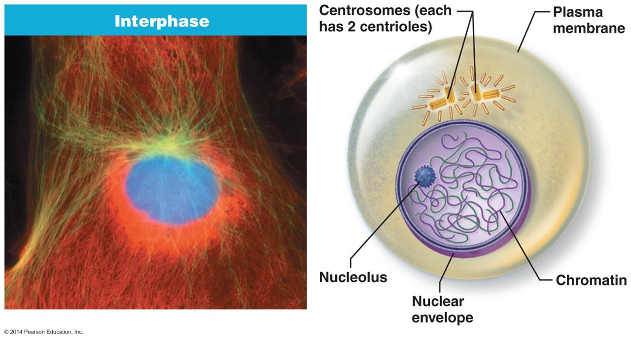
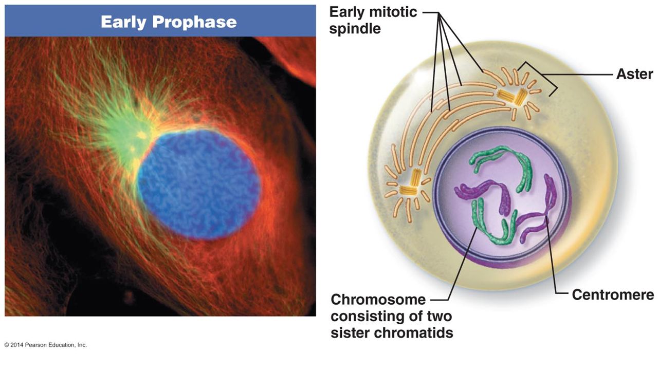
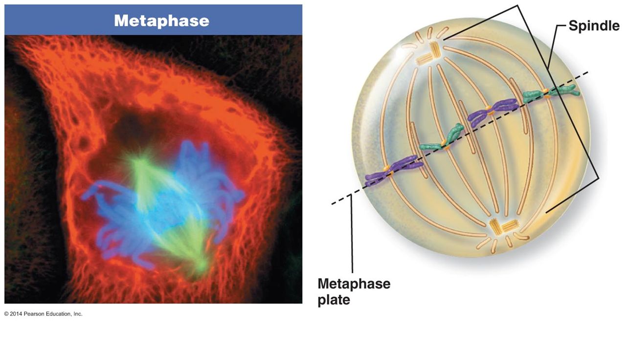
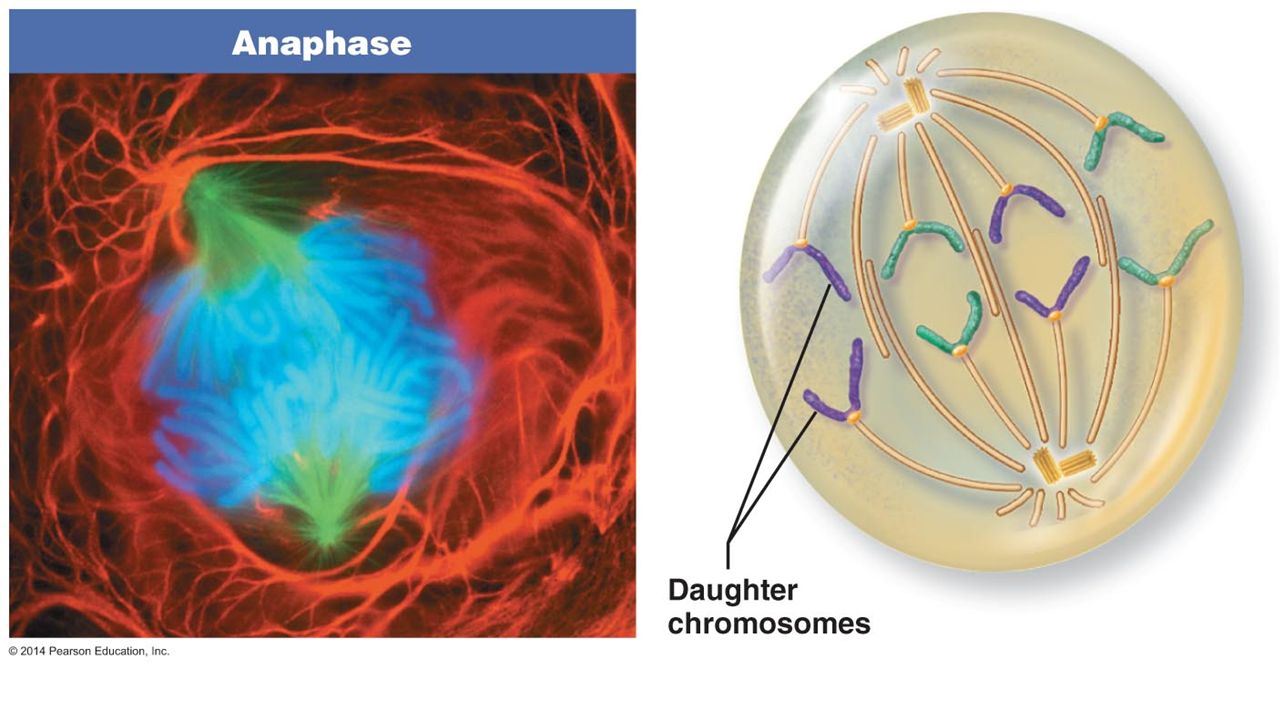
Lab Exercise 3 (p. 24) Activities 6 and 7 Identifying Mitotic Stages and Creating Figures Observe whitefish blastulae (embryo) cells in various stages of mitosis. Interphase-cell conducts normal life process and replicates DNA in preparation for mitosis Mitosis consists of the following stages: Prophase-chromosomes condense, nuclear envelop disintegrates, mitotic spindles form from centrioles Metaphase-chromosomes are aligned along metaphase plate by spindle apparatus Anaphase-sister chromatid separate at centromere and chromosomes are pulled to opposite poles toward newly forming cells. Cytokinesis begins. Telophase-new nuclear envelops form as chromosomes uncoil. Cytokinesis is completed. Create mitotic figure using pipe cleaners to represent chromosomes
 Activities 6 and 7 Identifying Mitotic Stages and Creating Figures Observe whitefish blastulae (embryo) cells in various stages of mitosis. Interphase-cell conducts normal life process and replicates DNA in preparation for mitosis Mitosis consists of the following stages: Prophase-chromosomes condense, nuclear envelop disintegrates, mitotic spindles form from centrioles Metaphase-chromosomes are aligned along metaphase plate by spindle apparatus Anaphase-sister chromatid separate at centromere and chromosomes are pulled to opposite poles toward newly forming cells. Cytokinesis begins. Telophase-new nuclear envelops form as chromosomes uncoil. Cytokinesis is completed. Create mitotic figure using pipe cleaners to represent chromosomes.")
25
Interphase

26
Prophase

27
Metaphase

28
Anaphase

29
Telophase

30
Practice

31
For Review Complete pages 367-373 (all) Complete pages 25-27 1-6
 Complete pages")
Similar presentations


 Spring 2010 Prof. AnnMarie Armenti, MS.>")

 Microscope>")


, as well as the cell.>")


